E.P.
Tikhonova, S.V. Lipnyagova, T.Yu. Kuzmina, S.V. Levitsky Department of Infectious Diseases and Epidemiology Krasnoyarsk State Medical University Municipal Clinical Hospital No. 6, Krasnoyarsk
The problem of opisthorchiasis in our region continues to be very relevant for both infectious disease specialists and general practitioners. In the structure of parasitic morbidity, opisthorchiasis in the adult population is 84.5%. Early diagnosis at the first examination of the patient presents great difficulties due to the peculiarities of the clinical picture of the disease, often the lack of data from laboratory and instrumental studies. The purpose of our message was to remind about this helminthiasis and present an interesting case from practice.
Opisthorchiasis
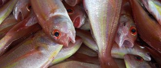
(code according to ICD10-B66.0) – natural focal biohelminthiasis, characterized by damage to the hepatobiliary system and pancreas. Humans are parasitized by the cat (Siberian) fluke Opisthorchis felineus and Opisthorchi viverrini (squirrel fluke). The definitive hosts are humans, cats, dogs, and civets (family Viverridae of the order of carnivorous mammals). Intermediate hosts are various species of mollusks of the genus Codiella and others; additional hosts are fish of the carp family - ide, dace, chebak, European roach, roach, tench, rudd, carp, bream, silver bream, podust, asp, bleak.
Source of infection
are people infected with opisthorchiasis, as well as domestic and wild carnivores whose diet includes fish. Human infection occurs through consumption of raw or undisinfected fish containing viable metacercariae by heating, freezing, or salting.
The world's largest natural focus of opisthorchiasis is located in the Ob-Irtysh basin; the highest incidence rates of the population are recorded here (up to 500 per 100 thousand). In the basins of the Northern Dvina, Kama, Volga, Ural, Don, Dnieper, Desna, Southern Bug, Northern Donets and Neman rivers there are also natural foci of opisthorchiasis, but they are less intense.
The main foci of opisthorchiasis in the Krasnoyarsk region are the five territories of the Chulym region, where up to 70% of all cases of opisthorchiasis in the region are registered annually. An increase in the incidence of opisthorchiasis was noted in 19 territories. The incidence has increased not only in the endemic areas of the Chulym basin, but also in a number of territories of the region due to population migration and an increase in imported cases.
Every year, from 20,050 to 26,100 cases of parasitic diseases are registered in the Krasnoyarsk Territory. In the general structure of infectious and parasitic diseases, they occupy second place and account for 3.6%. Opisthorchiasis is also detected in a number of European countries - France, Bulgaria, Holland, Italy, Switzerland, Sweden, Poland and Romania.
The main role in the pathogenesis of opisthorchiasis is played by:
– allergic reactions
(especially pronounced in the early phase of the disease), which arise as a result of the release of metabolic products by helminths;
– mechanical impact of helminths
, which consists of damage to the walls of the bile and pancreatic ducts and gallbladder by suckers and spines covering the surface of the helminth’s body. The accumulation of parasites causes a slowdown in the flow of bile and pancreatic secretions;
– neuro-reflex influences
through helminth irritation of the nerve elements of the ducts, resulting in pathological nerve impulses transmitted primarily to the stomach and duodenum;
– occurrence of conditions ( biliary dyskinesia
, accumulation of parasites, eggs, desquamated epithelial cells in them, temporary and complete cessation of bile flow), favorable for the addition of a secondary infection of the biliary tract;
– glandular proliferation
epithelium of the bile and pancreatic ducts, which should be considered as a precancerous condition.
What is opisthorchiasis
Opisthorchiasis is a parasitic disease characterized by damage to the intrahepatic bile ducts, gallbladder, and pancreatic ducts.
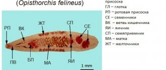
It is caused by flat, lanceolate parasites, a class of flukes (in Russia most often the helminth Opisthorchis felineus, or cat fluke). This helminth changes hosts three times - mollusks, freshwater fish of the carp family (bream, ide, dace, sabrefish, roach, carp, etc.) and the final host - predators, cats, dogs, humans that feed on fish. They release eggs in their feces. When they enter a body of water, the mollusks swallow the eggs and the worm’s development cycle begins.
Children suffer from opisthorchiasis less often than adults; it is more often found in men 15-50 years old. A person becomes infected by eating raw, lightly salted, dried or insufficiently heat-treated river fish of the carp family containing infective larvae. In the stomach and the initial part of the duodenum, the cyst shell dissolves, and its contents, the larva, pass into the gallbladder, common bile duct and smaller ducts. In them the parasite matures into a mature individual. The full development cycle takes 4-4.5 months, and the fluke can be a parasite in the human body for up to 25 years.
Treatment
Opisthorchiasis therapy is carried out in several stages.
This is the only way to get rid of the symptoms of the disease and fully recover. First, the patient takes choleretic drugs and antispasmodics under strict medical supervision. If necessary, antibacterial drugs may be prescribed. Physiotherapy serves as an auxiliary method.
The goal of everything is to normalize the secretion of bile and get rid of the inflammatory process in the gastrointestinal tract.
Next, the main treatment begins, which involves getting rid of the parasite. The patient is prescribed anthelmintic therapy, where the drug Praziquantel and its analogues have proven to be very effective.
Monitoring of effectiveness is carried out by three-time examination of stool and contents obtained through duodenal intubation.
At the last stage of treatment of opisthorchiasis, restorative therapy is carried out using prokinetics, hepatoprotectors, sorbents, as well as probiotics and prebiotics.
All treatment is carried out against the background of diet number 5, which excludes the appearance of fried, smoked or spicy foods on the table.
If you suspect chronic opisthorchiasis, or in the case of an already confirmed diagnosis, you can get an in-person or telemedicine consultation with an infectious disease specialist, gastroenterologist at the New Hospital in Yekaterinburg.
If indicated, hospitalization is possible in the gastroenterology department, where they have been successfully treating chronic opisthorchiasis for a long time.
Using the most modern diagnostic and therapeutic tools, we help patients get rid of this unpleasant disease and eliminate a number of symptoms associated with it.
Why is opisthorchiasis dangerous?
When infected with opisthorchiasis, a reaction first occurs to the release of parasitic toxins:
- increased vascular permeability;
- swelling of soft tissues and internal organs;
- rashes and skin itching;
- allergic reactions.
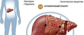
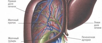
Since the main site of parasitism of the liver fluke is the gallbladder, the bile ducts, to which the helminth is attached, inflammation develops in their walls (cholecystitis, cholangitis), the movement of bile slows down and is blocked. There may be blockages of small ducts by helminths and the formation of stones.
At the same time, the outflow of pancreatic juice from the pancreas is disrupted with the expansion of its ducts and the proliferation of connective tissue (fibrosis). The most severe forms occur with destruction and cirrhosis of the liver, peritonitis (inflammation of the peritoneum). The long-term, long-term presence of the liver fluke in the biliary system can result in liver cancer, gall bladder cancer, and pancreatic cancer.
Medicine for opisthorchiasis in adults
For the purpose of deworming, a doctor may prescribe one of the following drugs (the active ingredient in all of them is praziquantel, which helps get rid of the fluke):
- cysticide;
- biltricide;
- cesol;
- azinox;
- bithionol;
- chloroxyl
Important! This group of drugs cannot be used in the early stages of gestation. When prescribed to lactating women, you should refrain from breastfeeding for two days after taking praziquantel tablets.
Symptoms of opisthorchiasis
2-4 weeks after infection, body temperature rises to 37.8-39 degrees, then it drops to 37.1-37.3 degrees and lasts 7-20 days. Fever is accompanied by chills, weakness, abdominal pain, and diarrhea.
For more severe cases, the following are added:
- muscle and joint pain;
- urticaria, with the appearance of papules (red nodules) on the skin, and then blisters of various sizes;
- cough;
- nausea, vomiting;
- enlarged lymph nodes;
- jaundice;
- pain in the right like hepatic colic or shingles;
- disorders of the heart and nervous system.
Symptoms and forms of the disease
The disease occurs in two forms: acute and chronic. The intensity of the course can be different and depends on the degree of invasion, the state of immunity, and the duration of infection of the body.
Symptoms of acute opisthorchiasis:
- high body temperature;
- aching pain in muscles and joints;
- spasms in the right hypochondrium;
- loss of appetite, loss of sleep;
- heartburn, nausea, vomiting;
- allergic rashes;
- eczema.
Symptoms of chronic opisthorchiasis:
- urticaria, itching on the skin;
- signs of dyspeptic syndrome;
- spasms in the gallbladder;
- girdle pain in the lower back;
- arthralgia, Quincke's edema;
- irritability, fatigue;
- styes, boils near the eyes;
- tremor of the tongue, fingers, hands;
- other diseases of organs and systems.
The incubation period ranges from 7 to 31 days. Acute opisthorchiasis can last 4-10 weeks, chronic - up to 20 years.
With an erased form, the disease may be asymptomatic and there will be no signs of intoxication of the body.
Diagnosis of the disease opisthorchiasis
To make a diagnosis, take into account:
- Staying of a person in the center of opisthorchiasis.
- Eating half-raw, frozen, lightly salted fish.
- Patient complaints (fever, abdominal pain, diarrhea).
- Instrumental diagnostic data (ultrasound, cholangiography, tomography): signs of duodenitis, biliary dyskinesia, cholecystitis, cholangitis, pancreatitis, hepatitis.
- Laboratory diagnostic data: increased liver enzymes, bilirubin in a biochemical blood test, increased leukocytes, eosinophils, ESR in a clinical blood test.
To confirm opisthorchiasis, prescribe:
- microscopy of duodenal contents;
- feces on worm eggs;
- molecular genetic study of feces and bile for the presence of helminth DNA using PCR;
- blood test for antibodies to opisthorchiasis proteins.
Folk remedies for opisthorchiasis
Traditional medicine recipes aim to reduce the appearance of sexually mature worms, create conditions in which parasites cannot live and reproduce, and in some cases are aimed at destroying flukes. The advantage of this therapy is that the plants contain substances that help restore the epithelial lining of the digestive organs and restore their normal functioning. It remains to figure out how to get rid of parasites using specific means.
ethnoscience
Proper nutrition
Nutrition should be done in a gentle manner, but have a stimulating effect on the secretory activity of the digestive system. It is not recommended to eat very hot or cold foods. Meals should be fractional, often, but in small portions.
What foods should be included in the diet:
- fruit and berry juices;
- compotes, jelly;
- tomato juice without added salt;
- weak tea, coffee with milk only;
- infusion of rose hips;
- stale bread;
- low-fat, mild cheese;
- low-fat dairy products;
- vegetable puree soup;
- a spoonful of butter per day;
- unhealthy cookies;
- steamed one-egg omelette;
- honey, fruit jam;
- porridge cooked in water;
- lean meats;
- lean fish and poultry;
- soaked herring;
- apples, pears, plums, peaches, apricots.
Food should be steamed or baked. Avoid adding excess salt and spices. Do not fry foods, do not use oil in the cooking process.
What should be excluded from the diet:
- baked goods;
- confectionery;
- soups with meat or fish broth, mushroom soups;
- fatty types of meat and fish products;
- boiled or fried egg;
- pickled, salted vegetables;
- sausages;
- canned food;
- ice cream, chocolate;
- alcoholic drinks.
Birch tar
This method is considered quite effective. It does not require significant costs or any special training.
Advice! It is better to take the medicine in the morning before breakfast.
The essence of the method: you need to take two drops of birch tar and mix with two tablespoons of boiled water. Every day the dosage should be increased by 1-2 drops until it reaches ten drops per day. Then the quantity is reduced in reverse order. The treatment course lasts about three weeks. Aspen bark
Aspen bark contains phenols, which have a toxic effect against parasites. The anthelmintic effect is based on this. The dose of toxic compounds is so small that it has no side effects on the human body. There are a number of studies that prove the therapeutic effectiveness of this drug. Aspen bark can be prescribed to both adult patients and children. To enhance the effect and speed up the achievement of a positive result, it is recommended to supplement treatment with choleretic preparations.
Recipes:
- Preparation of the decoction. Take 100 g of crushed bark and add 1 liter of cold water. Next, bring to a boil over medium heat and cook for 15 minutes. Then remove, cool and leave to infuse for about 4 hours. Take 50-100 ml 30 minutes before meals.
- Preparation of tincture. You need to pour 100 g of dry matter into 1 liter of vodka (you can take medical alcohol and dilute it with water). Leave to infuse in a dark and warm place for about 3 weeks. You need to drink 1-2 tbsp. l. before meals. Cannot be used to treat opisthorchiasis in children.
The course of treatment takes 3-4 weeks. If there is a need for repeated therapy, then it is necessary to take a break for a month.
Herbs
A complex of medicinal herbs can rid the human body of the fluke present in it, help the body recover as a whole, improve its functioning, remove toxic substances produced by helminths, and erect a protective barrier. As a rule, the preparations contain components that have anti-inflammatory, reparative, choleretic and antiparasitic effects.
Herbal treatment
List of plants from which you can independently or on the recommendation of a doctor make an individually selected collection:
- plantain leaves;
- calendula flowers;
- St. John's wort inflorescences;
- tansy;
- wormwood leaves;
- thyme leaves;
- clover flowers.
To prepare the decoction you will need 2 tbsp. l. collection They need to be poured with 1.5 cups of boiling water and allowed to brew. It is recommended to consume 150 ml per day. You can undergo treatment for as long as you like. A lasting therapeutic effect will be achieved after the first month of therapy.
Pumpkin seeds
When using pumpkin seeds to treat opisthorchiasis, you can not only get rid of parasites, but also satisfy your hunger.
Recipes:
- Millet porridge is cooked in water. Take the herring and remove all the bones. Then the herring and pumpkin seeds are passed through a meat grinder. When ready, mix with millet porridge. The dish is consumed in the morning on an empty stomach. After this, it is recommended to take a laxative or perform a cleansing enema. The duration of the course is as follows: three doses of porridge, with a 7-day break between them.
- Take crushed pumpkin seeds, 3 yolks, 2 tbsp. l. flower honey and a little water. All ingredients are mixed until a homogeneous consistency is obtained. The medicine is taken on an empty stomach 20 minutes before the morning meal. The course is 10 days, then a 7-day break. Thus, it is necessary to repeat the treatment three times.
Garlic and cloves
Garlic is rightfully considered a universal product. It helps in the fight against helminths, bacterial and viral infections, and helps restore the immune barrier.
Getting rid of flatworms can be done at home. For this you will need cloves and young garlic.
The essence of the technique: 5 cloves of garlic are crushed manually or with a special device and poured with 1.5 glasses of low-fat milk. The resulting liquid must be poured into an enamel pan, put on fire and brought to a boil. After this, 5-6 inflorescences of dried cloves are added. When ready, remove the mixture from the heat and cool. Before you start taking it, you must pass it through several layers of gauze or through a sieve. The medicine is taken 30 minutes before meals. The course of treatment is two weeks. Black Walnut
Black walnut is able to fight the proliferation of helminths, destroy laid eggs and remove waste products and decay products of worms from the body.
Method of preparation: nuts are collected unripe while their shell is soft. Take any container and completely fill it with whole fruits. After this, the bottle must be filled with vodka or diluted medical alcohol. Place in a dark place and leave for about 3 weeks. Chanterelles
Another effective remedy in the fight against opisthorchiasis can be a tincture of chanterelle mushrooms.
Appearance of the parasite
Method of preparation: wash the mushrooms and grind through a meat grinder. To complete the recipe you only need 4 tbsp. l. chanterelles, to which you need to add 300 ml of vodka. The tincture should be kept for 3 weeks.
The course of therapy is one month. Drink chanterelle tincture 2 tsp. before bedtime. Shake the mixture before drinking.
Helminthiasis
Parasitic worms found in the body of humans, animals and plants cause diseases called helminthiasis. Human parasitic worms disrupt the functioning of internal organs and the entire body as a whole, causing poor absorption of nutrients, weakness and diseases of the host body. There are three groups of worms that are classified as helminths:
- Nematodes or roundworms (pinworms, roundworms, Trichinella, Filaria, Intestinal eel);
- Trematodes or flatworms (Opisthorchis, Chinese fluke, Paragonimus, Schistosomes);
- Cestodes or tapeworms (Bovine and pork tapeworms, broad tapeworms, echinococcus, alveococcus).
Helminths have a similar morphology and are multicellular organisms that are visible to the naked eye. In their life cycle, helminths necessarily go through a number of stages: egg, larva and adult. During their life cycle, some worms change hosts. Such helminths are called biohelminths. Geohelminths are parasitic worms that do not require a change of hosts, and their life cycle occurs in the body of a single host. NEMATODOSIS Ascariasis Etiology, pathogenesis .
The causative agent is roundworm, which parasitizes in the adult stage in the small intestine. The lifespan of roundworms is about a year. In the migratory stage (the first 6–8 weeks after infection), roundworm larvae have a mechanical and sensitizing effect, causing hemorrhages and eosinophilic infiltrates in the tissues of various organs. In the intestinal phase (8 weeks after infection), adult roundworms cause general toxicoallergic and neuroreflex reactions of the body and a variety of local mechanical effects.
Symptoms, course
. The migration phase often occurs under the guise of acute respiratory infections and bronchitis; volatile eosinophilic infiltrates in the lungs are possible. In the intestinal phase, a gastrointestinal form is distinguished (salivation, nausea, loss of appetite, cramping pain around the navel, sometimes disorders of stool and gastric secretion); hypotonic (decreased blood pressure, weakness) and neurological (dizziness, headache, fatigue, sleep disturbance, vegetative-vascular disorders) forms.
Complications
. Ascariasis intestinal obstruction, a characteristic feature of which is the release of roundworms with vomit and palpation of a moving “tumor” - a ball of helminths; ascariasis appendicitis; perforated peritonitis; ascariasis of the liver with the development of jaundice, purulent angiocholitis, liver abscess, subphrenic abscess; ascariasis of the pancreas with symptoms of acute pancreatitis; crawling of roundworms into the respiratory tract with the development of asphyxia.
Diagnostics.
The diagnosis of the early phase of ascariasis is based on the detection of nematode larvae in the sputum and antibodies in the blood, and the late intestinal phase - ascaris eggs in the feces.
Prevention
.
Mass examination of the population and treatment of all those infected with ascariasis. Protection of the soil of vegetable gardens, orchards, and berry fields from contamination with feces. Thorough washing and scalding of vegetables and fruits with boiling water. Personal hygiene measures .
Hookworm disease (hookworm and necatoriasis)
Etiology, pathogenesis.
The causative agents are hookworm and necator, parasitic in the small intestines of humans, most often in the duodenum. Infection occurs when the larvae actively penetrate the skin or when they are ingested with contaminated vegetables, fruits, or water. The larvae migrate through the systemic and pulmonary circulation, lasting 7–10 days. In the small intestine, the larvae turn into sexually mature individuals and after 4–6 weeks begin to lay eggs. The lifespan of hookworms ranges from several months to 20 years. During the migration period, the larvae cause toxic-allergic phenomena. Adult helminths are hematophagous. When fixed to the intestinal mucosa, they injure tissue, lead to the formation of hemorrhage, erosion, cause bleeding, anemia, support the state of allergies, gastrointestinal dyskinesia and dyspepsia.
Symptoms, course
. Soon after infection, skin itching and burning, urticarial rashes, asthmatic phenomena, fever, and eosinophilia occur. In the late stage, nausea, drooling, vomiting, abdominal pain, intestinal dysfunction (constipation or diarrhea), and bloating appear. Sometimes pseudoulcer syndrome occurs (hunger pain in the epigastric region, hidden bleeding). The development of hypochromic iron deficiency anemia is characteristic; its severity depends on the intensity of invasion, nutritional deficiency, and individual characteristics of the organism. With low intensity, the invasion can occur subclinically, manifesting itself as eoeinophilia.
Diagnostics
. The diagnosis is confirmed by the detection of eggs in the stool and occasionally in the duodenal contents.
Prevention
. In areas of hookworm infection, you should not walk barefoot or lie on the ground without bedding. It is necessary to thoroughly wash and scald fruits, vegetables, and berries with boiling water; before eating them, you should not drink unboiled water.
Strongyloidiasis
Etiology, pathogenesis.
The causative agent is the intestinal eel, parasitizing in the intestinal wall (mainly the duodenum), sometimes in the ducts of the liver and pancreas, and during the migration period - in the bronchi and lung tissue. The penetration of Strongyloides larvae into the human body can occur actively through the skin (when walking barefoot, etc.) and through the mouth (when eating contaminated fruits, vegetables, and also when drinking water). Adult parasites, localized in the intestinal wall, injure the intestinal crypts (Lieberkühn's glands), solitary follicles and contribute to ulceration of the mucous membrane, and the larvae, migrating, damage the tissue of the liver, lungs and other organs. Of great importance is the toxic-allergic effect of adult parasites and their larvae on the human body, as well as secondary infection.
Symptoms, course
. Signs of allergy often occur: skin itching, burning, urticaria, eosinophilia, sometimes “volatile infiltrates” in the lungs, etc. In the late stage, the symptoms of gastroduodenitis, enterocolitis, and sometimes cholecystitis predominate. The typical triad is chronic recurrent urticaria, persistent enterocolitis and long-term high eosinophilia. Dizziness, headache, insomnia, and increased excitability are common. Sometimes the disease manifests itself only as eosinophilia and skin itching. The course is long, many years (until specific treatment), since with this helminthiasis (especially with constipation), autoreinvasion is possible. There are indications of a connection between hyperinvasion and the use of corticosteroids and immunosuppressive drugs. In severe forms of the disease, ulcerative enterocolitis develops with dehydration, cachexia, anemia and death.
Diagnostics
. The diagnosis is based on clinical data and the detection of acne larvae in feces (when examined using the Berman method) and in duodenal contents, less often in sputum and urine.
Trichocephalosis
Etiology, pathogenesis.
The causative agent is the whipworm, which parasitizes the human colon. The lifespan of the parasite is about 5 years. Whipworms injure the intestinal mucosa, are hematophagous and promote inoculation of microflora, causing reflex reactions in other organs of the abdominal cavity. Their metabolic products sensitize the body.
Symptoms, course
. Worries include drooling, decreased (less often increased) appetite, pain in the right half of the abdomen and epigastrium, nausea, constipation or diarrhea. There may be inflammatory phenomena in the intestines. Headache, dizziness, restless sleep, irritability are often observed; moderate hypochromic anemia and slight leukocytosis are possible. With intense invasion in children, cases of rectal prolapse (due to persistent diarrhea), epileptiform seizures, and Meniere's syndrome have been described. At low intensity, whipworm infestation is asymptomatic.
Diagnostics.
The diagnosis is made when whipworm eggs are found in the stool.
Prevention
. Mass examination of the population and treatment of all infested forms. Protection of the soil of vegetable gardens, orchards, and berry fields from contamination with feces. Thorough washing and scalding of vegetables and fruits with boiling water. Personal hygiene measures.
Trichostrongylidosis
The causative agents are small helminths from the family Trichostrongylidae. The main source of invasion is sheep, goats, and cattle. A person becomes infected through contaminated food or water.
Symptoms, course
. Characterized by nausea, abdominal pain, unstable stools, dizziness, and headache. Sometimes hypochromic or normochromic anemia and loss of strength develop. The diagnosis is based on the detection of trichostrongylid eggs in the stool.
Prevention.
Compliance with personal hygiene measures.
Trichinosis (trichinosis)
Etiology, pathogenesis
. The causative agent is Trichinella. In the larval stage, it enters the human body with the meat of pigs or wild animals (boar, bear, badger, etc.). After digestion of meat, muscle Trichinella are released from capsules, penetrate into the villi tissue, quickly grow, develop, and already 80–90 hours after infection, females lay numerous larvae, which are spread throughout the body. The place of further development of young Trichinella are the striated muscles, especially the intercostal, masticatory, muscles of the diaphragm, tongue, pharynx, and eyes. Here, after 2–3 weeks, trichinella coil up, become encapsulated, and some of them become calcified. The duration of the intestinal stage of Trichinella is about 2 months. Muscular trichinella remain viable for up to 20 years or more. The breakdown and metabolic products of Trichinella sensitize the body, causing eosinophilia, capillaropathy, muscle infiltrates, edema and other painful phenomena. The mechanical effects of adult Trichinella in the intestines and their larvae in muscles and other tissues, as well as secondary infection, are important.
Symptoms, course
. The incubation period is from 3 to 45 days. Early symptoms are characteristic: facial puffiness, accompanied by conjunctivitis, fever, muscle pain, eosinophilia. Various skin rashes are common, as well as gastrointestinal disorders, hypotension, muffled heart sounds, palpitations, headache, and insomnia. In mild forms, only eyelid swelling and eosinophilia are noted. The duration of the disease is from 2–3 days to 8 weeks or more. Sometimes a recurrent course of trichinosis is observed. Complications: myocarditis, meningoencephalitis, thrombosis of arteries and veins, pneumonia, nephritis, etc.
Diagnostics.
The diagnosis is based on epidemiological (consumption of undercooked pork and other meat, group diseases), characteristic clinical data (eosinophilia, swelling of the eyelids, fever, muscle pain). Serological reactions with trichinosis antigen are used. Confirmation of the diagnosis can be the detection of Trichinella and characteristic infiltrates in biopsied pieces of muscle (trapezius, deltoid, gastrocnemius) or in the remains of meat that caused the disease. A biopsy is performed on the 9th–10th day of illness.
Prevention.
You should not eat raw or undercooked pork, as well as the meat of wild boar, bear, badger, etc.
TREMATODOSIS Clonorchiasis
Clonorchiasis is a helminthiasis of the liver and pancreas caused by the fluke fluke. It is found among the population living in the Amur basin. Pathogenesis, clinical picture, diagnosis, treatment and prevention are the same as for opisthorchiasis.
Metagonimiasis
Metagonimiasis is a helminthiasis caused by a small trematode. The pathogen parasitizes in the small intestine of humans, dogs, cats, and pigs. Human infection occurs through consumption of raw fish. In the intestine, larvae hatch from the metacercariae of Metagonimus, which reach sexual maturity in the thickness of the mucous membrane after 2 weeks and emerge into the intestinal lumen. As a result of mechanical and toxicoallergic effects, enteritis develops.
Symptoms, course
. Abdominal pain, drooling, persistent diarrhea. Sometimes it occurs subclinically.
Diagnostics
. The diagnosis is based on the detection of Metagonimus eggs in the stool.
For prevention, see Diphyllobothriasis.
Nanophyetosis
Nanophyetosis is an intestinal helminthiasis that occurs among the population of the Far East. The causative agent is nanophyetus, which parasitizes the intestines of humans, dogs, cats, minks, and badgers. Infection occurs by eating raw fish. Helminths have a mechanical and toxic-allergic effect on intestinal tissue.
Symptoms, course.
Pain and rumbling in the abdomen, diarrhea, constipation, drooling and nausea at night.
Diagnostics
. Diagnosis is made based on the detection of nanophyetus eggs (resembling diphyllobothrid eggs) in the stool.
Treatment
. Male fern extract in a dose of 3–3.5 g.
Prevention
. You should not eat raw, uncooked or insufficiently salted and dried fish, as well as “live” pike caviar.
Opisthorchiasis
Etiology, pathogenesis
. The causative agent is the cat fluke, or Siberian fluke, which parasitizes the bile ducts of the liver, gallbladder and pancreatic ducts of humans, cats, dogs, etc. The parasite lives in the human body for 20–40 years. Infection occurs when eating raw (thawed, frozen), lightly salted and insufficiently fried carp fish (ide, chebak, dace, etc.). Opisthorchises injure the mucous membranes of the pancreatic and bile ducts, create obstacles to the outflow of bile, and contribute to the development of cystic enlargements and liver tumors. They have toxic and neuro-reflex effects. In the early stage, pronounced allergization of the body is observed (eosinophilic-leukemoid blood reactions).
Symptoms, course
. The incubation period is about 2 weeks. In the early period there may be fever, pain in muscles and joints, vomiting, diarrhea, pain and enlargement of the liver, less often the spleen, leukocytosis and high eosinophilia, allergic skin rashes. In the chronic stage, the most common complaints are pain in the epigastric region, right hypochondrium, radiating to the back and left hypochondrium. Sometimes there are attacks of pain such as gall bladder colic. Frequent dizziness and various dyspeptic symptoms. Muscle resistance in the right hypochondrium, liver enlargement, occasionally icteric sclera, enlarged gallbladder, and symptoms of pancreatitis are detected. Most often, with opisthorchiasis, the phenomena of angiocholitis, cholecystitis, biliary dyskinesia, chronic hepatitis and pancreatitis develop, and less often - symptoms of gastroduodenitis and enterocolitis. In some cases, a picture of cholangitic cirrhosis of the liver occurs. Opisthorchiasis can be asymptomatic.
Diagnostics
. The diagnosis is based on the detection of helminth eggs in the stool and duodenal contents.
Prevention
. Explaining to the population the dangers of eating raw, thawed and frozen (stroganina), lightly salted and insufficiently fried fish.
Fascioliasis
Etiology, pathogenesis.
Pathogens: liver fluke and giant fluke. The main source of human invasion is various farm animals. Human infection usually occurs in the warm season when fasciola larvae are ingested in water, sorrel, lettuce and other greens. The lifespan of helminths in the body is about 10 years. Trauma and toxic-allergic damage to the hepatobiliary system are important. Fasciolae can be carried into other tissues and organs.
Symptoms, course
. The disease is characterized by eosinophilia, allergic phenomena, disorders of the liver and gall bladder, reminiscent of the symptoms of opisthorchiasis (jaundice and attacks of gall bladder colic are more common).
Diagnostics
. Diagnosis of the early stage of fascioliasis is difficult, since helminth eggs are released only 3–4 months after infection. Immunological methods are used. In the late stage, the diagnosis is based on the detection of fasciola eggs in the duodenal contents and feces.
Prevention
.
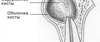
Prohibition of drinking water from stagnant bodies of water; Thoroughly washing and scalding the greens with boiling water. CESTODOSES Alveococcosis Etiology, pathogenesis.
The causative agent is the larval stage of alveococcus. Infection occurs after oncospheres enter the mouth after contact with contaminated skins of foxes, arctic foxes, dogs, with the water of stagnant reservoirs and by eating wild berries collected in endemic areas. Clusters of larvae (usually in the liver) infiltrate and grow into tissues, disrupt the blood supply to organs, cause tissue degeneration and atrophy, and have a toxic-allergenic effect.
Symptoms, course
. The disease develops slowly and remains asymptomatic for a long time. There is a progressive enlargement of the liver, heaviness and pressure appear in the right hypochondrium, and a dull aching pain. After a few years, the liver becomes lumpy and very dense. Jaundice may develop. The spleen often becomes enlarged. Possible ascites. When the nodes disintegrate, body temperature rises, sweating, leukocytosis, eosinophilia, and increased ESR are observed. Hyperproteinemia and hypergammaglobulinemia are characteristic. Necrotization and invasion into the inferior vena cava can lead to profuse bleeding. With metastases to the lungs, symptoms of pneumonia, bronchitis, and hemoptysis may occur. Brain metastases mimic the clinical picture of a brain tumor.
Diagnostics.
Diagnosis is based on clinical data. Serological tests with alveococcal antigen are performed. To clarify the localization, X-ray and ultrasound examination, liver scanning, peritoneoscopy, and computed tomography are used. Test puncture is strictly prohibited due to the risk of contamination of other organs. Differentiate from tumors, echinococcosis and cirrhosis of the liver.
Hymenolepiasis
Etiology, pathogenesis
. The causative agent is the dwarf tapeworm. Infection occurs through ingestion of parasite eggs that come into contact with the patient and with contaminated feces, household items (chamber pots, toilet seats, etc.) and restroom walls. The local effect of larvae and adult forms on the intestinal mucosa is expressed in the destruction of villi, proliferative inflammation (sometimes with ulceration) of the mucous membrane with copious secretion of mucus. Toxic-allergic effects are also observed.
Symptoms, course
. Decreased appetite, nausea, abdominal pain, diarrhea or constipation, dizziness, headache, irritability, increased fatigue, restless sleep. Sometimes weight loss, moderate anemia, and eosinophilia are observed. Hymenolepidosis can be asymptomatic.
Diagnostics _
. The diagnosis is made based on the detection of dwarf tapeworm eggs in the feces.
Prevention
. Maintaining hygiene of the body, clothing, home, and public hygiene. Examination of all members of the patient’s family for helminths and simultaneous treatment of all infested ones.
Diphyllobothriasis
Etiology, pathogenesis
. The causative agent is the broad tapeworm. Its lifespan is tens of years. Human infection occurs when eating fresh, insufficiently salted caviar and raw fish (pike, perch, omul, grayling, etc.). The tapeworm, attaching to the intestinal mucosa with its bothria, injures it. Large accumulations of the parasite can clog the intestinal lumen. Helminth metabolic products sensitize the body. Absorption of vitamin B12 by the broad tapeworm directly from the digestive tract leads to hypovitaminosis B12 and the development of anemia.
Symptoms, course
. Characterized by nausea, unstable stools, and the release of fragments of strobili during defecation. Patients sometimes complain of weakness, dizziness, and abdominal pain. Occasionally, pernicious anemia develops, close to Addison-Birmer anemia.
Diagnostics
. The diagnosis is made by detecting tapeworm eggs and strobila fragments in the feces.
Prevention
. You should not eat raw, uncooked or insufficiently salted and dried fish, as well as “live” pike caviar.
Taeniasis
Etiology. The causative agent is the pork tapeworm, which can parasitize humans not only in the sexually mature stage, but also in the larval stage, causing the disease cysticercosis. The adult helminth parasitizes the small intestine for many years. Humans become infected with taeniasis by eating raw or half-raw meat containing taeniasis.
Symptoms, course.
The same as with teniarhynchosis, however, the segments of the parasite do not actively come out of the anus (occasionally, tender segments come out with feces, which the patient usually does not notice).
Diagnostics
. The diagnosis is made on the basis of repeated examination of stool for the presence of helminth segments and mucus from the perianal folds (by scraping) for the presence of tapeworm eggs.
Prevention.
You should not eat undercooked or undercooked pork.
Diphyllobothriasis
Diphyllobothriasis is a helminthic disease affecting the digestive organs. The causative agent is the broad tapeworm. This is the largest of the human helminths, its length can reach 10 and sometimes 20 meters. The parasite consists of a head, neck and body. The head is oblong oval in shape, flattened on the sides and has on its narrow sides two longitudinal suction slits (bothria), with which the tapeworm is attached to the intestinal wall. The body consists of many segments, and the width is much greater than the length, which is due to the name of the parasite (wide tapeworm). The number of segments can reach 3000-4000 pieces. The tapeworm lives in the upper parts of the small intestines, feeds on the entire surface of the body, while absorbing various nutrients, including vitamins Bi2 and folic acid. The wide tapeworm is hermaphrodite. During the day, up to 2 million eggs are released into the external environment with feces. The number of parasites can reach up to 100 copies. The lifespan of parasites in the human body reaches 28 years.
For the development of a wide tapeworm, like opisthorchiasis, the presence of three hosts is necessary.
The definitive host is humans, domestic and wild animals. All of them secrete eggs, which end up in water bodies with meltwater. Diphyllobothriasis cannot be contracted through water.
The intermediate host is Cyclops (crustaceans). The eggs are swallowed by crustaceans (cyclops) and larvae develop in their bodies. Cyclops are swallowed as food by freshwater predatory fish.
Additional hosts are fish of predatory species: pike, burbot, perch, ruff; pike caviar is especially dangerous.
Having attached themselves to the wall of the intestines, the parasites injure the intestinal mucosa with bothria and can be one of the reasons for its necrosis. Sometimes intestinal blockage occurs.
Diphyllobothriasis occurs in mild or severe form, which is associated with the intensity of invasion, the presence of concomitant diseases and the general condition of the body. Sometimes the disease is asymptomatic.
In mild cases, patients complain of general weakness, poor appetite, nausea, pain and rumbling in the abdomen, intestinal disorders, and decreased ability to work.
In severe cases, intestinal obstruction occurs. In 2-3% of patients, a severe form of anemia (anemia) occurs. Patients complain of weakness, drowsiness, dizziness. Bright red spots and cracks appear on the tongue. The skin becomes pale with a yellowish tint; The liver and spleen may enlarge. Body temperature reaches 36-38 degrees.
The diagnosis of these diseases is established based on the detection of tapeworm and opisthorchid eggs in the feces.
Prevention
To prevent the development of opisthorchiasis, you must adhere to simple rules:
- do not eat raw or undercooked fish;
- fry fish products well for at least 20 minutes;
- when cooking fish, after the water boils, cook for another 20 minutes;
- when making salted fish, observe the salt norm per 1 kg of raw product, salt for at least 3 weeks;
- You can disinfect a raw product by deep freezing it;
- Observe personal hygiene rules after cutting fish.
Prevention
Now it is clear how to eliminate opisthorchiasis in adults. The choice of treatment method remains with the patient, but consultation with a doctor should not be neglected. The risk of developing serious complications if the approach to treatment is incorrect or therapy is not started in a timely manner is too high.