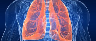
Structure of the human intestine
The concept of “intestine” combines two functionally different sections of the entire digestive system: the small and large intestines. Based on the name, it is not difficult to guess that the difference between the two sections is precisely the size of the intestinal lumen.
In turn, the small intestine has three sections. The first, located immediately after the stomach, is the duodenum. Its name is directly related to its size - its length is about 12 fingers. After it is the jejunum. Its diameter is slightly smaller than that of the first section. It lies in the peritoneum in loops. This section of the intestine is up to 2 meters in length. The small intestine ends with the ileum. The small intestine has smooth muscle throughout its entire length.
Its inner surface is lined with villi, which are responsible for the complete absorption of nutrients from the food bolus. It is in the small intestine that the processes of assimilation of food components take place, saturating the circulatory system with vitamins, minerals and other substances important for health. Enzymes released into the lumen of the duodenum from the pancreas and liver are responsible for the processing of food. In the following sections of the small intestine, broken down fats and proteins are processed by microorganisms that inhabit the inner surface of the hollow organ. These are mainly from the families of lactobacteria and bifidobacteria.
The large intestine is separated from the small intestine by a sphincter, which will not allow the food bolus to reverse. This department is responsible for the absorption of liquid from incoming products and the formation of feces from fibers and food fragments that the body does not need. It is in the large intestine that there is a section called the cecum, which has a process in the form of a conditional “dead end”. This dead-end branch was called the appendix.
The entire large intestine wraps around the small intestine along the edge of the abdominal cavity. Due to their location, they are divided into: ascending, transverse and descending colon. The large intestine ends with the rectum. It has a sphincter at the end called the anal ring.
Publications in the media
The gastrointestinal tract (GIT) performs not only a digestive, but also an immune function, in particular, it participates in the implementation of the body’s defense reactions against pathogenic, conditionally pathogenic microorganisms and many inorganic substances.
Local intestinal immunity
About 80% of all immunocompetent cells of the body are localized in the intestinal mucosa; about 25% of the intestinal mucosa consists of immunologically active tissue and cells; Each meter of intestine contains about 1010 lymphocytes [1].
The immunocompetent (lymphoid) tissue of the gastrointestinal tract is represented by organized structures (Peyer's patches, appendix, tonsils, lymph nodes) and individual cellular elements (intraepithelial lymphocytes, plasma cells, macrophages, mast cells, granulocytes). The population of lymphoid tissue cells is heterogeneous and consists of many groups, subgroups and clones of cells with different functional properties and specificity of receptors for antigens [2, 3].
The epithelium of the gastrointestinal tract delimits the tissues of the macroorganism from a huge number of living and non-living antigens - substances that carry signs of foreign genetic information. Oral exposure to an antigen (including microbes and their toxins) usually creates, on the one hand, local “mucosal” IgA protection (secretory immunity) and a cell-mediated reaction, but, on the other hand, systemic tolerance or hyporeactivity - suppression of subsequent the production of antigen-specific antibodies of classes G and M and the development of cell-mediated immunity. In relation to pathogenic and opportunistic microorganisms, the local intestinal immune system must show adequate protective properties, and in relation to normal flora - at least tolerance, and in the best case - actively participate in the processes of adhesion, survival and reproduction of representatives of normal flora.
Specific immune mechanisms are developed by the intestines to protect against potentially dangerous microorganisms throughout life. Undifferentiated lymphocytes, mostly producing secretory IgA or IgM antibodies, are present in the stratum propria or Peyer's patches. Stimulation of B and T lymphocytes in the presence of a foreign antigen occurs following their exit from the mesenteric nodes into the thoracic duct, the bloodstream and return to the intestine, where they also accumulate in the own layer of the mucous membrane. Activated cells produce specific antibodies of the IgA and IgM classes, which are secreted on the surface of the mucous membrane 4–8 days after stimulation. Immunoglobulins form complexes with antigens, neutralize toxins, prevent contact of microorganisms with “target” cells of the macroorganism, and promote the rapid removal of microorganisms from the gastrointestinal tract due to agglutination.
The main function of intestinal antibodies is immune rejection at the mucosal surface. It is known that IgA predominates among immunoglobulins in all secretions and in the intestinal lamina propria. Secretory IgA, which plays the role of the main “cleaner” and immunomodulator of the gastrointestinal mucosa, is retained near epithelial cells as a result of interaction with the glycocalyx, largely due to the presence of normal flora. IgA occupies a favorable position that prevents the absorption of antigens. The two-dimensional IgA molecule can function as an agglutinin, reducing bacterial adherence to enterocytes [4].
In the intestinal mucosa there are also cells that produce immunoglobulins of other classes, but there are much fewer of them. Thus, the ratio of plasma cells producing IgA, IgM, IgG is 20:3:1, respectively [2].
The most important property of the intestinal local immune system is the phenomenon of lymphocyte recycling. Sensitized by antigens (both food and infectious), lymphocytes of Peyer's patches migrate to the mesenteric lymph nodes, and from there through the lymphatic vessels through the thoracic duct and the circulatory system they are sent to the own layer of the intestinal mucosa, mainly as cells secreting IgA. This mechanism ensures the formation of lymphocyte clones and the formation of specific antibodies in areas of the mucous membrane remote from the site of primary sensitization. In the process of sensitization of plasma cells followed by cloning of lymphocytes that produce antibodies with certain properties (similar to those that acted as the matrix), not only native immunoglobulin molecules, but also active Fc- and F(ab')2-fragments are involved.
Cellular immunity of the intestine, in contrast to the system of antibodies secreted by it, has not been studied enough. It is known that systemic cellular immune responses are rarely detected after oral exposure to antigens. Obviously, when healthy people receive harmless antigens (for example, normal flora antigens), cellular immunity reactions do not develop in the intestinal mucosa [2].
The local intestinal immune system works as follows. Microorganisms that enter the intestinal lumen or mucous membranes are recognized by memory immunoglobulins (IgG), after which the information is transmitted to the immunocompetent cells of the mucous membrane, where plasma cells responsible for the synthesis of IgA and IgM are cloned from sensitized lymphocytes. As a result of the protective activity of these immunoglobulins, the mechanisms of immunoreactivity or immunotolerance are activated. The immune system “remembers” normal flora antigens, which is facilitated by genetic factors, as well as class G antibodies transmitted from the mother to the fetus during pregnancy, and immunoglobulins entering the baby’s gastrointestinal tract with breast milk. As a result of lymphocyte recycling and cloning, the immune response covers all mucous membranes of the gastrointestinal tract.
The regulation of immune responses of the intestinal mucosa is a complex process that can change in various situations, such as: the presence or absence of mucosal damage, maintenance of biofilm integrity and functionality, the presence of acute or chronic infections, the maturity of the immune system, the nutritional status and genetic potential of the individual . Changes in immunological reactivity may occur as a result of mucosal damage, although in this situation it is difficult to distinguish between primary and secondary effects.
The role of intestinal microflora in immune reactions
The intestinal microflora protects humans from colonization by exogenous pathogens and inhibits the growth of pathogens already present in the intestine through competition for nutrients and binding sites, as well as the production of certain pathogen growth-inhibiting substances. In addition, bacteria are involved in the implementation of immunological defense mechanisms [5].
It is known that one of the functions of normal flora is immunotropic, which consists in stimulating the synthesis of immunoglobulins, potentiating the mechanisms of nonspecific resistance, systemic and local immunity, properdin, complement, lysozyme, as well as stimulating the maturation of the system of phagocytic mononuclear cells and the intestinal lymphoid apparatus [6, 7]. Normoflora activates not only local intestinal immunity, but also the immune system of the whole organism, which is confirmed in experiments on germ-free animals [8]. The main activities of indigenous (normal) microflora in ensuring a normal immune response: changing the immunogenicity of foreign proteins by proteolysis; decreased secretion of inflammatory mediators in the intestine; decreased intestinal permeability; direction of antigen to Peyer's patches. The same effects are realized in probiotic preparations [5].
In the intestine, bacteria are the most important component of the biofilm: glycocalyx - mucus - IgA - normal flora. Biofilm covers the intestinal mucosa from the inside, occupies all the bulges formed by enterocytes, and protects the mucous membrane from dehydration, physical and chemical aggression, as well as from attacks by microorganisms, bacterial toxins, and parasites [9].
Against the background of a decrease in bifidobacteria and lactobacilli, the permeability of the intestinal epithelial barrier to food macromolecules and the deficiency of secretory IgA increases [10]. In turn, deficiency of secretory IgA can lead to the development of intestinal diseases and frequent sinubronchial infections, and ultimately to a predisposition to atopy and autoimmune diseases [11].
Animal studies have shown that when biocenosis is disrupted in the gastrointestinal tract, autoimmunization to a complex antigen of the intestinal wall develops, and the use of immunobiological drugs prevents this process [12].
Dysbiosis as immune dysfunction
The immune system regulates the balance of the intestinal biocenosis, i.e. the mechanisms of self-regulation of normal flora are controlled by the local intestinal immunity. Since any microorganism is an antigen, there must be mechanisms for the rejection of foreign microorganisms, as well as tolerance and the creation of favorable conditions for normal flora.
It is known that IgG, that is, immunoglobulins that provide immunological memory, is transmitted through the placenta from the mother to the fetus. Antibodies of classes M and A do not pass through the placenta, which explains the lack of protection of the newborn against gram-negative microorganisms (enterobacteria, salmonella) [13]. In addition, it has been proven that the first microorganisms that enter the intestines appear there during and after the birth of a child and attach to certain receptors [14]. The process of specific adhesion of opportunistic and pathogenic microorganisms to the gastrointestinal mucosa can be blocked, among other factors, by the presence of IgA and lysozyme, which, in turn, promote adhesion to the receptors of representatives of bifido- and lactoflora [15].
Confirmation of the role of IgA in preventing colonization of mucous membranes by foreign microorganisms is the fact that 99% of normal flora bacteria are not covered with secretory immunoglobulins. On the contrary, enterobacteria, staphylococci, and other opportunistic and saprophytic microorganisms are completely covered with IgA [8]. This phenomenon is based on the phenomenon of immunological tolerance to normal flora.
In newborns and young children, transient immune deficiency is a biological pattern, mainly related to humoral immunity [13]. Children of this age group, much more often than children older than one year, experience persistent disturbances of the intestinal biocenosis, which is partly due to a deficiency of the immune system.
The physiological insufficiency of the local intestinal immune system in the first three months of a child’s life is compensated by the intake of IgA and other protective factors with human milk. When breastfeeding, a child receives up to 1.5 g of IgA every day. In children who are on artificial or early mixed feeding, i.e., deprived of the protective factors of human milk, food allergies and intestinal dysbiosis are much more likely to be observed, which is noted by most researchers in this area.
The penetration of infectious agents into the mucous membranes of the gastrointestinal tract and other organs causes a response from the local immune system in the form of an increase in the concentration of IgA, which is produced with the participation of normal flora. Accordingly, a situation may arise when a microbiological imbalance of one type will contribute to the aggravation of microecological disorders. Thus, a decrease in the amount of normal flora entails IgA deficiency, resulting in increased colonization of the mucous membranes with opportunistic pathogenic flora (OPF).
Congenital and transient anomalies of the local intestinal immune system reduce the body's resistance not so much to aggressive virulent microorganisms, but to UPF. The stability of intestinal dysbiosis is associated with them [16].
Almost 100% of people with acquired immunodeficiencies (as a result of radiation exposure and other immunosuppressive factors) have disturbances in the composition of the intestinal microflora, while they experience not only an increased increase in the UPF, but also a sharp decrease in the normal flora [8], that is, the protective function is also impaired local immunity, and immunological tolerance, which may indirectly indicate that the local immune system contributes not only to the elimination of foreign microorganisms, but also creates optimal conditions (and not just immunological tolerance) for normal flora.
Considering the significant interaction between the intestinal biocenosis and the local intestinal immune system, it is advisable to consider dysbiosis not only a microbiological, but also an immunological problem, which should be reflected in therapeutic tactics.
Immunocorrection for intestinal dysbiosis
The development of dysbiosis indicates a lack of functioning of the local intestinal immune system. Fully supporting the thesis about the secondary nature of biocenosis disorders (dysbacteriosis is always secondary and causally determined), we can assume that one of the reasons for the development of any dysbacteriosis is immunological dysfunction and, above all, insufficiency of humoral immunity.
In children in the first months of life, transient immune deficiency is a biological pattern, mainly related to humoral immunity. That is why, in children of this age group, persistent and transient disturbances of the intestinal biocenosis without any other visible reasons occur much more often than in children older than one year. Then, the causes of immune dysfunction can be chronic sluggish parasitic or microbial infections, acute intestinal infections, acute respiratory diseases, childhood infections, vaccinations; unfavorable environmental factors; stress; the use of antibiotics, etc. Very often, manifestations of dysbacteriosis are observed some time after the listed effects.
The main remedy for the immunocorrection of dysbacteriosis is a complex immunoglobulin preparation (CIP), developed by employees of the Moscow Research Institute of Experimental Medicine named after. G. N. Gabrichevsky [17]. The material for obtaining CIP is donor plasma from several thousand donors, so we can talk about herd immunity. KIP, unlike normal human immunoglobulin, contains immunoglobulins of three classes: 50% IgG, 25% IgM, 25% IgA. CIP is characterized by an increased content of antibodies to enterobacteria (Shigella, Salmonella, Escherichia, Proteus, Klebsiella, etc.), Pseudomonas aeruginosa, staphylococci, and rotaviruses. Thus, the CIP includes immunoglobulins of 3 classes to the main types of pathogenic and opportunistic flora. Specific antibodies contained in the CIP neutralize the effect of enteropathogenic microorganisms, which is achieved by the presence in the preparation of antibodies of the same specificity, but of different classes, promoting agglutination, neutralization and precipitation of infectious agents.
The drug is a lyophilized mixture in vials. 1 standard dose contains 300 mg of protein and trace amounts of preservatives. When administered orally, CIP is partially broken down in the stomach and duodenum into active components: Fc- and F(ab')2-fragments, which retain the serological and antigen-binding activity of immunoglobulins [18]. These fragments have too large a molecular weight to penetrate into the systemic circulation through the intestinal mucosa, so CIP has mainly a local effect in the lumen, on the mucous membranes and in the layer of the mucous membrane, penetrating into the bloodstream in micro quantities by pinocytosis, etc. The action of CIP occurs throughout the gastrointestinal tract, but especially in the large intestine, where a large amount of lymphoid tissue (Peyer's patches) is concentrated.
To understand the mechanism of action of CIP, one should recall the basic principles of classical immunology [13, 16]. It is known that the most abundant IgG (75%) in the blood serum of any person has the simplest structure among antibodies and is the main carrier of immunological memory. Specific monoclonal immunoglobulins are formed in lymphoid tissue; they are synthesized by lymphocytes that have undergone differentiation due to antigen-sensitized antibodies. Despite the short lifespan of class G immunoglobulins (21–28 days), due to the differentiation of lymphocytes, immunological memory is preserved for quite a long time (often lifelong). Immunoglobulin molecules in all people have a similar structure (for example, IgG to Klebsiella is the same in everyone), and therefore are not perceived by the immune system as foreign proteins. “Foreign” antibodies introduced into the body, having reached the intestinal lymphoid tissue, are included in the formation of immunological memory along with their own, which are produced as a result of contact with the antigen. The phenomenon of lymphocyte recirculation promotes the formation of specific antibodies in areas of the mucous membrane distant from the site of primary sensitization. Therefore, immunoglobulins administered enterally not only perform the function of an immune response in the intestine, but also act as a matrix from which plasma cells with desired properties are cloned. The local intestinal immune system acquires the ability to resist those microorganisms to which antibodies are contained in the CIP. Passive immunization of a child receiving breast milk is carried out similarly through the immunoglobulins contained in it. Thus, immunocorrection with a complex immunoglobulin preparation is physiological. CIP stimulates the mechanisms of development of one's own local humoral immunity, which is especially important for children deprived of mother's milk.
In addition to its effect on intestinal immunity, KIP has a direct antimicrobial effect due to the content of antibodies of classes M and A. These immunoglobulins, by binding to complement, cause lysis of bacteria. Therefore, CIP can be used without the addition of other antibacterial drugs [19].
To correct microbiological disorders, CIP is prescribed in a course of 5–10 days, 1 dose 1 time per day (in the morning 30 minutes before meals). A five-day course is recommended for the following types of dysbiosis:
- dysbiosis with the absence of UPF in the study, compensated;
- dysbacteriosis with the amount of UPF ≤ 50%;
Extended instrumentation courses (ten-day or two five-day courses with an interval of 5 days between them - 5+5 scheme) are shown:
- for any decompensated dysbacteriosis;
- with dysbacteriosis with the amount of UPF > 50%).
In the described situations, prolonged courses turned out to be more effective than the traditional five-day course, which was confirmed by a special study [20].
In addition to CIPs, there are suppository forms in vials, as well as combinations of CIPs with interferon (Kipferon). Kipferon in suppositories has a local effect in the distal parts of the rectum and a general immunostimulating effect due to absorption in the hemorrhoidal plexus of the rectum (inferior vena cava system).
CIP in suppositories is used in children with the following indications: constipation accompanied by the development of rectal fissures; symptoms of colitis; prevention and treatment of respiratory infections in children over 1 year of age; and also together with CIP in vials, used per os, to enhance the immunostimulating effect in children with severely weakened immunity.
The course of treatment for KIP in suppositories is 5–10 days, 1/2–1 suppository once at night, after bowel movement. The child’s well-being improves during treatment or at the end of the course. The effect of using instrumentation in candles is confirmed by laboratory studies.
In addition to correcting dysbiosis, CIP is used in combination with traditional etiotropic and pathogenetic therapy for the treatment of acute intestinal infections of established or unclear etiology, especially in young children [21, 22]. In patients, on days 2–3, intoxication decreases, the frequency of stools decreases, its consistency improves, pathological impurities disappear, and on days 5–6, stool normalization occurs. A study of the intestinal microflora shows the sanitization of the body from the pathogen, while, unlike the use of antibiotics, a decrease in the amount of normal flora is not observed. Suppositories with CIP are indicated for the treatment of acute intestinal infections in a selected group of children (for vomiting, intolerance to oral administration, etc.).
Safety of instrumentation use
TPIs should be used with caution in children with an allergy to protein, a history of reaction to the administration of immunoglobulins, as well as in other situations fraught with the development of adverse reactions during use, and contraindications to the use of immunoglobulins.
The technology for obtaining CIP, including alcohol fractionation of serum with subsequent precipitation of the immunoglobulin fraction with polyethylene glycol, eliminates the possibility of transmission of hepatitis B viruses, HIV and other pathogenic microorganisms with the drug. In addition, donor or placental blood from which plasma is obtained for the preparation of CIP, as well as batches of the finished drug, are carefully checked. Therefore, fears of infection through the use of CPIs are not justified [23].
Clinically significant allergic reactions when taking CIP were observed extremely rarely. In some cases (especially when used together with bacteriophages), a short-term deterioration in well-being and an increase in symptoms that existed before treatment were noted, which is apparently associated with lysis of the UPF. Some children experienced a decrease in appetite while taking CIP, but it always recovered quickly and independently.
The use of CPI in prolonged courses did not increase the incidence of side effects compared to traditional regimens. To be on the safe side, in some cases, antihistamines can be prescribed simultaneously with taking CIP.
Human intestines: functions
Contrary to popular belief, the intestines are not only involved in the formation of feces. This organ is responsible for immunity and the general condition of the body. The child’s intestines ensure the development of all organs, systems and tissues of the body. Throughout its entire length, the small intestine is intertwined with a large number of blood vessels, due to which nutrients quickly enter the bloodstream through its walls.
It depends on the intestines whether the beneficial flora for absorption will be able to identify vitamins and minerals in the food bolus or not. Dysfunction of the long digestive organ threatens developmental delays, general weakness of the body, and decreased immunity.
The intestines are able to filter pathogenic flora and toxins, preventing them from entering the circulatory system and poisoning the body. At the first manifestations of the presence of a pathogen, a protective function is activated, accelerating peristalsis. Due to this, poisoned food does not linger in the body and leaves the lumen in an unprocessed form through the anus.
The more beneficial microflora in the intestines, the higher the body’s immunity and resistance to pathogens. When the balance of beneficial bacteria is disturbed, the pathogenic flora, having gained quantitative superiority, provokes the development of diseases that cause digestive disorders.
Microflora
The intestinal tract is inhabited by the following bacteria:
- lactobacilli;
- bifidobacteria;
- bacteroides;
- enterococci;
- coli;
- Proteus;
- staphylococci;
- fungi.
The first three names refer to the main group of microorganisms present in the intestines. In addition to beneficial bacteria, the microflora also consists of opportunistic microorganisms. Under the condition of strong immunity, these bacteria do not cause any disturbances in the body, but when the immune forces are weakened, these same microorganisms get out of control, begin to actively multiply and can cause serious abnormalities in the body.
Interesting! The human intestine is inhabited by microorganisms that are seventy times more numerous than the number of inhabitants of the globe.
Bacteria present in the intestines are divided into two main groups: anaerobes (do not require oxygen) and aerobes (live on oxygen). The overwhelming number of microorganisms in the intestinal tract are anaerobes: lactobacilli, bifidobacteria, bacteroides. And, for example, E. coli and enterococci are aerobes.
How to understand that the intestines hurt: signs
Abdominal pain can indicate various diseases, because the abdominal cavity includes not only the intestines. Other organs are also located here. Some symptoms can only be read correctly by a specialist. It is especially difficult to isolate diseases of the colon, and not confuse them with pathologies of the stomach, pancreas or gall bladder. Due to the close proximity of the transverse intestine to these organs and similar symptoms, it may take longer to make a diagnosis.
Infants often (up to 20% of all newborns) experience intestinal obstruction. Pathology is usually concentrated in the small intestine. In the first month of life, a digestive problem may remain undetected due to the specifics of the intestines, when colic and baby crying at this age are most often considered as normal, and the volume of stool and its regularity do not have clearly defined normal limits. For example, a healthy breastfed baby can have a bowel movement every 4-5 days, which will not raise questions from the pediatrician. And only after one or two months, surgical intervention may be required, when, with the introduction of the first complementary foods, stools will remain as rare, and the digestion process will bring severe pain to the child.
Signs of clearing the intestines at all ages:
- pain occurs 15-30 minutes after eating;
- the pain is periodic (it increases, lasts for a while, then subsides);
- pain can be compared with the intake of certain foods (milk, grain products, fermented milk products, fish);
- diarrhea;
- constipation.
In addition, blood fragments in the stool will indicate problems with the intestines. If the blood is dark and coagulated, this will indicate that there is bleeding in the upper intestines. If the blood is bright and fresh, you need to look for pathology in the rectum or at the anal ring.
Features of the small intestine
The main purpose of the small intestine is to digest food.
This part goes from the stomach to the large intestine. The main purpose of this area is to digest food.
The structure of the organ has its own characteristics: it is covered with a thin serous membrane - the peritoneum, which passes into the mesentery, which attaches the small intestine to the posterior wall of the abdominal cavity.
The mesentery fixes the intestines in the desired position and holds its loops. The mesentery itself includes blood vessels, lymphatic vessels, and nerves.
Diagnostics
A surgeon at the Yusupov Hospital conducts a visual, manual and instrumental examination, collects anamnesis and refers for further examination.
Intestinal obstruction due to intestinal volvulus can be confirmed or refuted using:
- X-ray contrast study with barium;
- ultrasound diagnostics;
- endoscopy;
- laparoscopy;
- laboratory diagnostics.
There are no specialized laboratory tests, but a general analysis and blood biochemistry can show the depth of intoxication and the inflammatory process.
Treatment
Since the patient's condition with intestinal obstruction quickly deteriorates, only surgical treatment of intestinal volvulus is used
. The operation is abdominal, the surgeon manually straightens the formed intestinal loop; depending on the stage of development of the process, excises the affected area or the entire swollen area; returns the functional state.
For successful treatment and recovery after surgery, doctors at the Yusupov Hospital use a nasointestinal tube.
It is administered during surgery, after all major surgical procedures. The probe is passed through the esophagus and stomach into the operated section of the intestine and remains there until innervation, motility and blood circulation are restored, maintaining the correct position and free lumen of the intestine. Under medical supervision, it is removed after 3–7 days.
Drug treatment is symptomatic
: pain relief, relief of spasms and urge to vomit, restoration of water-salt balance.
Siphon enemas are also used for therapeutic purposes.
Violations
Poor functioning of the digestive organ can be associated with several factors. The more factors that affect the intestines at the same time, the more severe the pathology and the more difficult it is to treat. The following reasons play a role in the development of intestinal tract diseases:
- genetic predisposition;
- weakened immunity;
- poor nutrition;
- bad habits;
- passive lifestyle;
- some medicines;
- intestinal infections.
The following symptoms unite intestinal diseases:
- Abdominal pain. The pain syndrome can be intense aching or even sharp paroxysmal. In some cases, it appears in episodes or is associated with food intake. In some diseases, patients can name a clear localization of pain, while in other disorders, the pain outbreak is diffuse. For example, when the small intestine is damaged, discomfort occurs in the umbilical region. Diffuse pain is more characteristic of intestinal bloating due to stretching of the walls by gases.
- Flatulence. This symptom occurs due to excess accumulation of gases. The cause of this condition may be fermentation processes, intestinal atony or decreased motor function.
- Decreased appetite. In fact, patients develop a fear of eating. This is explained by the fact that after a meal the intestines begin to actively contract and secrete digestive juices, which provokes painful attacks.
- Constipation or diarrhea.
Intestinal diseases usually develop against a background of weakened immunity
Heart attack
A heart attack is the death of the intestinal wall. Impaired blood flow can occur due to blockage or spasm. The insidiousness of this pathology lies in the difficulty of diagnosis. Without an angiographic study, it is almost impossible to make a diagnosis.
The pathology manifests itself in the form of sudden cramping pain in the abdomen, nausea, vomiting, and diarrhea. Given the fact that most often the disease is detected in late stages, treatment is mainly surgical. It is advisable to use conservative therapy until signs of peritonitis develop.
Dyskinesia
The pathology is based on deterioration of intestinal tone and motility. Organic damage is not detected during examination, but functional activity is significantly reduced. Dyskinesia causes indigestion. Pathology often develops against the background of neurological disorders. This is why dyskinesia is most often diagnosed in women.
Dyskinesia is divided into hypertonic and hypotonic types. In the first case, persistent spastic contractions of the intestine are observed. They can cause chronic constipation and painful colic. The pathology causes acute cramping pain in the lower abdomen and iliac regions.
The painful outbreak subsides for some time after defecation, and after eating it returns again. Chronic intoxication of the body leads to mental and physical decline in performance. With hypertensive dyskinesia, there may be no stool for several days, and then a large amount of feces is released.
With hypotension, on the contrary, peristalsis is weakened. Patients are bothered by dull painful cramps in the abdomen, a feeling of fullness, and bloating. Feces pass with great difficulty and in small quantities. This causes poisoning of the body.
Endometriosis
A benign neoplasm occurs due to the entry of endometrial cells of the uterus into other organs. Hormonal changes, hereditary predisposition, and weakened immunity play a major role in the formation of the disease. When the external intestinal muscles are affected, nausea and abdominal pain occur during menstruation. If the sigmoid colon is involved in the process, the pain attack is localized in the left lower abdomen.
The following symptoms are typical for endometriosis:
- pain in the depths of the pelvis and in the anus during menstrual periods;
- constipation or diarrhea;
- painful bowel movements;
- the appearance of blood and mucus in the stool;
- increased bowel movements during menstruation.
In women, intestinal endometriosis can cause pain during intercourse, as well as prolonged and heavy menstruation. Drug treatment is aimed at normalizing hormonal levels, since intestinal endometriosis is only a secondary process.