Neck[edit | edit code]
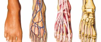
Neck muscles: a) front view;
b) rear view The seven cervical vertebrae with the muscles and ligaments attached to them provide the neck with great flexibility. The vertebrae, muscles and ligaments work as a unit to support the head and allow it to perform a variety of movements. The first and second cervical vertebrae have a specific shape and special names - atlas and axial vertebra, or axis. Atlant
is a bony ring that supports the skull.
The axial vertebra
has a bony outgrowth, or tooth, on its upper surface that acts as an axis on which the atlas rotates. The axial and five remaining cervical vertebrae also have posterior processes, which serve to attach thick occipital-spinous ligaments. The vertebral bodies are oval in shape and connected to each other by the posterior and anterior longitudinal ligaments. The spinous and transverse processes of the vertebrae are also the sites of attachment of ligaments that ensure the integrity of the spine. Between the vertebrae there are “intervertebral discs.” They provide mobility to the cervical spine, allowing the neck to tilt forward, backward, and to the sides.
The muscles in the neck area are located in the form of two triangles - anterior and posterior. The boundaries of the anterior are the lower jaw, sternum and sternocleidomastoid muscle, and the main muscles are the sternocleidomastoid and scalene muscles.
The boundaries of the posterior triangle are the clavicle, sternocleidomastoid and trapezius muscles. Its main muscles are the trapezius muscle, as well as the longissimus, semispinalis and splenius capitis muscles.
The neck muscles allow flexion (tilt the head forward), extension (tilt the head back), lateral flexion and extension of the neck (tilt the head to the right and left), as well as rotation. Since the neck muscles are paired, they are all involved in lateral flexion and extension. For example, the right sternocleidomastoid muscle is responsible for lateral flexion to the right, and the left muscle of the same name is responsible for extension from this position. Flexion of the neck is limited not only by the tightness of the muscles in the back, but also by the elasticity of the ligaments in this area, the strength of the flexor muscles, the relative position of the vertebrae, the elasticity of the intervertebral discs and the purely physical circumstance that the chin rests on the chest. Similarly, neck extension is limited by the degree of muscle tightness and elasticity of the ligaments of the front of the neck, the strength of the extensor muscles, the relative position of the vertebrae and the elasticity of the intervertebral discs. Finally, with lateral bending, the limiting factors are not only the tightness of the muscles and ligaments, but also the position of the transverse processes of the vertebrae, which abut each other.
When doing stretching exercises, we rarely think about the neck muscles until we feel stiffness in this area. It is commonly thought that this may be caused by sleeping in an unusual position (such as in an airplane seat) or sitting at a desk for long periods of time, but in fact it can be caused by any type of physical activity, especially those that require the head to remain in a fixed position at all times. Thus, neck stiffness may be detrimental to sports in which it is important to hold your head correctly (such as golf) or make rapid movements of the head to observe the movement of an object (such as tennis). Stiffness and rigidity usually occur as a result of prolonged maintenance of a fixed position, as well as after intense training.
Since all the major neck muscles are involved in turning the head, stretching them is not difficult. When choosing specific exercises, the first thing you need to do is determine what is more difficult for you to do - flexion or extension. Then you can add side bends. In other words, to work the extensor muscles, start with them first, and then, as your mobility increases, add those responsible for bending and rotating.
If done incorrectly, stretching the neck muscles can be dangerous. Some exercises are performed in a position where the head lies with the back of the head on the floor and the body is raised almost perpendicular to the floor. This will create a lot of tension at the flexion point, especially in people with low neck mobility, which can lead to either vertebral damage or excessive compression of the intervertebral discs. A slipped disc puts pressure on the spinal cord and can damage it. Also, when performing neck stretches, be careful not to make any sudden or rapid movements, as this may result in a whiplash injury. In the worst case, the consequence may be a rupture of the vertebral artery or a fracture of the vertebrae, as a result of which bone fragments penetrate into the medulla oblongata, causing death.
Remember also that stretching too hard does more harm than good. The result may be the opposite - muscle tightening. Therefore, always start with the least restrained mice and move on to others only after several weeks of training, when the stiffness of a particular area is eliminated. This also means that both agonists (muscles that perform a movement) and antagonists (muscles that perform the opposite movement) need to be stretched. Remember that even if you experience tightness on only one side, you need to stretch the muscles on both sides to maintain balance. To stretch a specific muscle, you need to perform a movement that is the opposite of what it is usually involved in. For example, if you want to stretch the left scalene muscles, you need to tilt your head back and to the right. If the muscles are too tight, then you should start with simple movements (for example, to work the right scalene muscles, first you just need to tilt your head to the left). When the muscle relaxes slightly, you can simultaneously add opposite movements.
Features of the functioning of the neck muscles during pain in the cervical spine
- A decrease in the activity of the deep neck flexors (DNF) occurs in parallel with an increase in the activity of the superficial muscles, even with minimal load and performance of non-functional tasks. This change in activation can be seen during the performance of functional or cognitive tasks, which is independent of the cause or duration of neck pain.
- Patients with neck pain have difficulty relaxing the superficial neck flexors, even after stopping physical activity.
- These changes in muscle function occur soon after the onset of pain and do not disappear even after the pain resolves.
- Changes in the functioning of the cervical spine affect the function of the cervical spine as a whole and can lead to overload in its individual segments.
- Research suggests that both cervical flexors and extensors lose their strength and endurance in the presence of neck pain, which negatively impacts their joint performance and performance of functional tasks.
- The superficial neck flexors fatigue more quickly.
- The endurance of the neck muscles decreases with both maximum and low-intensity contractions.
- Anticipatory muscle activity is lost, this affects the activation time and is clearly noticeable when a person makes rapid movements with his hands. For example, when a patient without neck pain quickly raises their arm, the neck muscles are activated within 50 ms of the deltoid muscle activation; Patients with neck pain exhibit significant deficits in the rate of activation of both superficial and GFS, which can lead to increased stress on the cervical spine.
- Patients with neck pain may experience wasting/atrophy of the neck muscles.
- With long-standing pain due to whiplash, fatty infiltration is observed in the deep flexors and extensors of the cervical spine.
- Research shows that people with a history of head and neck cancer who suffer from temporomandibular joint and neck pain have several active trigger points indicating peripheral and central sensitization.
Neck muscles[edit | edit code]
Neck muscles
- Sternocleidomastoid muscle
- Longus capitis muscle
- Rectus anterior capitis muscle
- Longus colli muscle
- Anterior scalene muscle
- Scalene medius muscle
- Posterior scalene muscle
- Sternohyoid muscle
- Omohyoid muscle
- Sternothyroid muscle
- Thyrohyoid muscle
- Digastric muscle of the neck
- Stylohyoid muscle
- Mylohyoid muscle
- Geniohyoid muscle
Objective assessment
You can read more about the examination of the cervical spine here.
Craniocervical flexion test
This test assesses motor control and isometric endurance of the longus capitis and colli muscles in comparison with the sternocleidomastoid and scalene muscles.
The patient is positioned in a supine position. A pneumatic cuff (biofeedback) is placed in the suboccipital region and inflated to 20 mmHg.
- The patient is instructed to slowly press the chin into the neck, as if performing a nodding movement with the head. This action causes the pressure in the pneumatic cuff to increase - in the first stage the pressure should increase by 2 mmHg. (superficial muscles are relaxed).
- Next, the patient should stay in this position for 10 seconds.
- He then relaxes (the cuff pressure decreases to 20 mm Hg) and again makes the head movement described above, increasing the cuff pressure to 24 mm Hg. and holding this position for 10 seconds. The patient should repeat this action until the pressure in the pneumatic cuff reaches 30 mm Hg.
Cervical flexion requires activation of the GFS. The superficial flexors should not be involved during this movement. With each stage, the range of movements should increase.
People who do not have neck pain can hold the muscle contraction for 10 seconds at stage 3 (26 mmHg) or higher. Those who experience neck pain usually reach stage 1 or 2 before they lose neutral position or begin to use the superficial muscles.
Endurance testing of neck flexors
- The patient is positioned supine with knees bent. He needs to perform craniocervical flexion (CCF). The person then raises the back of the head 2.5 cm, keeping the chin pressed into the neck.
- This is a good test to use to evaluate people with neck pain. This test has been shown to be a reliable tool for measuring the rehabilitation progress of patients with neck pain.
- Various studies have recorded the following rates for this test: Asymptomatic men - average 25 seconds, asymptomatic women - 20 seconds (Olson, 2016).
- Asymptomatic group - mean = 39 seconds, neck pain group - mean = 24 seconds (Harris, 2005).
- Male patients with neck pain have greater neck flexor endurance than females.
- Although retention times can vary, numerous studies have shown that men have longer retention times than women and that this should be taken into account when interpreting the test.
Endurance testing of neck extensors
- The patient is placed in a prone position on his stomach, with his head extending beyond the edge of the couch (forehead resting on the chair), arms along the body. Then he is asked to perform the CSC (press his chin into his neck) and hold this position, the chair is removed. The task is to keep the head with the chin pressed into the neck in a horizontal position.
- When comparing the neck flexor endurance test with the neck extensor endurance test, it can be concluded that the extensor muscles have significantly greater endurance than the flexor muscles in the neck pain population. These results are the same for both patients with acute and chronic neck pain, regardless of age.
Muscle synergies - what kind of beast?
If we consider the remaining category of muscles (which move bones in relation to each other), then everything becomes more interesting. What's most important to us? So that after any movement or position of the head our vertebrae fall into place. And nothing was pinched, hurt or pinched. Right? Right! And for this we need to remember that the muscles on the right and left are not antagonists to each other! Consistent tension of the right and left muscles has nothing to do with the correct restoration of the position of the vertebra.
And we need muscle synergies! These are joint coordinated contractions of various muscles and muscle groups that ensure the implementation of a targeted motor act. Simply put, these are coordinated movements of opposing muscles, the correspondence of tension and relaxation of the muscles involved in the movement. It is this consistency that ensures that the vertebra returns to its original position after you tilt or turn your head. And as you already understand, muscle pumping is definitely not needed for this. We need to teach the muscles to work correctly and this is very far from the initial “strengthen and pump up”.
How to cope with a spasm
If there were no injuries to the problem, then it can be assumed that the spasms appeared due to pinched nerve root, which is located in the cervical region. If you take an ordinary x-ray, you can detect the presence of a gap between the vertebrae. If no pathologies were found during such studies, this does not mean that you don’t have to worry about the cause of pain.
On the contrary, it can be assumed that the changes affected the muscle itself or that the nerve was pinched in another part. By the way, it may be pinched both in the area of the face and in the area of the jaw joint.
To make a diagnosis and prescribe treatment, you need to see the full picture, no matter who you turn to, a neurologist, chiropractor or massage therapist.
It is also necessary to evaluate the condition of the main muscles that are present in the cervical region:
- Long muscle.
- Staircase.
- The muscle that is responsible for lifting the scapula.
- Trapezoidal.
When nerves are compressed in the cervical region, there may be a decrease in muscle strength. Treatment, including medicinal and non-medicinal, is prescribed individually.
Today there are many techniques that can not only relieve muscle spasms, but also affect the cause of pain. You need to understand that spasm of ten neck muscles can lead to torticollis, therefore it is impossible to confirm this or that diagnosis without diagnostics.
Rehabilitation
Muscular performance or muscle strength is “the ability of a muscle to generate force regardless of action, load, or intensity.” For a muscle to control a segment of the body, it must have sufficient strength. To train muscle performance, a muscle must perform a repeated action against some resistance (either a body segment or an added weight), around an axis of rotation in the same plane. This type of training has been shown to improve proprioception of a dysfunctional segment, such as when people with neck pain perform a controlled head lift exercise.
What will the specialist do at the appointment?
Approximate actions of a doctor, if there is a suspicion that the sternocleidomastoid muscle of the neck is damaged, would be as follows:
- To begin with, the specialist must study the patient’s complaints, which will suggest the direction in which to look for the cause.
- The doctor evaluates the movements that the patient can and cannot perform.
- Testing and diagnostics.
- Prescription of treatment.
- After treatment, a re-examination should be carried out.
- Treatment can be cyclical and may not help immediately, but only after several courses.
Training your neck
Training the muscles of this department is important both for health and for the aesthetic side of the issue. Sometimes you have to bend your neck, sometimes you have to stretch it, but in any case, it does not remain motionless. If you turn to wrestling, you will notice that many of its types require developing the neck muscles. Otherwise, it will be difficult to achieve something, for example, in martial arts, opponents can strike in the face or head.
In addition, if you are working on developing other muscles, then you should not forget about the neck muscles either. Otherwise, there will be an imbalance in development, which may negatively affect the assessment of athletes.
Separately, it is worth mentioning the aesthetic side, because the neck muscles are always in sight, no matter what clothes you are wearing. Correctly developed muscles look attractive in both men and women. Also, training will be simply irreplaceable for those who have a sedentary job and spend a lot of time at the computer. This makes it possible to prevent the occurrence of diseases such as cervical osteochondrosis. But many people neglect seemingly simple exercises that do not require much time.