Read about the diagnosis and treatment of cholelithiasis at the link.
Get help right away if you develop signs and symptoms of a serious complication related to gallstones, such as:
- Abdominal pain is so severe that you cannot sit still or find a comfortable position
- Yellowing of the skin and whites of the eyes (jaundice)
- High temperature with chills
Number to call an ambulance in Moscow – 103
How does the liver work?
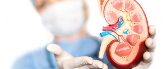
The liver is the largest gland in humans. It is located in the upper floor of the abdominal cavity on the right. It consists of two main lobes - right and left, which in turn are divided into 8 segments.
In an adult, it weighs about 1/40 of the total body weight (approximately 1.6 kg in men and 1.2 kg in women). The liver has a dual blood supply: approximately 80% of the blood entering the liver comes from the portal vein (venous blood collected from all unpaired abdominal organs), the rest (oxygenated arterial blood) comes from the hepatic artery. Having entered the portal of the liver, both vessels give rise to multiple branching smaller vessels. At the exit from the liver, 3-4 hepatic veins are formed, which flow into the inferior vena cava.
The main structural unit of the liver is the hepatic lobule, which consists of liver cells - hepatocytes. This cell is one of the most important biochemical laboratories of the body. The hepatocyte takes part in the metabolism of proteins, carbohydrates, fats, bile acids, and vitamins. Neutralizes toxic substances coming from the intestines, followed by the release of associated products into the intestinal lumen. An important function of the hepatocyte is the synthesis of bile, which is involved in digestion.
Stages of gallstone disease
Gallstone disease has several stages:
- Pre-stone (also called physicochemical). It is the initial stage at which the first changes occurring in the composition of bile are noted. There are no special or obvious symptoms. The development of the disease can only be determined using a biochemical examination.
- Hidden form. At this stage, stones are just beginning to appear. There is also no clear clinical picture. The presence of pathology can be determined using instrumental diagnostic procedures.
- More severe stage. Symptoms of the disease begin to appear.
What is bile and why is it needed?
Bile is a secretion produced by hepatocytes. The color of bile ranges from light yellow to dark green. Bile contains bile pigments - bilirubin and biliverdin, cholesterol, fatty acids, lecithin, mucin.
Bile is the main participant in digestion. It neutralizes hydrochloric acid that enters the duodenum from the stomach, thereby creating favorable conditions for the action of pancreatic juice and parietal digestion in the jejunum.
Bile acids emulsify fats, allowing them to be absorbed in the intestines.
Bile has a bactericidal effect that prevents the proliferation of pathogenic microflora.
Types of gallstones
Types of gallstones that can form in the gallbladder include:
Cholesterol stones in the gall bladder. The most common type of gallstones, called cholesterol gallstones, are often yellow in color. These gallstones are composed primarily of undissolved cholesterol, but may contain other components.
Pigment stones in the gall bladder. These dark brown or black stones form when your bile contains too much bilirubin.
How are the bile ducts arranged and how do they work?
The bile ducts are a system of ducts that originate from hepatocytes and gradually gather into interlobular, segmental and right and left lobar ducts, which unite into the common hepatic duct.
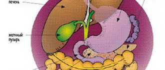
One of the stages of bile removal is its accumulation in the reservoir - the gallbladder. The main function of the gallbladder is to concentrate bile and release it into the lumen of the duodenum during digestion. The volume of this organ is about 80 ml.
The gallbladder is located on the lower surface of the liver. It is a hollow muscular organ, externally covered with a serous membrane and internally lined with mucous membrane. The structure of the gallbladder is divided into the bottom, body and neck of the bladder, which passes into the cystic duct.
What do these diseases have in common and how to treat them?
Cholecystitis and pancreatitis have similar symptoms
Both the gallbladder and the pancreas have similar functions - they produce enzymes that contribute to the normal digestion process. The first organ is responsible for dosing the bile produced by the liver so that it arrives in the right quantities, and the second organ produces pancreatic juice. If the functions of one of the organs are impaired, disturbances in the functioning of the other are observed.
Pancreatitis and cholecystitis, as is customary, are treated with medications. Therapy is often supplemented by the use of traditional medicine, as well as the prescription of a variety of diets. The main requirement for food intake in these diseases is frequent split meals. It promotes more effective action of medications used during therapy.
It is recommended to limit the consumption of foods that are too fatty and eat more healthy foods, such as steamed vegetables. It is also useful to drink a glass of kefir before bed. Typically, diseases are treated with the complex use of various drugs aimed at combating the main symptoms of the disease. Properly thought out treatment helps restore the functions of the gallbladder and pancreas. The main medications used for pancreatitis:
Why do gallstones form?
The exact cause of cholelithiasis has not yet been fully elucidated. Only a few factors are known that increase the likelihood of its occurrence.
- Genetic: family predisposition increases the risk of cholelithiasis by 4-5 times;
- Congenital anomalies of the biliary tract;
- Gender - the risk of developing gallstones in women is approximately 2-3 times higher, which is associated with the influence of estrogens on the ability to form gallstones;
- Age - the maximum frequency of clinical manifestations of cholelithiasis is recorded at the age of 40-69 years;
- Dietary (high-calorie diet, poor in plant fiber, as well as with an excess of simple carbohydrates, animal proteins, foods containing cholesterol);
- Use of medications (oral contraceptives, fibrates, ceftriaxone, somatostatin, nicotinic acid);
- Concomitant diseases and conditions (obesity - a tendency to form gallstones in persons with a body mass index of more than 30 is 3 times higher, type 2 diabetes mellitus, hypothyroidism, cirrhosis of the liver, etc.);
- Pregnancy - the risk of developing cholelithiasis increases during pregnancy, especially with repeated pregnancies (the likelihood of stone formation increases by 10-11 times). During pregnancy, bile heterogeneity, or the so-called biliary sludge, develops in 20-30% of patients, stones - in 5-12% of cases.
There is a so-called 5F rule that describes patients at greatest risk of developing stones:
1. Female (female);
2. Fat (overweight/obesity);
3. Forty (age 40 years or more);
4. Fertile (fertile women - that is, pregnant and giving birth);
5. Fair (light-skinned blondes).
Causes of cholelithiasis
It is not clear what causes gallstones to form. Doctors believe that gallstones can occur in the following cases:
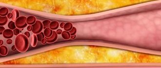
Your bile contains too much cholesterol . Normally, your bile contains enough chemicals to dissolve the cholesterol released by your liver. But if your liver produces more cholesterol than bile can dissolve, the excess cholesterol can turn into crystals and eventually stones.
Your bile contains too much bilirubin . Bilirubin is a chemical that is produced when red blood cells are broken down in the body. Under certain conditions, the liver produces too much bilirubin, including cirrhosis of the liver, bile duct infections, and some blood disorders. Excess bilirubin promotes the formation of gallstones.
Your gallbladder is not emptying properly. If the gallbladder does not empty completely or often enough, the bile can become very concentrated, which contributes to the formation of gallstones.
How are gallstones formed?
The process of stone formation is quite well studied. The key links are:
- An increase in the lithogenicity of bile (the ability to form stones) means an increase in the content of cholesterol and bilirubin in it.
- Impaired contractile function of the gallbladder.
- Increased pressure in the bile duct system.
Mucin is a natural gel, constantly secreted by the wall of the gallbladder; when lithogenicity increases, it begins to “absorb” cholesterol crystals, thereby forming a “fossilization nucleus.” The formed suspension, with impaired contractile function, continues to attach salt crystals encrusted with calcium salts. Thus, gallstones can grow up to 5-7 mm per year.
Types of stones
Stones may differ in their chemical composition. Depending on this they are:
- Bilirubin. Bilirubin is a product formed as a result of the breakdown of hemoglobin. They are relatively small (up to 1 cm), but can be present in considerable quantities.
- Cholesterol. Despite the fact that cholesterol is present in bile, if it accumulates excessively, stones will appear. This component is ingested along with consumed products. When the process of its absorption is difficult, it begins to accumulate and turn into stones. They usually have an oval or round shape, not exceeding 1.5 cm in diameter.
To size:
- Small ones. They reach 3 cm in diameter.
- Big ones. They reach more than 3 cm in diameter. They impede the outflow of bile, which is why attacks often occur.
How does gallstone disease occur?
The main factors influencing the course of the disease are the nature of nutrition and the size of the stones. Gallstone disease can occur both in the form of chronic stone carriage and in acute, and sometimes in complicated, form.
Where does pain come from with gallstone disease?
By themselves, gallstones do not cause any pain and can be an accidental finding during medical examination. When food enters the duodenum, the mechanoreceptors are irritated and cause contraction of the gallbladder. The stones wedge into the cervix, causing a prolonged spasm, which manifests itself as pain. When taking antispasmodic drugs, the calculus can “move” back from the cervix and the attack of hepatic colic is stopped. If the stone remains in the cervix, then inflammation begins to develop against the background of obstruction.
Chronic calculous cholecystitis is an asymptomatic stone carrier. The size of the stones can vary from 2 – 3 mm to 5 cm. This stage is characterized by an almost complete absence of clinical manifestations of the disease. Stones can remain in the lumen of the gallbladder for years without causing any symptoms. However, if there is an error, discomfort may occur in the right hypochondrium, which can be relieved independently or while taking antispasmodic drugs.
Hydrocele of the gallbladder - occurs against the background of obstruction of the neck of the gallbladder by a calculus, cutting off the latter from the entry of bile. The bladder is filled with mucus secreted by the mucous membrane and is not involved in digestion.
Acute calculous cholecystitis - occurs with prolonged obstruction of the neck of the gallbladder by a calculus. The pain syndrome persists against the background of ineffective conservative treatment. The patient is bothered by vomiting, which does not bring relief; pain can be felt behind the sternum - cholecystocardial syndrome, in the right shoulder blade. Body temperature rises and chills appear. Inflammatory changes begin to develop in the wall of the gallbladder, which progress from catarrhal, phlegmonous and gangrenous forms. Against the background of the latter, perforation of the gallbladder or abscess may develop.
Choledocholithiasis and acute biliary pancreatitis. Choledocholithiasis develops as a result of migration of biliary sludge or small stones from the gallbladder into the common bile duct. Once in the common bile duct, small stones freely descend to the sphincter of Oddi, which is a release mechanism - a circular muscle cuff. Sludge and stones “wedge” into the sphincter, causing its long-term spasm, causing a violation of the outflow of bile - obstructive jaundice. The patient's skin and sclera become a rich yellow color. This condition requires emergency decompression of the bile ducts - restoration of bile outflow.
An increase in pressure in the common bile duct promotes the reflux of bile into the main pancreatic duct and intraductal activation of pancreatic juice enzymes - a trigger for the development of acute biliary pancreatitis.
Mirizzi syndrome . A fairly rare complication of gallstone disease. Develops as a result of prolonged stone bearing. First, a stricture forms, then a bedsore between the gallbladder and the common bile duct.
Mirizzi syndrome presents significant difficulties for surgical treatment. This is due to the fact that in the human body there is no plastic material to close the defect of the common bile duct. Surgical treatment of Mirizzi syndrome is often accompanied by the formation of an anastomosis between the small intestine and the common bile duct. When a stone migrates into the small intestine, acute intestinal obstruction may develop, requiring emergency surgical treatment.
In the photographs, in the lumen of the resected section of the small intestine there is a calculus that caused intestinal obstruction.
Clinical picture and symptoms of the disease
Most often, the first sign that forces the patient to see a doctor is pain in the right hypochondrium, of varying intensity. Cutting or stabbing in nature, the pain is most often constant, often radiating to the right shoulder blade, lower back, and forearm. In some cases (with cholecystocoronary Botkin's symptom), it can radiate beyond the sternum, resembling an attack of angina. It should be borne in mind that the intensity of pain is in no way an indicator of the severity of the process. For example, in some cases, severe pain may disappear, but mild pain does not mean a mild form of the disease.
Often the patient’s condition worsens after eating spicy or fatty foods; their consumption increases the need for bile to process food, which leads to contraction of the gallbladder. In any form of the disease, there is an increase in body temperature. In the form of short rises, the temperature rises to 37-38°C, and the patient often experiences pain. However, during an acute attack accompanied by chills, the temperature can rise to 38-40°C.
How to diagnose gallstone disease?
The gold standard for diagnosing gallstone disease is ultrasound examination of the abdominal cavity. This method allows you to identify stones in the gallbladder. Assess their size, shape and location. An important indicator is the thickness of the gallbladder wall. Normally, it does not exceed 2-3 mm. Thickening of the wall is an absolute sign of inflammatory changes - acute cholecystitis. An important criterion is the diameter of the common bile duct. If there are stones, its diameter increases from 3 – 6 to 20 mm.
Magnetic resonance imaging allows you to see the intra- and extrahepatic bile ducts, as well as the main pancreatic duct. This diagnostic method is used when there is a suspicion of the presence of stones in the common bile duct. This is necessary for treatment planning.
Multislice computed tomography is indicated for the development of acute biliary pancreatitis. This diagnostic method allows you to identify inflammatory changes in the pancreas and surrounding tissues.
Gastroduodenoscopy - this diagnostic method is especially valuable for complicated cholelithiasis, as it allows not only to evaluate the stomach, duodenum, but the flow of bile through the major duodenal papilla. If necessary, gastroduodenoscopy can easily be transformed from a diagnostic procedure into a therapeutic one. Using special instruments, you can “paint” the bile ducts and remove stones.
In the figure, the arrow indicates a stone in the common bile duct. Using a duodenoscope, a dissection of the sphincter of Oddi was performed in the duodenum. A radiopaque substance was introduced to allow visualization of the calculus. Using a special basket, the stone is captured and removed.
Gallstone disease and consequences of cholecystectomy: diagnosis, treatment and prevention
Gallstone disease (GSD) is a disease of the hepatobiliary system caused by impaired metabolism of cholesterol and/or bilirubin, characterized by the formation of stones in the gall bladder and/or bile ducts with the possible development of dangerous complications.
Ultrasound reveals gallstones in 10–15% of apparently healthy adults, the frequency of detection of which increases with age. Most gallstones form cholesterol by depositing in supersaturated bile, especially at night, when the concentration of bile in the gallbladder is at its highest.
Between meals, bile is concentrated in the gallbladder, which acts as a reservoir for bile acids (cholic and chenodeoxycholic).
Bile acids form the outer layer of the micelle, which contains fat-soluble cholesterol (cholesterol) in the center. Phospholipids concentrated in the center of the micelle increase its ability to keep cholesterol from crystallization.
Bile acids are necessary for the emulsification of fats, for their breakdown (hydrolysis) under the influence of lipase (mainly pancreatic) into triglycerides and fatty acids, the latter are absorbed (absorbed) together with bile acids mainly in the ileum.
A deficiency of bile acids involved in the enterohepatic circulation (eg, in terminal small intestinal disease) or an imbalance between the concentration of phospholipids and cholesterol (lithogenic bile) in the bile can lead to the precipitation of cholesterol crystals from the supersaturated bile, which then nucleate to form cholesterol bile stones
Factors predisposing to the formation of cholesterol gallstones include gender (women), obesity, diet (low dietary fiber), liver cirrhosis (30%), terminal ileal disease (Crohn's disease, etc.) or ileal resection , consumption of medications (oral contraceptives containing mainly estrogens, clofibrate).
Pigment stones consist of bilirubin with the formation of calcium-insoluble precipitates (calcium bilirubinate). Black, small, dense pigmented stones account for 70% of all radiopaque gallstones.
Brown pigment stones, also consisting mainly of calcium bilirubinate, are soft, predominantly intrahepatic and are found extremely rarely.
The following factors predispose to the formation of black pigment stones, consisting predominantly of calcium bilirubinate: chronic hemolysis (sickle-shaped and spherical red blood cells, for example, in sickle cell anemia, implanted artificial heart valves); cirrhosis of the liver; biliary tract infection (E. Coli, Clostridium Sp.). Infection of bile with microorganisms producing β-glucuronidase leads to an increase in the content of poorly soluble direct unconjugated bilirubin in the bile.
Brown pigment stones are usually formed in patients with sclerosing cholangitis and with biliary invasions (opisthorchiasis, clonorchiasis, giardiasis, etc.).
Along with cholesterol (single) and pigment (purely pigmented black and brown), mixed, usually multiple, gallstones are more common. It is extremely rare to find stones consisting of calcium carbonate and phosphorus.
Modern classifications provide for the identification of at least three stages of cholelithiasis. The first of them is physical and chemical. At this stage, the liver produces bile oversaturated with cholesterol, with a decrease in the content of bile acids and phospholipids (lithogenic bile).
Patients have no clinical symptoms of the disease; the diagnosis is based on the results of a study of gallbladder bile (portion B). A violation of the micellar properties of bile is revealed; cholesterol “flakes”, crystals and their precipitates are found in it. There are no gallstones.
The first stage of cholelithiasis can be asymptomatic for many years.
Therapeutic and preventive measures in this preclinical stage of cholelithiasis include a general hygienic regime, systematic physical activity, rational fractional nutrition with the exception of nutritional excesses (high-calorie and cholesterol-rich foods, especially in cases of obesity and hereditary predisposition).
Preventive measures also include adequate treatment of patients with gastrointestinal dysfunction (intestinal dysbiosis, colitis, etc.).
The second stage of cholelithiasis (latent asymptomatic stone carriage) is characterized by the same physicochemical changes in the composition of bile as the first stage, but with the presence of stones in the gall bladder. The process of stone formation at this stage is associated not only with physicochemical changes in bile, but also with the addition of gall bladder pathogenesis factors (stagnation of bile, damage to the mucous membrane, increasing the permeability of the bladder wall for bile acids, inflammation) and disturbances in the enterohepatic circulation of the bile acids
Most stones located at the bottom of the gallbladder do not manifest themselves in any way. The advancement of stones into the cystic duct and its blockage lead to the development of cholecystitis, which stops if the obstruction of the duct is eliminated, or progresses with the development of complications.
The third stage of cholelithiasis is clinical, complicated (calculous cholecystitis, acute, chronic, etc.). Clinical manifestations of cholelithiasis depend on the location of gallstones, their size, localization and activity of inflammation, the functional state of the biliary system, as well as damage to other digestive organs.
A stone lodged in the neck of the gallbladder obstructs its outlet, causing biliary (hepatic) colic. Subsequently, obstruction of the cervix may turn out to be temporary, and the stone returns to the gallbladder or penetrates the cystic duct, where it stops or passes into the common bile duct. If the size of the stone (up to 0.5 cm) allows, it can enter the duodenum and appear in the stool; the stone can also stop in the common bile duct, more often in its distal part, causing complete or intermittent obstruction (valve stone) with the corresponding clinic. In this case, the bile always becomes infected, and cholelithiasis is accompanied by inflammation (choledochitis, cholangitis).
Acute calculous cholecystitis (ACC) usually occurs when a stone enters the cystic duct, which leads to stagnation and infection of bile, swelling of the gallbladder wall with hemorrhages and ulceration of the gallbladder.
Signs of acute cholecystitis:
- Fever and constant pain in the right upper quadrant of the abdomen, in contrast to chronic cholecystitis.
- The main reason for the development of the disease is strangulation of a stone in the cystic duct (duct obstruction).
- Typical pain (with acute cholecystitis in less than 50% of patients).
- Pain that often occurs soon after eating and increases over the course of an hour, in contrast to attacks of pain with biliary colic, which are short-lived and disappear on their own.
- Fever joins the pain syndrome usually 12 hours after the onset of the attack and is associated with bacterial inflammation (invasion), as a result of which the pain becomes constant.
- Murphy's sign is usually positive, but it is not related to a specific test.
- An X-ray of the abdominal cavity taken in the supine position reveals calcified stones in the gall bladder and sometimes gas inside the biliary tree in some patients (15%) (the formation is associated with infection of the latter by Clostridium Welchii).
Complications of acute cholecystitis:
- chronicity (50%);
- cholangitis caused by the presence of stones in the bile ducts (10%);
- dropsy, empyema or gangrene of the gallbladder (1%);
- biliary peritonitis (0.5%) with mortality in 50% of cases.
Chronic calculous cholecystitis is usually characterized by recurrent attacks of biliary colic, less often by constant pain in the right upper quadrant of the abdomen. Patients show significant differences in the degree of thickening and fibrosis of the gallbladder wall and inflammatory infiltrate.
Biliary colic sometimes occurs suddenly, “for no reason” or after eating, and is combined with low-grade fever, nausea, and sometimes vomiting. The pain intensifies with movements and deep breathing. Severe pain usually disappears quickly.
An attack is provoked by fatty foods, spices, smoked foods, hot seasonings, severe physical stress, working in an inclined position, as well as infection. In women, colic sometimes coincides with menstruation or occurs after childbirth. The pain often radiates to the right scapula and subscapular region. Sometimes the pain radiates to the lumbar region, to the region of the heart, simulating an attack of angina. The pain varies in intensity: from strong cutting to relatively weak, aching. However, exacerbation of cholecystitis, especially non-calculous, is not always accompanied by typical attacks of biliary colic. The pain may be dull, constant or intermittent. Vomiting with cholecystitis does not bring relief.
The undoubted signs of calculous cholecystitis include:
- pain in the right upper quadrant of the abdomen - acute, episodic (less than 60 s) and cramping (from 1 to 72 hours);
- pain-free intervals (from several weeks to several months);
- intolerance to fatty and fried foods (often);
- flatulence - increased gas evolution (often);
- flatulence - bloating (often);
- positive palpation and percussion symptoms such as Murphy, Kera, etc. (sometimes absent even with exacerbation of chronic calculous cholecystitis);
- gallstones and a thickened wall of the gallbladder, always detected by ultrasound;
- non-functioning gallbladder, determined using oral cholecystography.
There is constant dull and variable pain in the right upper quadrant of the abdomen without irradiation. Cholecystolithiasis with a normal gallbladder wall, a functioning gallbladder even in the presence of dyspeptic disorders that occur in chronic calculous cholecystitis, are more characteristic of stone carriers rather than cholecystitis.
Along with calculous cholecystitis (acute, chronic), not stones are detected in the gallbladder, but sediment (sludge), associated with an increased content of mucin in it, on the matrix of which bile components crystallize. The formation of sediment in the gallbladder occurs when it is slowly or incompletely emptied. This condition is often associated with prolonged fasting or insufficient stimulation of gallbladder motility by cholecystokinin produced in the intestines. Although bile sludge is a reversible stage in the pathogenesis of gallstone disease, if appropriate therapy is prescribed, stones will inevitably form as it progresses, which leads to the appearance of corresponding symptoms.
Treatment includes: fractional hypocaloric nutrition aimed at reducing body weight if there is obesity; drug decontamination if a syndrome of excessive microbial contamination of the initial parts of the small intestine is detected; oral administration of bile acid preparations (chenodeoxycholic and ursodeoxycholic) for 1.5-2 months, endoscopic papilosphincterotomy, if a stenotic process is diagnosed at this level of the common bile duct.
Choledocholithiasis—stones in the common bile duct—is manifested by pain and jaundice. Choledocholithiasis occurs when a gallstone passes from the bladder into the common bile duct. Secondary stone formation in the common duct is possible, especially in the presence of stasis caused by duct obstruction.
The presence of stones in the common bile duct should be suspected in any patient with calculous cholecystitis whose serum bilirubin level exceeds 50 mmol/l and the alkaline phosphatase level is three normal levels. The level of aminotransferases may increase 2–10 times compared to normal, especially with acute obstruction. Once the obstruction is relieved, aminotransferase levels usually return to normal quickly, while bilirubin levels often remain elevated for 2 weeks, and elevated alkaline phosphatase levels persist even longer.
Symptoms, often intermittent, include colicky pain in the right hypochondrium, fever, chills and jaundice with a characteristic increase in alkaline phosphatase and transaminases in the blood serum. Choledocholithiasis, if it is not eliminated immediately, is almost always accompanied by ascending cholangitis, an infection of a confined space that can lead to sepsis.
Cholangitis is characterized by pain in the upper abdomen, often on the right, jaundice and fever, often accompanied by chills. Bacterial cholangitis is one of the most dangerous complications of cholelithiasis; it is usually associated with subhepatic cholestasis, which often occurs with calculous obstruction of the main bile duct. The severity of cholangitis depends on a number of factors, primarily on the duration of cholestasis and the level of cholemia. With a short-term, but repeatedly recurring violation of the outflow of bile, chronic cholangitis develops, in which (usually following a quickly passing attack of biliary colic) mild chills occur with an increase in temperature to subfebrile levels, the urine becomes dark in color and sometimes jaundice occurs. These symptoms usually last no more than 2–3 days. Blood tests in some cases reveal slight neutrophilic leukocytosis, a moderate increase in ESR, transient hyperbilirubinemia, and a short-term and slight increase in alkaline phosphatase levels.
Such exacerbations of cholangitis are more often associated with the passage of a stone through the common bile duct, less often due to the valve mechanism in choledocholithiasis and sometimes, possibly, papillitis (odditis). Between episodes of cholestasis, there may be no symptoms of cholangitis. This form of cholangitis is classified as chronic; its course is largely determined by the frequency of relapses and the duration of cholestasis, as well as the nature of the inflammatory process (catarrhal, purulent).
Diagnostic tests:
- The most effective non-invasive method for identifying gallstones is ultrasound. Often, asymptomatic stone carriers (silent stones) are diagnosed using this method. Occasionally, gallstones are not detected even with careful examination. In some patients, the gallbladder cannot be visualized due to intestinal gas, fibrosis of the gallbladder, or its unusual anatomical location. Ultrasound also turns out to be an informative method for detecting obstruction of the common bile duct, diagnosing acute and chronic cholecystitis and assessing gallbladder function, which is determined before and after the use of cholekinetics.
- CT has an advantage over ultrasound when it comes to identifying stones in the common bile duct.
Blood test:
- In chronic cholecystitis without exacerbation and asymptomatic cholecystolithiasis, the peripheral blood picture is normal.
- With choledocholithiasis, there is an increase in alkaline phosphatase (3 norms or more) and GGTP (3 norms or more).
Neutrophilic leukocytosis is characteristic of AC and cholangitis. The content of serum cholesterol is not important for the diagnosis of cholecystitis and cholangitis, but cholesterol naturally increases with primary biliary cirrhosis and sclerosing cholangitis.
Other studies:
- An abdominal x-ray has diagnostic value in the presence of acute abdominal pain. It may reveal calcifying stones in the gall bladder, bile ducts and intestinal lumen, an enlarged liver and the presence of air in the biliary tract (biliary enteric fistula, cholangitis caused by clostridia or occurring after surgery).
- To assess the function of the gallbladder in a patient with chronic cholecystitis who is undergoing conservative treatment, “oral” cholecystography can be performed.
- Isotope scanning with the Hida radiopharmaceutical has some diagnostic value only in acute illness, when it is possible to assess the function of the gallbladder, including determining its shutdown as a result of duct obstruction.
- ERCP is used for diagnosis of cholestatic syndrome and for the treatment of biliary obstruction (benign stricture in the area of the papilla of Vater, choledocholithiasis, etc.).
- Percutaneous transhepatic cholangiography is indicated in cases where there are dilated intrahepatic bile ducts identified by echohepatography, and it is not possible to perform ERCP or the condition of the intrahepatic bile ducts needs to be clarified (filling of the bile ducts with contrast can be blocked by a tumor or stricture that has arisen at one or another level of the biliary tract ).
- Carrying out intravenous cholangiography and a test with bromsulfalein to assess the state of the excretory function of the biliary system of the liver should be considered outdated methods.
Stasis of bile in the gallbladder (hypomotor dyskinesia of the gallbladder), caused by its hypokinesia, is considered typical for patients with cholelithiasis. It is also observed in a number of conditions that are risk factors for the development of cholelithiasis: during pregnancy, long-term use of anticholinergic and antispasmodics, after vagotomy, diabetes, obesity, etc.
Against the background of bile stasis in the gallbladder, ultrasound often reveals a sediment accumulating in it, known to surgeons as “putty,” found during cholecystectomy. On ultrasound, the sediment has the appearance of a “cloud” with multiple small echo-positive inclusions. Unlike gallstones, echogel is not detected, but a clear boundary between such a “cloud” and echo-negative bile can be determined. The “cloud” may include cholesterol crystals, bilirubinate particles, and calcium carbonate particles. Obviously, these patients may subsequently develop cholecystolithiasis. Along with microliths, changes on ultrasound of the gallbladder may be associated with the presence of polyps in it. True polyposis of the gallbladder can be differentiated from cholesterol parietal microlites by prescribing ursodeoxycholic acid (ursofalk) at a dose of 7.5 mg/kg for 3 months and performing dynamic ultrasound of the gallbladder. If, while taking the drug, the initially identified signs of the parietal “deposit” disappear, then polyposis is excluded and cholelithiasis is diagnosed at the stage of microlith formation.
When bile stasis in the gallbladder, one should keep in mind cholesterosis, characterized by the deposition of cholesterol in the CO of the bladder. Cholesterosis often develops in obese women with severe disorders of fat metabolism and high cholesterol levels in the blood.
In cholesterosis, lipids (mainly cholesterol) are found predominantly in the endothelial cells of the mucous membrane, from which then, if they are significantly deposited, a cholesterol polyp can develop. On ultrasound, this formation looks like a polyp, but histological examination does not determine the true structure of the polyp.
The presence of a correlation between cholesterosis, cholesterol levels in the blood and bile, and the development of atherosclerosis, including atherosclerosis of the aorta, coronary and cerebral arteries, has not been confirmed. The presence of malignant degeneration in cholesterosis has also not been established.
Stasis of bile in the gallbladder is usually clinically manifested by constant dull aching pain in the right hypochondrium, aggravated by shaking when driving, walking quickly, carrying a heavy weight in the right hand, or bending forward. Diagnosis is sometimes difficult, although duodenal probing reveals cholesterol crystals in portion B.
The choice of adequate treatment for cholelithiasis is often determined jointly by the therapist, surgeon and patient.
When determining the indications for cholecystectomy, we used the international recommendations presented in the table.
Treatment of the consequences of cholecystectomy
In patients who have undergone cholecystectomy, most often symptomatic manifestations are associated with dysfunction of the sphincter of Oddi of the pancreatic or biliary type. Preventive measures should be aimed at normalizing the chemical composition of bile, restoring the patency of the sphincter of Oddi, decontaminating the duodenal mucosa and restoring its motor-evacuation function.
As a symptomatic remedy, the drug Odeston (hymecromone) is prescribed, 200 mg 3 times a day before meals, the course of treatment is 1–2 weeks. The drug has a choleretic and antispasmodic effect on the sphincter of Oddi, but does not weaken intestinal motility.
For cholestasis and cholangitis in the absence of infection of the biliary tract and restoration of bile outflow, along with the elimination of stricture, we have accumulated positive experience in using the herbal preparation hepabene (1-2 capsules after dinner), and in case of infection of the biliary tract - an antibacterial drug from the macrolide group - clarithromycin (clacid, Klabaksa), for purulent infection - the drug Meronem.
Absolute indications for surgery:
- acute cholecystitis;
- chronic cholecystitis with a normal history (recurrent biliary colic) and non-functioning gallbladder (according to ultrasound or cholecystography);
- stones of the common bile duct: a) in persons under 70 years of age - ERCP; sphincterotomy; according to indications - cholecystectomy; b) in people over 70 years of age and in the presence of a high surgical risk, endoscopic sphincterotomy gives less mortality, but the risk of relapse of choledocholithiasis remains;
- gangrene of the gallbladder - urgent cholecystostomy (safer than cholecystectomy), cholecystectomy is possible in the future, but often you can limit yourself to spontaneous closure of the wound;
- intestinal obstruction caused by a gallstone is an operation to eliminate intestinal obstruction followed by cholecystectomy.
Relative indications for surgery: chronic calculous cholecystitis, if the symptomatic manifestations of the disease are associated with the presence of stones in the gall bladder. In this case, it is necessary to exclude gastric and duodenal ulcers, irritable bowel syndrome, chronic pancreatitis, and urinary tract diseases, which may have symptoms simulating chronic cholecystitis.
Currently, along with standard laparotomic cholecystectomy, laparoscopic cholecystectomy is being widely introduced into practice, the advantage of which is a short stay for the patient in the hospital (less than 48 hours) and earlier restoration of working capacity (after 5–7 days). Laparoscopic cholecystectomy, if performed by a highly qualified specialist and according to strict indications, allows you to quickly and much less traumaticly remove stones from the gallbladder even before the symptoms of inflammation increase. The advantage of cholecystectomy compared to conservative methods of treating cholecystolithiasis (lithotripsy, stone dissolution) is to eliminate the risk of recurrent stone formation.
Prevention of gallstone disease
The first stage of cholelithiasis can be diagnosed if appropriate biochemical studies of bile are carried out, mainly portion C. Lithogenic bile is characterized by oversaturation of bile with cholesterol, a decrease in the concentration of bile acids and phospholipids in it, as well as hypokinesia of the gallbladder and damage to the liver parenchyma. In order to prevent the progression of cholelithiasis (transition to the second stage - latent, asymptomatic stone carriage), it is recommended to change your diet and lifestyle. The effectiveness of preventive measures depends on their strict compliance by the patient.
In the second stage (latent stone carriage), the goal of prevention is to prevent the formation of complications of cholelithiasis and the consequences of cholecystectomy. Adequate tactics for patient management at this stage are determined jointly by the therapist and the surgeon.
For primary prevention of cholelithiasis it is recommended:
- In order to eliminate stagnation of bile and improve its quality, eat food at least 5 times a day (first breakfast at about 8 o'clock, second at 11 o'clock, lunch at 14 o'clock, afternoon snack at 17 o'clock and dinner at 21 o'clock). For patients predisposed to the development of cholelithiasis, as well as at the first stage of the disease, there are no prohibited foods and dishes. Leisurely eating at a strictly defined time stimulates the secretion of digestive juices (including bile) and the motor-evacuation function of hollow organs (including the gallbladder). The diet should include meat, fish, fats, vegetables, fruits and their juices. A decrease in working capacity and an increase in body weight (obesity, obesity) should not be allowed; a sufficient night's sleep, daily formed stools and sufficient and painless urination are necessary; It is not recommended to smoke and drink alcoholic beverages even in so-called “small” quantities.
- If there is a tendency to constipation or difficult, prolonged or painful defecation, it is necessary first of all to exclude organic pathology (hemorrhoids, peptic ulcer, diverticular colonic disease, polyposis of the rectum and colon, colorectal cancer, etc.), then make appropriate changes to the regimen nutrition and lifestyle (consume at least 0.5 kg of vegetables and fruits daily, increase fluid intake to 1.5-2 liters per day, eat only “dark” varieties of bread, increase daily physical activity (brisk walking, swimming, etc.) ...), after meals, take the drug allohol in the amount of 3-4 tablets per day. The so-called “morning block” is recommended: in the evening, soak 4 to 10 prunes in boiling water (prepare an infusion), and in the morning drink it and eat the fruits Then have breakfast with the obligatory consumption of a glass of any juice and a small amount of salad from fresh vegetables or fruits.
- For idiopathic functional constipation, along with compliance with the appropriate dietary and physical regimens, it is possible to prescribe laxatives for a short time, mainly mucofalk or laminarid (4 teaspoons of granules per day) or Forlax (2 sachets per day), or lactulose (30 ml of syrup or 20 g granules per day). Other laxatives are used less frequently.
- If the liver is involved in the process (fatty hepatosis, reactive hepatitis with weak activity, etc.), along with appropriate nutritional and physical regimens, drugs with a hepatoprotective and choleretic effect are prescribed for a long period (hepabene 2 capsules per day for a year).
- Lithogenicity of bile is successfully eliminated with long-term (many months) use of chenodeoxycholic acid (henosan, henofalk, henochol, etc.) in combination with ursodeoxycholic acid (ursofalk, ursosan, etc.) at the rate of 5-10 mg/kg body weight. For example, before going to bed, take 1-2 capsules of henochol and 1-2 capsules of ursofalk for 6 months.
- Prevention of the formation of brown and black pigment stones in the gallbladder and ducts includes treatment of patients with chronic hemolysis (splenectomy, sanitation of the biliary tract, etc.), adherence to an appropriate diet, and maintaining a healthy lifestyle. In some cases, it is possible to additionally prescribe choleretics and cholekinetics, which stop the process of stone formation.
- Considering the cholekinetic role of intestinal hormones formed in the mucous membrane of the duodenum (cholecystokinin, secretin, etc.), as well as the involvement in the inflammatory and, as a consequence, in the atrophic process of its mucosa due to excessive bacterial growth, it is advisable to conduct a course of antibacterial therapy (clarithromycin 500 mg 2 times a day + metronidazole or tinidazole 500 mg 2 times a day for 5 days). Antihelminthic and antiparasitic therapy is carried out according to indications.
- Cholelithiasis often occurs in the presence of a hereditary predisposition (burden), liver diseases (fatty hepatosis, hepatitis, cirrhosis, etc.), disorders of the digestive and absorption processes caused by pancreatitis, duodenitis, enteropathies, intestinal motor-evacuation disorders, including constipation. This disease often occurs in women. The occurrence of cholelithiasis is promoted by pregnancy, excess body weight, as well as a number of diseases and bad habits (alcohol abuse, smoking). In this regard, the prevention of cholelithiasis consists of normalizing the functional state of the liver and biliary system, including the gallbladder, duodenum, pancreas, and adequate treatment of existing diseases. Particular attention should be paid to the preservation of physical and mental activity, appetite, normalization of body mass index (20-25), relief of symptoms of certain diseases, including hemolytic anemia (for example, splenectomy for Minkowski-Schoff disease, etc.).
With the constant use of hepabene (a herbal preparation that has a hepatotropic and choleretic effect) 2 capsules after dinner, we, using dynamic ultrasound cholecystography, revealed the restoration of impaired contractility of the gallbladder, a decrease in the volume of residual bile and the time of contraction of the gallbladder. In almost 100% of cases, as a result of long-term use of hepabene, not only the contractility of the gallbladder was restored, but also the lithogenicity of bile disappeared. In the presence of sludge and polypous changes in the wall of the gallbladder, it is recommended to prescribe ursodeoxycholic acid (ursofalk, ursosan) for 3 months at the rate of 7.5 mg per 1 kg of body weight.
In the vast majority of patients at the first stage of cholelithiasis, such therapy stops the progression of the disease and stone formation.
Treatment of gallstone disease
Treatment of gallstone disease is only surgical. Conservative treatment, taking choleretic drugs promotes the migration of stones from the gallbladder into the common bile duct and can cause acute biliary pancreatitis and obstructive jaundice.
Depending on the access and equipment used, cholecystectomy can be performed openly, laparoscopically through punctures in the anterior abdominal wall, and robotically using the DaVinci apparatus. Each method has its own indications and limitations.
Open cholecystectomy, this type of surgical intervention is indicated if there are contraindications for laparoscopic cholecystectomy. Such as severe cardiovascular or respiratory failure, abdominal adhesive disease, the presence of internal biliodigestive fistulas, Mirizzi syndrome.
Laparoscopic cholecystectomy is the gold standard in the treatment of gallstone disease. This method is low-traumatic and provides excellent visualization and functionality. Modern high-tech equipment reduces the risk of intraoperative complications.
One of the options for laparoscopic surgery is robotic cholecystectomy using the DaVinci device. Allows you to isolate extrahepatic bile ducts with pinpoint accuracy, thereby protecting the patient from a dangerous complication - intersection of the common bile duct.
Special categories of patients
Elderly and senile patients require special attention. Many concomitant diseases can cause refusal of planned surgical treatment in favor of surgical treatment “for health reasons” in other clinics. Our clinic has accumulated extensive experience in providing care to patients of the older age group. After preoperative evaluation and preparation, older patients can expect to undergo surgical intervention.
Patients suffering from diabetes with a high body mass index require increased attention. Against the background of diabetes mellitus, the body's reparative capabilities are significantly reduced, which, along with severe pain and prolonged bed rest, can lead to a number of undesirable complications.
In both groups of patients, laparoscopic cholecystectomy is the operation of choice, because minimally invasive access can significantly reduce surgical trauma and significantly speed up the process of patient activation. As a rule, you can get out of bed 3-4 hours after surgery. Thanks to early activation, the risk of developing cardiovascular complications and abdominal adhesive disease is significantly reduced. The use of special extended instruments allows laparoscopic surgery to be performed on patients with a body mass index of more than 40 kg/m2.
How is laparoscopic cholecystectomy performed?
The photographs show in detail the stages of laparoscopic cholecystectomy.
The picture shows the stage of mobilization of the neck of the gallbladder, its isolation from the adhesions with the duodenum.
In this image, using a laparoscopic instrument, the peritoneum in the area of the neck of the gallbladder is opened and the cystic duct is mobilized.
In this image, the cystic duct is highlighted and a clip is applied to it.
After applying two clips to the remaining part of the cystic duct and one to the outgoing part, it can be crossed.
After crossing the cystic duct, the cystic artery is isolated and clipped.
Now that the cystic artery has been crossed, all that remains is to separate the gallbladder from the bed and remove it from the abdominal cavity.
This image shows the separation of the gallbladder from its bed in the cervical region. Seemingly simple to perform, it requires very careful work to avoid liver damage.
The final stage of the operation. The gallbladder is separated from the bed and removed from the abdominal cavity.
What do gallbladder stones look like?
The photographs show removed gallbladders with extracted stones. The appearance of stones is varied. They differ in shape, size, appearance and structure.
Transvaginal laparoscopic cholecystectomy using the NOTES method
Rice. 6. Scheme of laparoscopic transvaginal cholecystectomy using NOTES technology
Rice. 7. View of the anterior abdominal wall during laparoscopic cholecystectomy using SILS technology.
Since 2007 in France, and since 2008 in the Russian Federation, a unique method of removing the gallbladder has been practiced - transvaginal cholecystectomy using NOTES technology. The operation is performed without any punctures of the abdominal wall, therefore, there are no postoperative scars. The essence of the surgical intervention is access to the affected organ through a small puncture (1 cm) of the posterior vaginal fornix. The intervention is performed using laparoscopic instruments and optics introduced into the peritoneal cavity through this access. After removing the gallbladder through the same access, one suture is placed on the puncture. The synthetic suture material used in this case dissolves within 3-4 weeks.
The advantages of this method are:
- in the absence of pain after surgery;
- the patient's motor activity is not impaired;
- short rehabilitation period, patient hospitalization lasts only one day;
- excellent cosmetic effect;
After 7-10 days after surgery, a person can begin to work, and they can play sports on the 14th day. Among the restrictions in the postoperative period should be the need to exclude intimate relationships for a month. At the same time, the genital organs (uterus, appendages, etc.) are not affected during transvaginal cholecystectomy, so their functionality remains unchanged.
Complications after laparoscopic cholecystectomy
The most common intraoperative complications are the intersection of the cystic artery and the common bile duct.
Bleeding from the transected cystic artery is stopped by clipping and does not cause technical difficulties for an experienced surgeon.
Damage to the common bile duct requires reconstructive intervention, which is associated with significant difficulties. In the long term, such a surgical injury can lead to a significant decrease in quality of life and disability.
Performing cholecystectomy using minimally invasive and high-tech equipment can significantly reduce the risks of intraoperative complications.
Diagnosis and treatment
If you notice symptoms of the disease, you should immediately consult a doctor. Initially, a physical examination and medical history are prescribed (they are preliminary procedures). Based on the examination, the specialist can refer the patient for certain examinations, namely: blood test (clinical or biochemical), abdominal ultrasound, cholecystography. For maximum clarity, MRI or CT is prescribed.
The disease can be managed using medical and conservative methods. Initially, they resort to therapeutic methods.
- Taking medications. Many doctors prescribe Urolesan.
- Using a shock wave, which will destroy the formed stones. It is created due to the action of special medications. The procedure is practically painless. Stones are passed out of the body during bowel movements.
- Special diet. During the entire treatment course, you should adhere to fractional meals. You need to eat small portions 5-6 times a day. It is worth giving up fried and spicy foods, sweets, and spices. You need to add more dairy and plant products to your diet. Wheat bran is especially useful.
In advanced cases, surgery cannot be avoided.
When choosing a particular treatment method, it is worth taking into account the patient’s age and the presence of other diseases.
Postoperative period
In case of an uncomplicated course of the postoperative period, early activation of the patient is practiced, 3-4 hours after the operation. Eating is allowed on the second day. Discharge from the hospital occurs within 3-4 days, and depends on the individual threshold of pain sensitivity. Removal of sutures is not required, as skin wounds are sutured with cosmetic sutures and absorbable sutures.
A return to normal lifestyle and physical activity is possible within 10–14 days, after complete healing of skin wounds.
Appearance after laparoscopic cholecystectomy:
Single-port laparoscopic cholecystectomy SILS
The use of the NOSE technique is technically impossible in cases where patients have undergone many surgical interventions on the pelvic organs in the past. Therefore, an equally effective method of minimally invasive cholecystectomy was developed, which has been used in America since 2008, and since 2009 such operations have been performed by domestic surgeons. We are talking about removing the gallbladder through a puncture in the navel area (SILS technology).
Single-port laparoscopic cholecystectomy
consists of performing an operation using a special device (port), which is a soft plastic apparatus that is inserted into the abdominal cavity through a puncture. Port diameter 23-24 mm. It is through this that laparoscopic instruments are inserted; the diameter of the laparoscope does not exceed 5 mm. Upon completion of the operation, a cosmetic suture is applied to the small wound. Surgical intervention using SILS technology (single-port surgery), in contrast to conventional laparoscopic access (multi-puncture), has a number of advantages:
- fewer punctures on the abdominal wall;
- less pain after surgery;
- shorter rehabilitation period;
- better cosmetic effect;
The advantages of this method of surgery are especially noticeable when there are multiple stones in the gallbladder - with the usual method, the surgeon must increase the puncture to remove stones and the diseased organ.
The choice of the appropriate surgical treatment method depends on the individual characteristics and health status of the patient. Visiting a clinic with qualified and experienced specialists guarantees the highest possible treatment results.
What questions can you ask the doctor at your appointment?
- Do I need surgery?
- Is it possible to dissolve stones?
- Why is it better not to dissolve stones?
- Is it possible to simply remove the stones and leave the bubble?
- How will my life change after cholecystectomy?
- Will there be dietary restrictions?
- Will it hurt after the surgery?
- What kind of anesthesia will it be?
- When can I eat fatty heavy foods and alcohol?
- Are dressings necessary after discharge from the hospital?
You can contact the clinic of coloproctology and minimally invasive surgery for diagnosis and treatment of gallstone disease. Thanks to modern operating rooms and highly qualified staff, the clinic provides all types of treatment for gallstone disease, including robotic surgeries.
Treatment under the compulsory medical insurance policy is possible.
Sign up for a consultation and receive quality treatment as soon as possible.
Prevalence of the disease
Every decade, the number of patients with cholelithiasis is steadily growing. Since the middle of the last century, the number has doubled every ten years. Currently, more than 10% of the world's population suffers from this pathology. In our country, the disease has been diagnosed in 15 million patients; in the United States, more than 30 million patients are registered. Most often, residents of developed countries are susceptible to the disease, and the risk of the disease increases with age. Among people over 45 years of age, every third person develops the disease. The number of operations performed for cholelithiasis in America in the 70s of the last century exceeded 250 thousand; in the 80s, over 400 thousand were performed. By the end of the century, the number of operated patients had already reached 500 thousand. As you can see, the incidence rate is steadily increasing and today in America the number of surgical interventions on the biliary tract and cholecystectomies reaches 1.5 million cases annually; this figure exceeds data for all other abdominal operations, including appendectomy.