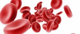
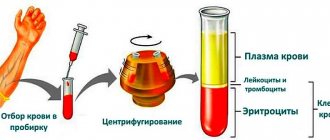
The abbreviation ESR stands for “erythrocyte sedimentation rate,” measured in millimeters per hour. (In old reference books this analysis was called ROE). The research is based on a process caused by gravitational forces. Since red blood cells are heavier than other blood elements, they are the first to precipitate. Their counting in a vertically placed test tube after an hour is the basis of the analysis.
The speed at which red blood cells sink to the bottom depends on many factors, both physiological and pathological. ESR values differ depending on age and gender. Changes in indicators are possible due to dietary habits, the patient undergoing a course of treatment with certain types of medications, corticosteroids, and anti-inflammatory drugs.
Knowing what a high ESR indicates is especially important, since exceeding the standards most often indicates the development of a particular disease. High ESR numbers may indicate the presence of oncopathology, inflammatory process, infectious, rheumatological, or anemia in the body. The indicator will exceed the norm in case of myocardial infarction, trauma, allergies, pregnancy.
Despite the fact that ESR is a nonspecific indicator, the diagnostic value of the study is enormous. This is due to its high sensitivity. A change in this indicator is noted already in the early stages of the development of the pathological process, when other tests are not yet informative and remain normal. An increased ESR in a blood test is a reason to continue the examination and clarify the situation.
Indications for testing
Blood is one of the most informative resources of the human body. By sending it for laboratory testing, the vast majority of diseases can be diagnosed with high accuracy. Clinical analysis contains many indicators, each of which reflects a specific process and function and serves as an important diagnostic criterion. However, despite the abundance of examinations, the most common and in demand at the MedArt clinic is a general blood test (ESR).
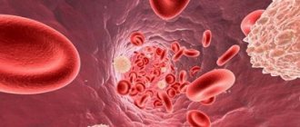
Erythrocyte sedimentation rate is the most important indicator, often confirming the presence of inflammation or other pathology (in the acute and latent stages). The mechanism of this analysis is quite simple, so you don’t need to spend several days to get the result. Red blood cells are much heavier than plasma and other cellular elements, and therefore, by placing blood in a vertically placed test tube, after a certain period of time a specific sediment will form at the bottom of the container, and a translucent liquid will appear at the top.
This is a completely natural phenomenon that occurs as a result of gravity. Red blood cells can stick together, forming entire colonies that settle to the bottom much faster than individual elements. This is due to the larger mass, which may indicate a problem.
ESR norm after 50 years: how to reduce an increased ESR value using folk remedies?
You can use folk remedies to reduce elevated ESR levels in the blood after 50 years in women and men:
- Drink an infusion of anti-inflammatory herbs : chamomile, coltsfoot, calendula, linden, horsetail, sea buckthorn, 2-3 r. per day, a few sips at a time. Drink no more than 1 glass per day.
- Eating vegetables and fruits rich in vitamin C (green onions, citrus fruits, kiwi) will boost your immunity.
- Drink 100 ml of freshly squeezed beet juice before bed for 10 days in a row, it lowers the erythrocyte sedimentation rate.
- Add boiled beets to salads every day.
- Walk outside more often, in the fresh air - lack of oxygen affects the ESR rate after 50 years.
Attention . Beetroot has a laxative effect on stool, it is especially useful for those people who have frequent constipation, and for those who often have diarrhea, you should not get carried away with beetroot salads.
Beetroot in any form can reduce the ESR content that is higher than normal after 50 years: juice, salad, beetroot soup
How to prepare for the test
ESR is included in the list of standard indicators that are displayed in all blood tests (general and clinical). However, special attention is paid to it in the following situations:
- Diagnosis confirmation
- Preventive examination
- Evaluation of the effectiveness of prescribed treatment
- Infectious and inflammatory pathologies
- Autoimmune disorders
- Tumors (malignant and benign) of any location
Many pathologies of internal organs are asymptomatic, and often identifying an ESR deviation from the norm becomes a reason to begin a more detailed diagnosis, thanks to which it is possible to identify the problem in the early stages and begin effective treatment. Most often, after any abnormalities are found, an additional biochemical analysis is prescribed, which allows a more detailed study of the bloodstream.
Normal ESR after 50 years: symptoms indicating a low level of ESR in the blood
Indirect signs by which you can find out that the ESR norm after 50 years in women and men is underestimated:
- Weakness and dizziness are common
- Shortness of breath and dry cough appeared
- Sometimes I feel nauseous to the point of vomiting
- Nosebleeds
- Rapid breathing
- Losing weight for no apparent reason
- Bruises on the skin from small touches
If, after 50 years, women and men often feel dizzy and constantly feel weak in the body, it is worth taking a blood test and checking the ESR level in the blood
How the research is carried out
The accuracy of this diagnostic method depends on many nuances: proper preparation for the test, the professionalism of the laboratory worker and the quality of the reagents. Subject to these conditions, you can guarantee the most reliable result. And if the person donating blood cannot influence the last 2 points, then the preparatory stage completely depends on him. Despite the fact that in this case no special and complex preparation is required, there are a number of mandatory general rules that are strongly recommended to be followed.
First of all, 1 day before the test, you must stop drinking alcoholic beverages, and also refrain from eating for 4-5 hours. Only drinking plain water is allowed. It is also recommended to quit smoking an hour before the test. Secondly, if the patient takes (on an ongoing basis or only at the moment) any medications, then the doctor must be informed about this in advance. Some medications can distort the results, which is why their use may be stopped and reinstated after donating blood. Thirdly, on the eve of the procedure you should not visit sports or gyms. You should also refrain from intense physical activity and avoid emotional stress.
If you doubt anything, just call us at this number +375(29) 666-30-96 or make an appointment with a doctor using our online form.
Classification
There are no clear digital gradations for dividing the increase in ESR by degree. Conventionally, moderate and high degrees are distinguished. According to the mechanism of occurrence they distinguish:
- True increase in ESR
. The cause is various inflammations, infectious, and oncological pathologies. Develops as a result of dysproteinemia, which increases erythrocyte aggregation. - False increase in ESR
. False acceleration of ESR is observed in anemia, azotemia, alkalosis, and high blood cholesterol levels. The cause of this phenomenon is various pathological processes in which the ESR increases due to changes in the number or shape of red blood cells, the protein-lipid composition of plasma, a shift in blood pH, and the presence of other chemical compounds.
How to prepare
The duration of the analysis does not exceed 5-10 minutes. As a rule, the procedure is accompanied by slight pain and discomfort in the puncture area, but the discomfort passes very quickly. If capillary blood is needed, then before piercing the third or fourth finger of the left hand, the skin in this place is treated with an alcohol cotton ball. After this, using a special medical blade, a small incision is made on the fingertip (its depth does not exceed 3 millimeters). The resulting drop of blood is disposed of with a sterile napkin, after which the laboratory assistant proceeds to collect the biomaterial. Having collected the required amount, the wound surface is lubricated with an antiseptic, and a cotton swab with alcohol is applied to the puncture site.
If the analysis involves taking biomaterial from a vein, then the patient’s forearm is tightened with a medical tourniquet or strap, after which he must work a little with his fist (clench and unclench) for better vascular filling. The site of the intended puncture is treated with an alcohol wipe, after which a needle is inserted into the selected vessel, to which a test tube is connected to collect the released blood. Having collected a sufficient amount of biomaterial, the needle is removed, and a cotton swab with alcohol is applied to the wound.
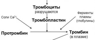
To calculate ESR, an anticoagulant is placed in the biological material to prevent clotting. Then it is sent to a vertical container for 60 minutes. Since the specific gravity of red blood cells exceeds the weight of plasma, gravity forces them down to the bottom of the container. Because of this, 2 visible layers are formed in the test tube: the upper (colorless plasma) and the lower (erythrocyte accumulations). Then the laboratory assistant takes measurements of the top layer. The indicator corresponding to the mark between the red blood cells and the plasma zone on the test tube scale is ESR (indicated in mm/h).
Today there are 2 main ways to detect ESR:
- Panchenkov's method. The capillary is divided into exactly one hundred compartments, and later 5% sodium citrate is added to it to the “P” level. Then the capillary is filled with biomaterial up to the letter “K”. The resulting mixture is mixed and installed vertically. The assessment takes place after 60 minutes.
- Westergren method. Here, venous blood is used, mixed with sodium citrate 3.8% in a ratio of 4:1. It can be mixed with Trilot B followed by the addition of sodium citrate or saline solution in an amount of 4:1. The study is carried out in test tubes equipped with a 200 mm scale. The result is assessed after 60 minutes. This technique is used everywhere, and its main distinguishing feature is the type of test tubes and measuring scale used.
Despite the coincidence of the results of these methods, the Westergren method is famous for its greater sensitivity to exceeding the ESR indicator, and therefore it is considered highly accurate and informative.
Two methods of performing a laboratory blood test for ESR
In Russia, two approaches to conducting this analysis are widespread. They differ in the degree of accuracy, the type of biological material being analyzed and the procedure for collecting it.
1.According to Panchenkov
To perform this test, capillary blood is usually taken from a finger. It is mixed with a special anticoagulant that prevents clotting in a ratio of 1 to 4. Then it is placed in a Panchenkov stand - a glass tube with a stand and a scale with 100 divisions. After an hour, when the blood is divided into plasma and heavier formed elements (erythrocytes, platelets and leukocytes), a measurement is taken.
This method is extremely common in the post-Soviet space.
2.According to Westergren
According to the method, blood is first taken from a vein. Then the analyzed material is mixed with an anticoagulant and placed in a special tube 300 mm high. Moreover, it is filled to ⅔. measurements are taken within an hour. It turns out that the result is assessed on a scale with 200 divisions - its accuracy is higher.
Westergren analysis complies with international standards.
Note! In 5% of the total population of the world, ESR indicators from birth differ from the standard. And this is not accompanied by any pathologies.
MCH norm indicators
The figure varies depending on gender and age. For newborns (up to 1 month), ESR ranges from 1 to 2 mm/h. These limits are explained by reduced protein concentration. From 1 month to six months it ranges from 12 to 17 mm/h. This sharp increase in the norm is explained by age-related processes that occur in the growing body. Then the data is stabilized - for a child under 10 years of age, normal limits are considered to be numbers from 1 to 10 mm/h.
Since blood viscosity has several gender differences, the ESR rate will be different for men and women. For representatives of the fair sex from 10 to 50 years old, the acceptable limits are 0-20 mm/h, and from 50 years old - from 0 to 30 mm/h. The number may change during pregnancy, which is normal, but requires monitoring by your attending physician. In men from 10 to 50 years old, this figure should be from 0 to 15 mm/h, and over 50 years old - from 0 to 20 mm/h.
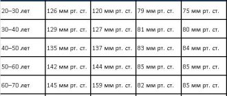
| Age, years | ESR norm |
| Baby up to 1 month | 1-2 mm/h |
| Child 1 month – 6 months | 12-17 mm/h |
| Child under 10 years old | 1-10 mm/h |
| Woman 10-50 | 0-20 mm/h |
| Woman over 50 | 0-30 mm/h |
| Male 10-50 | 0-15 mm/h |
| Man over 50 | 0-20 mm/h |
The final result is influenced by many factors: improper preparation, anxiety, taking medications and much more. In addition, the value may even depend on the time of day. As a rule, the maximum is determined around noon.
An increase in erythrocyte sedimentation rate (ESR) in women under 50 years of age is observed when the value is more than 15 mm/h, in women over 50 years of age - more than 30 mm/h; in men under 50 years old - more than 10 mm/h, over 50 years old - more than 20 mm/h.
Mechanism of occurrence
ESR depends on the intensity of the formation of erythrocyte agents, which is associated with the properties of the plasma and the charge of the erythrocyte membrane. The most important property of blood plasma that affects this indicator is its viscosity. It increases as the protein spectrum shifts towards coarsely dispersed proteins. This occurs primarily with an increase in the amount of fibrinogen, the main stabilizer of red blood cell suspension. An increase in other globulins in the plasma (γ-globulins, α2-globulins) also leads to a drop in the electrical charge of erythrocytes and promotes their aggregation. ESR also depends on the number, size, volume of red blood cells, and the concentration of hemoglobin in them. The fewer these cells, the faster they settle in the capillary.
On average, men have more red blood cells than women, so the ESR is higher in the latter. It also increases at low temperatures. As for physiological conditions, pregnancy is accompanied by a significant acceleration of ESR (up to 30–40 mm/h). In 1984, L. Wilson and co-authors found that in healthy elderly people this figure can reach 50–60 mm/h.
When the level of bile acids in the blood increases, the ESR slows down. Severe hypofibrinogenemia, for example in severe liver damage, can prevent an increase in ESR even with significant dysproteinemia. Its slowdown is facilitated by an increase in the partial tension of CO2 in the blood, as well as erythrocytosis.
When is accelerated ESR observed?
1. Inflammatory diseases:
- bacterial infections,
- immune inflammation,
- aseptic inflammation,
- viral infections.
2. Blood diseases:
- anemia,
- paraproteinemic hemoblastoses,
- other forms of hemoblastosis.
3. Malignant tumors.
4. Metabolic diseases:
- amyloidosis,
- diseases occurring with impaired fat metabolism.
Inflammatory diseases
An increase in ESR is associated with the development of dysproteinemia, the appearance in the bloodstream of tissue breakdown products, C-reactive protein, immune complexes and other components that change blood viscosity and the potential of the erythrocyte membrane. In bacterial infections, the severity of this process is often determined by the severity of the pathology.
With purulent inflammations and abscesses of various organs, the ESR is significantly accelerated. Sometimes its indications lag behind the clinical development of the disease, which is especially evident in acute inflammatory conditions. At the same time, there is no direct connection between the indicators of ESR, body temperature and leukocytosis. ESR increases relatively more slowly and also decreases to normal compared to the number of leukocytes and the clinical manifestations of the disease.
For example, in acute tonsillitis, the maximum acceleration of ESR is most often observed during the period of decreased body temperature and the reverse development of the inflammatory process in the tonsils. Nevertheless, the majority of such common inflammatory diseases as acute appendicitis, cholecystitis, pyelonephritis, pneumonia are characterized by a degree of ESR acceleration that correlates with the severity of the pathological process, although it occurs later than leukocytosis and fever appear.
In chronic inflammatory conditions, an increase in ESR is more often and more consistently recorded than fever and leukocytosis.
Sometimes chronic indolent infections of the biliary tract, urinary system, oral cavity and other localizations occur latently, and acceleration of ESR is one of the few or even the only symptom that allows one to suspect the presence of a chronic focus of infection. Encapsulation of an inflammatory focus, in which decay products do not enter the blood, is not always accompanied by an increase in ESR, in contrast to the rapid entry of necrosis products into the bloodstream. For active tuberculosis, an acceleration of ESR is typical, usually combined with moderate leukocytosis and lymphopenia.
Most specific bacterial infections are associated with an acceleration of ESR. Difficulties in diagnosis arise with latent infections that are not accompanied by a clear clinical picture. It is necessary to identify a latent infection that affects the teeth, tonsils, paranasal sinuses, bile ducts, kidneys, and female genital organs. In addition to a persistent acceleration of ESR, moderate leukocytosis can be detected, sometimes with a shift in the leukocyte formula to the left. C-reactive protein appears in the blood serum, the amount of sialic acids increases, dysproteinemia is possible, mainly due to a moderate increase in globulins of various fractions. Sometimes functional disorders are detected on the part of the organs involved in the inflammatory process. Immune inflammation covers a large group of various diseases with primary or secondary immunopathological reactions. In some cases, exposure to an infectious agent that initially causes infectious inflammation subsequently triggers a chain of immunological phenomena.
A common cause of increased ESR is rheumatism. The level of this indicator correlates with the activity of the rheumatic process and the severity of the exudative phase of inflammation. A significant acceleration of ESR is typical for all systemic connective tissue diseases and is often consistent with the activity of the disease. With these diseases, cryoglobulins sometimes appear in the blood, which sharply increase blood viscosity and reduce ESR. Immune kidney diseases also lead to an increase in it. This is typical for nephrotic syndrome of various origins due to pronounced dysproteinemia and hypercholesterolemia, often hyperfibrinogenemia.
Significant acceleration of ESR has been described in sarcoidosis. Aseptic inflammation also leads to this. It occurs under the influence of various exogenous physical and chemical factors (irradiation, burns, injuries, exposure to acids and alkalis, etc.). In such cases, infection can quickly set in.
A typical example of aseptic inflammation is the so-called resorption-necrotic syndrome during acute myocardial infarction, in which, in particular, ESR increases 1–2 days after the onset of leukocytosis and fever. Moreover, the acceleration remains until the heart attack is completely healed.
The dynamics of ESR, as well as leukocytosis and fever, have a certain diagnostic and prognostic significance. Sometimes this suggests the addition of various complications.
Viral infections, unlike bacterial ones, are rarely accompanied by a significant increase in ESR. In acute viral infections of the respiratory tract, primarily with influenza, a moderate acceleration of ESR is delayed and is often recorded for the first time against the background of already decreasing body temperature and the reverse development of clinical manifestations of the disease. Viral pneumonia often occurs without acceleration of ESR.
Viral hepatitis is characterized by a moderate acceleration of ESR in the pre-icteric period, a decrease in ESR to normal and even lower as jaundice appears, and an increase again with the disappearance of jaundice with a gradual return to normal during recovery. Long-term persistence of a moderately accelerated ESR indicates the persistence of the virus or the addition of a bacterial infection of the biliary tract.
Infectious mononucleosis is accompanied by normal or slightly accelerated ESR in combination with leukocytosis and the presence of polymorphic cells with a large nucleus in the blood.
The bulk of acute viral infections occur with normal or even reduced ESR in combination with moderate leukopenia and relative or absolute lymphocytosis.
Blood diseases
An increase in ESR in anemia is a typical symptom associated primarily with a decrease in the number of red blood cells, although dysproteinemia that is often present also plays a role. There are calculations and nomograms that determine the proper ESR values depending on the number of red blood cells.
It is believed that if the ESR is increased compared to the calculated value, then this may be associated with other severe pathologies that cause anemia (infectious disease, tumor, collagenosis, kidney pathology, etc.). On the contrary, if the ESR is accelerated to a lesser extent, then some authors attribute this to favorable signs of the regenerative nature of anemia (for example, this situation is sometimes observed during reticulocyte crisis in patients with B12-deficiency anemia).
With microspherocytic anemia, there may not be a significant increase in ESR, since the morphological characteristics of erythrocytes in this condition prevent their agglomeration. To diagnose the type of anemia, it is necessary to take into account the medical history, clinical picture, changes in blood tests, bone marrow, and instrumental data (ultrasound, endoscopic examination of the gastrointestinal tract).
Acceleration of ESR is observed in 85% of patients with multiple myeloma due to hyperviscosity syndrome. This diagnosis is made when more than 10% of plasma cells are found in the bone marrow and the paraprotein is present in the serum and/or urine. In Bence-Jones myeloma, when only light chains are secreted, the ESR may remain within normal limits unless severe anemia is present. The second most common, but very rare paraproteinemic hemoblastosis is Waldenström's macroglobulinemia, for which, unlike multiple myeloma, osteolytic processes are uncharacteristic. But hepatosplenomegaly, lymphadenopathy and pronounced hyperviscosity syndrome are typical. The latter appears:
- bleeding of mucous membranes,
- hemorrhagic retinopathy,
- dilatation of retinal veins,
- Raynaud's syndrome,
- ulceration and gangrene of the distal limbs,
- paraproteinemic coma,
- macroglobulinemic retinopathy.
The criteria for the diagnosis of Waldenström's macroglobulinemia are the presence of a mixed cellular substrate in the bone marrow (plasma cells and lymphocytes), detection of monoclonal macroglobulinemia, fibrosis in the trephine.
Heavy chain diseases (HCDs) are B-cell lymphatic tumors. There are options: BTC-γ, BTC-α, BTC-δ, BTC-μ.
With BTC-γ, the average age of patients is 60 years, but can also occur in children. The clinical picture includes: fever, night sweats, weakness, weight loss, lymphadenopathy, splenomegaly, hepatomegaly, damage to the pharyngeal lymphoid ring, recurrent infections, damage to the thyroid gland, salivary glands, skin, subcutaneous tissue, autoimmune processes (25%) with the clinic rheumatoid arthritis, systemic lupus erythematosus, autoimmune hemolytic anemia, thrombocytopenia, thyroiditis, Sjogren's syndrome and others. The bone marrow is affected in 50% of cases. This nosological form does not have a specific histological picture. In the immunochemical diagnosis of BTC-γ, the secretion of fragments of heavy chains of the PIgG subclasses is determined, serum PIg is present in the urine (overload proteinuria), Bence Jones protein is usually absent.
BTC-α is more common in children and young patients under 30 years of age. There are 2 options: abdominal and pulmonary. In the first case, malabsorption syndrome (chronic diarrhea, steatorrhea, exhaustion, edema, hypocalcemia, hypokalemia, baldness, amenorrhea), fever, and attacks of abdominal pain are noted. The pulmonary form is accompanied by bronchopulmonary lesions and mediastinal lymphadenopathy. In the immunochemical diagnosis of BTC-α, fragments of α-chains are determined in blood serum and urine, in the contents of the duodenum and small intestine, and in saliva. Bence Jones protein is never detected in BTC-α. Benign monoclonal gammopathies have been described, which manifest themselves only as a syndrome of increased ESR and are determined biochemically. Sometimes it is possible for such gammopathy to exist throughout the patient’s life. But in some cases, the clinical picture of myeloma or another malignant process gradually develops, sometimes after decades.
As for other forms of hematological malignancies, an increase in ESR is typical for all acute and chronic leukemias, malignant lymphomas, including Hodgkin lymphoma. For diagnosis, in addition to examining the hemogram, sternal puncture and/or trepanobiopsy are necessary. To determine the various forms of lymphomas, it is necessary to conduct histological, enzyme-linked immunosorbent examination of biopsy material, and genetic studies. To clarify the localization of damage to the lymph nodes and internal organs, ultrasound of the abdominal cavity and CT scan of the chest are used.
Malignant tumors
The increase in ESR in this case is associated not only and not so much with the severity of anemia, but with dysproteinemia, an increase in the amount of fibrinogen and a change in the charge of the erythrocyte membrane. More often, persistent and significantly accelerated ESR is observed in cancer of the bronchus, bones, ovary, hypernephroma, sarcomas, and less often in gastrointestinal tumors. Although with cancer of the stomach, colon, pancreas, and liver, a significant increase in ESR is also sometimes encountered. Its magnitude depends on the histological structure of the tumor, but is largely determined by the size of the tumor and the presence of complications. In some cases, an increase in ESR is at the first stage the only manifestation of malignant growth, ahead of clinical symptoms.
With the development of the tumor, other peripheral blood parameters may also change: anemia is often observed (less commonly secondary erythrocytosis), in some cases neutrophilic leukocytosis, lymphopenia, monocytosis, and hyperthrombocytosis are observed. If a neoplasm is suspected and there are no clear clinical manifestations, a thorough and systematic examination is necessary: sternal puncture (detection of signs of hemoblastosis or anemia as the cause of an increase in ESR), ultrasound of the abdominal organs, CT and X-ray examination of the lungs, bronchoscopy, FGDS, FCS. You should consult an obstetrician-gynecologist and urologist.
Metabolic disorders
Most often, an increase in ESR is observed with tissue dysproteinoses. This process may be associated with some forms of hyperlipidemia and widespread atherosclerosis. The main reasons for an increase in ESR in metabolic diseases are dysproteinemia and hypercholesterolemia.
Amyloidosis leads to its acceleration due to severe dysproteinemia. The most common is secondary amyloidosis, which complicates the course of chronic suppurative lung diseases, tuberculosis, chronic osteomyelitis, ulcerative colitis, Crohn's disease, and systemic connective tissue diseases. Paraproteinemic hemoblastoses can lead to the occurrence of this lesion. Amyloid deposition is possible in all organs and tissues. Most often, parenchymal organs are affected: kidneys, liver, spleen, adrenal glands, less often - the gastrointestinal tract, cardiovascular system, lungs, thyroid gland, etc.
There are 4 stages of renal amyloidosis: latent, proteinuric, nephrotic and azotemic. In the first case, the symptoms of the underlying disease, potentially dangerous in relation to amyloidosis, dominate. Sometimes a slight proteinuria appears and a persistent, significantly increased ESR is recorded, which is often not explained by the activity of the underlying pathology. Already at this stage, an enlarged liver and spleen can be palpated, amyloid deposition in which is most common in secondary amyloidosis.
The advanced stage is manifested by complete nephrotic syndrome (proteinuria more than 3 g per day, hypoproteinemia, hyperlipidemia, edema). Symptoms of chronic renal failure subsequently arise and progress. In some patients, amyloid is deposited primarily in the liver. This is characterized by moderate pain in the right hypochondrium, flatulence, and sometimes jaundice. The liver can be enlarged very significantly and descend into the pelvis. In rare cases, a predominant deposition of amyloid in the adrenal glands is possible with a gradual development of the clinical picture of hypocortisolism up to the full-blown symptoms of chronic adrenal insufficiency: increasing weakness, adynamia, persistent decrease in blood pressure, nausea, vomiting, diarrhea, hyperpigmentation of the mucous membranes of the lips and skin on exposed parts of the body and in the area of folds, loss of sexual function. Any impact (intercurrent infection, trauma, etc.) can trigger an adrenal crisis. At the same time, all the listed symptoms increase. Abdominal pain, uncontrollable vomiting appear, dehydration rapidly progresses, convulsive syndrome, oliguria and acute renal failure occur. As a result, a coma develops and the patient dies. Even more rarely, predominant amyloid deposition is observed in the intestine. Clinically, this is manifested by pain, intestinal atony, and persistent diarrhea. Sometimes bleeding is possible.
A persistent and significant increase in ESR as an early symptom is characteristic of primary (idiopathic) amyloidosis.
The most typical lesions are the skin (itching, petechiae, pigment spots, urticaria, sometimes dense swelling that makes the face amicable), muscle and nervous systems (muscle pain, stiffness, muscle tightening, sometimes their atrophy, paresthesia, less often - polyneuropathic syndrome, epileptiform seizures , psychotic reactions). In contrast to secondary amyloidosis, damage to parenchymal organs is less common in primary amyloidosis. Selective deposition of amyloid in any organ is possible. Various forms of hereditary amyloidosis, caused by genetics, have also been described.
A persistent increase in ESR is typical for senile amyloidosis , which is manifested by a triad of Schwartz symptoms: damage to the heart (progressing heart failure), brain (various types of dementia) and amyloid deposition in the islets of Langerhans of the pancreas with the development of symptoms of diabetes. All forms of amyloidosis are characterized by hypercholesterolemia and hyper-β-lipoproteinemia, which occur in the nephrotic stage of renal amyloidosis. When the latter are affected, occasional small proteinuria is observed at an early stage, then it increases and usually exceeds 3 g per day. Proof of amyloidosis is possible by biopsy of various organs and tissues, followed by histological and histochemical examination of the biopsy.
Diseases that occur with impaired fat metabolism, in particular, widespread atherosclerosis, with hypercholesterolemia, can cause a persistent, often moderate, sometimes significant increase in ESR.
In patients with severe atherosclerosis, weight loss, accelerated ESR, sometimes predominant signs of atherosclerotic damage to the cerebral vessels are found (headache, noise in the head or ears, dizziness, syncope, sleep disorders, memory disorders, changes in the emotional sphere, mental disorders; cerebral disorders may occasionally occur vascular crises), and symptoms of damage to the coronary arteries, aorta, and arteries of the lower extremities can be moderately expressed.
Hypercholesterolemia accompanies a variety of diseases, especially common in some forms of liver cirrhosis and nephrotic syndrome of any nature. The increase in ESR in these cases is associated not only with it, but also with severe dysproteinemia. Hypercholesterolemia is typical of hypothyroidism, in which the ESR may increase. It is also typical for some hereditary forms of hyperlipidemia, which contribute to the development of atherosclerosis, for some lipidosis and glycogenosis, often accompanied by a moderate increase in ESR.
A significant acceleration of ESR occurs with generalized xanthomatosis, in particular with Buerger-Grütz syndrome, or hypercholesterolemic xanthomatosis (a disease of middle-aged people; typical are tuberous xanthomas on the face, limbs and mucous membranes, hepatosplenomegaly, chronic pancreatitis, central nervous system damage) and Harbitz-Müller syndrome, or familial hypercholesterolemia (external xanthomas, xanthomatous changes in blood vessels, which can lead to changes in various organs). This process is also described in Urbach-Wiethe syndrome, or mucocutaneous lipoid proteinosis (hyalinosis of the skin and mucous membranes). Typical are nodular deposits in the skin, oral cavity, and vocal cords (which leads to persistent hoarseness), and dysphagia. Possible damage to the central nervous system with epileptiform seizures and mental infantilism. Disorders of fat and carbohydrate metabolism are detected.
conclusions
To summarize, we can say that in order to get an idea of the possible scope of the “diagnostic search” with an increased ESR as a leading sign, it is necessary to obtain detailed anamnestic information. Examination of a patient with an incomprehensible acceleration of ESR requires attention to the main “hot” spots: the size and consistency of the lymph nodes, careful palpation of the spleen, kidneys, listening to the heart, lungs, etc. The doctor must have laboratory and instrumental data.
Increasing ESR
A similar result may be caused by the following pathologies:
- Infection or inflammation.
- Connective tissue diseases (RA, SLE, vasculitis, etc.).
- Burn disease.
- Neoplasms of different etiology and localization.
- Myocardial infarction. In the post-infarction period, the maximum occurs after about 7 days (in this case, you need to contact a vascular surgeon).
- Anemia. These diseases are characterized by a decrease in red blood cells and an increase in their sedimentation rate.
- Injury.
- Amyloidosis (a pathology characterized by the formation of a pathological protein - amyloid).
Despite the discrepancy between normal limits, if a complete blood count of ESR showed an increase in this indicator, this does not necessarily indicate the presence of a problem. This result also occurs in healthy individuals: in women during the menstrual cycle, during pregnancy, or in overweight individuals. This also occurs when taking a number of medications, so you should consult your doctor in advance.
Treatment
Patients are often interested in the question: when can an increase in ESR be considered safe? A deviation of no more than 5 mm/h is considered acceptable. After 3–5 days, you should re-take the test in the same laboratory department.
Consistently high results with double repetition of the analysis require additional diagnostics (leukocyte formula, C-reactive protein, biochemical blood test, etc.).
After the doctor has made a final diagnosis, he selects treatment methods. Treatment of infectious inflammatory diseases begins with identifying the pathogen. If a bacterial nature is detected, a test is performed to determine the sensitivity of the pathogen to known groups of antibiotics. After which preference is given to the antibiotic with the maximum effect on pathogenic bacteria.
When a fungal infection is detected, the most effective antimycotic drugs are similarly selected. Treatment of diseases of viral etiology consists of the use of a complex of drugs with antiviral and immunomodulatory activity. Mixed infection requires the use of several groups of drugs.
Decrease in ESR
A reduced erythrocyte sedimentation rate often signals the presence of water-salt metabolism disorders or active muscular dystrophy. This is often a symptom of erythrocytosis, leukocytosis, hereditary spherocytosis, hepatitis and DIC syndrome. In addition, a similar result is characteristic of polycythemia and the conditions leading to it (CHF or damage to the pulmonary system). Low ESR can also be a consequence of fasting, vegetarianism, taking a number of steroid hormones, and is also often detected in the 1st and 2nd trimester of pregnancy.
You can take an ESR test and undergo other hematological tests at our MedArt medical center. With the help of modern equipment, you can find out absolutely accurate indicators, and highly qualified workers will competently advise you on this or that issue.
What does a low erythrocyte sedimentation rate indicate?
This indicator may be below normal due to:
- Following a strict vegetarian diet;
- Violations of water-salt balance;
- Diseases of the nervous system;
- Fasting;
- Excessive physical activity;
- Taking certain steroid hormones;
- Congenital disorder of the hemoglobin protein structure;
- Pregnancy in the second and third trimesters.
Decreased values are much less common than increased values. Often they do not mean any serious violations. Yet sometimes it is a low erythrocyte sedimentation rate that helps the doctor identify the problem. Or at least understand in which direction to look for it, what studies to prescribe for the patient.
Make an appointment now!
Leave your contact details or call and our manager will contact you to make an instant appointment
Reasons for acceleration and deceleration of ESR
Analysis for ESR is an auxiliary test that gives a preliminary result. To make an accurate diagnosis, it is not enough and additional research is needed. An erythrocyte sedimentation rate that is different from normal most often means that various pathological processes are developing in the body.
ESR increases in any inflammatory diseases accompanied by a rise in temperature
The causes of elevated ESR are various diseases, including:
- Acute and chronic infections and inflammations.
- Pneumonia.
- Liver diseases.
- Tuberculosis, syphilis.
- Myocardial infarction.
- Kidney diseases.
- State of shock.
- Anemia.
- Skin infectious diseases.
- Lymphoma.
- Disorders of the thyroid gland.
- Autoimmune diseases: lupus erythematosus, rheumatoid arthritis, rheumatic fever.
- Fractures and bone injuries.
- Tissue necrosis.
- Surgical interventions.
- Infectious heart diseases.
- Consequences of taking certain drugs.
A very high erythrocyte sedimentation rate is observed in autoimmune diseases:
- Allergic vasculitis.
- Hyperfibrenogenemia.
- Giant cell arteritis.
A low level indicates conditions such as:
- hypofibrenogenemia;
- circulatory failure;
- polycythemia;
- hyperbilirubinemia;
- taking corticosteroids;
- reactive erythrocytosis;
- vegetarianism;
- loss of muscle mass, starvation.