Published: 04/20/2021 11:00:00 Updated: 04/20/2021
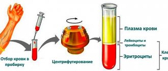
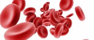
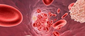
Erythrocytes are red blood cells containing hemoglobin. They carry oxygen and perform many important functions in the bloodstream.
We talked in more detail about red blood cells in the blood earlier.
In urine, red blood cells are contained in extremely small quantities. This is because these red blood cells are large enough that they normally do not pass through the kidney filter. If a lot of blood gets into the urine, it can be seen with the naked eye - the liquid changes color to pinkish or brown. A change in the color of urine is an alarming symptom.
However, a slight increase in red blood cell levels cannot be detected on your own. In this case, laboratory diagnostics will help. Red blood cells in the urine are determined as part of a general urine test.
What to do if you see blood in your urine?
Hematuria is defined as the presence of red blood cells in the urine. It can be described as “macrohematuria,” when blood in the urine is visible to the naked eye, or “microhematuria,” when red blood cells are detected under a microscope in urine tests.
Hematuria can originate from any part of the urinary tract, including the kidneys, ureters, bladder, prostate, and urethra. In many patients, during examination, serious causes leading to hematuria are not determined, but you need to know that hematuria can be a manifestation of infection, urolithiasis, and benign and malignant tumors of the urinary tract. Smoking, radiation, abuse of certain medications, severe pain, exposure to certain chemicals - all this can lead to hematuria.
Red blood cells in urine are normal
Normally, urine should be clear.
It becomes cloudy due to the admixture of red blood cells, leukocytes, and epithelial cells of the urinary tract. Urine can become reddish for two reasons: due to the admixture of blood (in this case, the level of red blood cells is tens of times higher than normal) or due to certain foods that can color urine (beets, blackberries, rhubarb). Therefore, if your urine has changed color, first remember if you have eaten meals with these foods in the last 24 hours - perhaps the reason for the changed color is not at all related to health problems. Normally, urine contains only a few red blood cells or none at all. The urine analysis of a healthy person contains no more than three red blood cells in the field of view of the microscope. The norm is the same for men and women.
For children, the normal limits are even slightly lower - 1-2 red blood cells per field of view. Exceptions are newborns, who during the first month of life can normally have up to 4 red blood cells in the field of view of the microscope. This is due to the functional immaturity of the glomerular membrane.
What are the common causes of hematuria?
Blood in the urine is often not a sign of illness. Studies have shown that 9 to 18 percent of healthy people may have clinically insignificant microscopic hematuria. However, hematuria is often a warning sign for immediate treatment. Below is a list of the most common causes of hematuria:
- Bladder cancer.
- Kidney cancer.
- Prostate cancer.
- Ureteral cancer.
- Urethral cancer.
- Urolithiasis disease.
- Urinary tract infection.
- Pyelonephritis (kidney infection).
- Kidney diseases (hydronephrosis, polycystic disease, tuberculosis).
- Benign prostatic hyperplasia.
- Radiation or chemical cystitis.
- Urinary system injuries.
- Prostatitis.
- Severe physical strain.
How to detect hematuria?
Visible hematuria often bothers patients and forces them to see a doctor.
Microhematuria is determined by microscopy of urine sediment.
What tests are needed to make a diagnosis?
Any patient with gross hematuria or significant microhematuria should undergo a comprehensive evaluation of the urinary tract. The first step is a thorough history and physical examination. Next, a laboratory urine test and examination of the urinary sediment under a microscope are performed. The urine is tested for protein (a sign of kidney disease) and urinary tract infections. The number of red blood cells in the urine (erythrocyturia) and the content of leukocytes in the urine (leukocyturia) are determined. A urine cytology test is needed to check for abnormal cells. Laboratory blood tests are performed to measure serum creatinine levels (kidney function tests).
Patients with significant protein in the urine or elevated creatinine levels should undergo additional testing to rule out kidney disease.
A complete urologic examination in patients with hematuria also includes x-rays of the kidneys, ureters, and bladder (an overview of the urinary system) to rule out masses and stones. Excretory urography is performed - a method for determining kidney function, based on the introduction of radiopaque agents into the bloodstream, followed by radiography and determination of the release of dye by the kidneys.
Many doctors may choose other imaging tests, such as computed tomography (CT) or multislice computed tomography (MSCT). These methods are preferred and more informative for assessing the condition of the kidneys, and are also the best methods for assessing urinary stones. Recently, many urologists have been using CT urography. This allows the urologist to look at the kidneys and evaluate the condition of the ureters as a result of a single x-ray. In patients with elevated creatinine levels or allergies to radiocontrast agents, magnetic resonance imaging (MRI) or retrograde pyelography is performed to evaluate the upper urinary tract. During retrograde pyelography, the patient is taken to the operating room, a radiopaque contrast agent is injected into the kidney through a ureteral catheter, followed by radiography.
Patients with hematuria undergo cystoscopy under local anesthesia using a rigid, or more often, flexible instrument - a cystoscope. After anesthesia, a cystoscope is inserted through the urethra into the bladder and the bladder and urethra are assessed for the presence of masses.
What to do if there was or is hematuria, but no cause was found as a result of the examination?
In at least 8-10 percent of cases, no cause for hematuria is found. Some studies have shown an even higher percentage of patients without a cause. Unfortunately, we have to admit that the same studies later showed that 3 percent of patients were later diagnosed with malignant tumors of the urinary system.
Thus, there is a risk of underexamining the patient or failing to determine the initial stages of some formations. There are no recommendations for subsequent comprehensive examination. Also, there is still no consensus among urologists on this topic.
It is still recommended to consider repeating urine and cytology tests at 6, 12, 24 and 36 months. In the case of repeated gross hematuria, a positive result of urine cytology, or the appearance of irritating urinary symptoms such as pain during urination or frequent urination, an immediate reassessment of the condition of the urinary system is carried out with cystoscopy and repeated radiological diagnostic methods. If none of these symptoms are detected within three years, no further urological examination is required.
General urine analysis (with sediment microscopy)
IMPORTANT!
The information in this section cannot be used for self-diagnosis and self-treatment.
In case of pain or other exacerbation of the disease, diagnostic tests should be prescribed only by the attending physician. To make a diagnosis and properly prescribe treatment, you should contact your doctor. We remind you that independent interpretation of the results is unacceptable; the information below is for reference only.
General urine analysis (with sediment microscopy): indications for use, rules for preparing for the test, interpretation of the results and normal indicators. Indications for the purpose of the study
A general urine test refers to routine laboratory tests aimed primarily at screening for diseases of the urinary system, since pathological processes in the kidneys and urinary tract affect the properties of urine.
With this simple diagnostic test, infectious and inflammatory diseases such as glomerulonephritis
(inflammation of the renal glomeruli),
pyelonephritis
(inflammation of the renal pelvis),
cystitis
(inflammation of the bladder).
Microscopy of urine sediment allows one to suspect injury or infarction of the kidney, urolithiasis, some neoplasms, renal amyloidosis (a systemic disease in which a specific insoluble protein is deposited in the kidneys, which impairs the functioning of the organ).
In addition to diagnosing kidney and urinary tract diseases, the results of a general urine test with sediment microscopy can provide information about your general health.
Urine is formed as a result of ultrafiltration of blood plasma through the glomeruli of the kidneys. With the development of various diseases, pathological metabolic products enter the blood and are excreted from the body, including through the kidneys.
Preparation for the procedure
Preparation for a general urine test begins the day before the collection of biomaterial. Certain foods, the amount of fluid you drink, taking medications and dietary supplements, and intense physical activity can distort the results of the study.
The day before collecting urine, you should avoid foods that can affect the color of your urine: for example, beets and blueberries give your urine a reddish tint; consuming large amounts of carrots or carotene supplements may change the color of your urine to orange.
On the eve of urine collection, it is not recommended to drink alcohol, coffee, dietary supplements or strong tea. If possible, you should limit your intake of diuretics (diuretics). It is necessary to exclude serious physical activity, as well as visiting a bathhouse or sauna.
Women during menstruation are not recommended to donate urine for testing, since even a small amount of blood will significantly distort the test result.
You should warn your doctor about the medications you are taking, as well as about invasive examinations (for example, cystoscopy) on the eve of the test.
Method of collecting urine for general analysis
- It is necessary to prepare in advance a disposable sterile container for collecting urine (can be purchased at a pharmacy or taken from the INVITRO medical office).
- Before collecting urine, hygienic treatment of the external genitalia should be carried out, without using antibacterial and disinfectants.
For children, you need to adhere to the following rules: girls are washed from front to back (from the pubis to the tailbone) so that bacteria that populate the intestines do not enter the urinary tract. Only wash the skin with soap, since contact with mucous membranes causes irritation, dryness and itching. In boys, the glans penis is fused with the foreskin (physiological phimosis), so it is not recommended to forcefully open the glans penis, as this leads to trauma to the delicate tissue. You just need to slightly pull the skin and rinse with water, but it is unacceptable to direct a stream of water into the opening of the urethra. - For general analysis, as a rule, the first morning urine sample is collected. First, release a small amount of urine into the toilet, then, without interrupting urination, place a container and collect approximately 50 ml of urine. In this case, it is necessary to ensure that the container does not touch the skin and mucous membranes.
- After collecting urine, close the container tightly with a screw cap.
- Special urinals have been developed for newborns and infants. You should not use urine squeezed out of a diaper or diaper - the results will be unreliable, since the diaper is a kind of filter for microscopic elements of urine, which are counted during the study.
- When taking the test during the day, it is not recommended to drink large amounts of water, tea, coffee or diuretics to stimulate urination.
The turnaround time for a general urine test is usually 1 business day. What may affect the results
Factors that may distort the results of the study:
- Violation of hygiene procedures and urine collection techniques.
- Drinking large or small amounts of water.
- Consuming foods, medications, or dietary supplements that change the color of urine.
- Menstruation.
- High blood pressure.
- Intense physical and psycho-emotional stress on the eve of urine collection.
- Visiting a bathhouse, sauna, hypothermia.
- Carrying out invasive procedures on the urinary tract a week before the test.
take a general urine test
at the nearest INVITRO medical office. A list of offices where biomaterial is accepted for laboratory research is presented in the “Addresses” section.
Urine examination includes the study of physical and chemical properties, as well as microscopy of sediment.
Physical properties: quantity, color, odor, transparency, relative density (specific gravity), urine reaction (pH).
Chemical properties: determination of protein, glucose, ketone bodies, urobilinogen, bilirubin, hemoglobin, nitrites, leukocyte esterase.
Microscopy: identification of erythrocytes, leukocytes, cells of flat, transitional and renal epithelium, cylinders, crystals, mucus, bacteria, fungi.
Normal indicators
| Index | Result |
| Quantity | 50 ml |
| Color | Colorless, light yellow, straw yellow, yellow, amber yellow |
| Smell | Odorless or non-specific |
| Transparency | Transparent |
| Relative density of urine (specific gravity) | 1003-1035 |
| Urine reaction (pH) | 5.0-8.0 (in children under 1 month – 5.0-7.0) |
| Protein | > 0.140 g/l |
| Glucose | > 2.8 mmol/l |
| Ketone bodies | > 1 mmol/l |
| Urobilinogen | > 34 mmol/l |
| Bilirubin | Not detected |
| Hemoglobin | Not detected |
| Leukocyte esterase | Not detected |
| Nitrites | Not detected |
| Red blood cells | Up to 2 cells per field of view |
| Leukocytes | Up to 5 cells in field of view |
| Epithelium | Up to 5 squamous epithelial cells per field of view |
| Cylinders | Not detected |
| Crystals | Little or no detectable amounts of urates, calcium oxalates, amorphous phosphates |
| Slime | In small quantities |
| Bacteria | Not detected |
| Fungi | Not detected |
Interpretation of indicators
It should be remembered that a general urine test is a screening test, so its results can be used when prescribing other laboratory and instrumental examinations to clarify the diagnosis.
Urine color
depends on the concentration of substances dissolved in it and ranges from transparent to amber-yellow.
Under normal conditions, urine is colored by products of pigment metabolism (in particular, bilirubin): urochromes, urobilinoids and other substances. When the level of bilirubin in the blood increases, more of it enters the urine and gives it a rich brownish or even greenish-brown color. When erythrocytes (red blood cells), myoglobin (the main protein of muscle tissue) or hemoglobin (the protein contained in red blood cells) enter the urine, its color changes to brownish-red and takes on the appearance of “meat slop.” Taking vitamins and nitrofuran drugs can give urine a lemon-yellow to orange color. When there are a large number of leukocytes (white blood cells), the urine becomes milky in color (a condition called pyuria).
Transparency.
Under normal conditions, urine is clear. Its cloudiness can be caused by the presence of salts, crystals, cellular elements (erythrocytes, leukocytes).
Smell
. Normally, urine has a weak, non-specific odor. The appearance of an ammonia odor may be a sign of a bacterial infection; a peculiar fruity odor (“rotting apples”) appears with an increase in the concentration of ketone bodies (which most often indicates diabetes mellitus - a disorder of glucose metabolism).
Relative density of urine
, or specific gravity, is determined using a urometer. The relative density of urine gives an idea of the concentrating ability of the kidneys and the dilution function, which are reduced, like the relative density of urine, in renal failure.
Urine reaction (pH)
- a pH value that reflects the ability of the kidneys to maintain the acid-base balance of the body. The kidneys are involved in the excretion of hydrogen ions and bicarbonates, maintaining a constant blood pH. The pH value of urine is greatly influenced by diet, metabolic characteristics, infectious and inflammatory processes in the kidneys and urinary tract.
Protein
in urine is a significant marker in the diagnosis of diseases of the kidneys, urinary tract and cardiovascular system; it is also important in the diagnosis of gestosis, a severe complication of pregnancy. The appearance of protein in the urine is called proteinuria. Normally, urine does not contain protein because the kidney filter prevents the release of protein molecules from the blood into the urine. There are several causes of proteinuria.
- Prerenal causes: intense physical and psycho-emotional stress, heart failure, hypertension, fever, gestosis during pregnancy, nephroptosis, forced prolonged standing (often with hairdressers, surgeons, military personnel), injuries, disturbance of the protein composition of blood plasma.
- Renal causes: glomerular damage (glomerulonephritis and glomerulopathy), tubular damage, nephrosclerosis.
- Postrenal causes: infectious and inflammatory processes in the urinary tract, neoplasms.
Urine glucose.
The appearance of glucose in the urine depends on its concentration in the blood. Normally, glucose is completely reabsorbed from primary urine into the bloodstream if its concentration in the blood has not reached the “renal threshold” - 8.8-10.0 mmol/l. An increase in the level of glucose in the urine is possible due to a number of physiological reasons: errors in the diet (abuse of sweet foods, especially on the eve of collecting biomaterial), prolonged fasting, stress.
The appearance of glucose in the urine serves as a signal indicating pathology of the kidneys, endocrine system, side effects of drugs, poisoning, and complicated pregnancy.
Ketone bodies
are a non-specific indicator. The appearance of an increased amount of ketone bodies in the urine is the result of accelerated fat metabolism or decreased carbohydrate metabolism. Most often, an increase in their level is observed during fasting, fever, vomiting, alcohol intoxication and diabetes mellitus.
Urobilinogen
in urine increases in diseases of the intestines, liver, and hemolytic conditions (destruction of red blood cells).
Bilirubin
appears in the urine in case of liver pathologies, infectious diseases and pigment metabolism disorders.
Hemoglobin
is determined by a large number of red blood cells in the urine, with myositis, extensive injuries to muscle tissue, and thrombosis of muscle vessels.
Nitrites
detected in urine when pathogenic microflora is activated in the urinary system.
Increased red blood cell
observed in the following cases:
- Inflammatory diseases of the kidneys and urinary tract of infectious and non-infectious origin (glomerulonephritis, pyelonephritis, nephritis, cystitis, prostatitis, tuberculosis).
- Urolithiasis disease.
- Traumatic damage to the kidneys and urinary tract.
- Fever.
- Arterial hypertension with involvement of the renal vessels.
- Various blood coagulation disorders (hemophilia, thrombocytopenia, overdose of anticoagulants).
- Poisoning with benzene derivatives, aniline, snake venom, poisonous mushrooms, with intolerance to anticoagulant therapy.
- Tumor diseases of the genitourinary system.
The presence of leukocytes
in an amount of more than five cells in the field of view is noted in the following cases:
- Inflammatory diseases of the kidneys and urinary tract of infectious and non-infectious nature (glomerulonephritis, pyelonephritis, tubulointerstitial nephritis, cystitis, urethritis, tuberculosis).
- Urolithiasis disease.
- Renal transplant rejection.
- Systemic inflammatory diseases of non-infectious etiology (nephritis with systemic lupus erythematosus).
Epithelium
. There are cells of squamous, transitional and renal epithelium. A large amount of epithelium indicates increased desquamation of cells of the mucous membrane of the urinary tract when they are traumatized (by a stone, during an inflammatory process).
Cylinders
are formed in the tubules of the kidneys and allow us to determine the level of their damage. Most often found in glomerulonephritis.
Crystals
are detected in the sediment of salts at a certain pH of urine. Most often (though not always) found in patients with urolithiasis.
Slime
Normally, it can be found in urinary sediment in small quantities. An increase in mucus content may be associated both with an inflammatory process in the urinary tract and with errors made when collecting urine for research.
Bacteria and fungi
Normally they are not found in urinary sediment. Their presence indicates the presence of an infectious process in the kidneys and urinary tract or errors made during the collection of biomaterial for research.
If the general urine analysis indicators deviate from the norm, the following instrumental examinations and laboratory tests may be additionally prescribed:
- Comprehensive ultrasound of the urinary system (kidneys, ureters, bladder).
- Comprehensive ultrasound of the abdominal organs (liver, gall bladder, pancreas, spleen) for suspected diseases of the liver, gall bladder, etc.
- Study of biochemical blood test parameters (total protein, protein fractions, C-reactive protein, ALT, AST, LDH, GGT, total and direct bilirubin, cholesterol, creatinine, glucose, electrolytes: potassium, sodium, chlorine, calcium).
- Determination of fasting blood glucose, conducting an oral glucose tolerance test, studying the level of glycated hemoglobin for diagnosing diabetes mellitus.
- Urine culture for microflora and determination of sensitivity to antimicrobial drugs.
- Urine culture for microflora and determination of sensitivity to an extended range of antimicrobial drugs.
If the indicators of a general urine test change, you may need to consult a pediatrician or general practitioner, as well as a nephrologist.
Sources:
- Pediatric nephrology: Textbook / ed. P.V. Shumilova, E.K. Petrosyan, O.L. Chugunova. – M.: MEDpress-inform, 2021. – 616 p.: ill.
- Grebenev A.L. Propaedeutics of internal diseases: Textbook. – 5th ed., revised. and additional – M.: Medicine, 2001. – 592 p.: ill.
- Danilova L.A. Tests of blood, urine and other biological fluids at different age periods. – St. Petersburg: SpetsLit, 2014. – 111 p.
- .
IMPORTANT!
The information in this section cannot be used for self-diagnosis and self-treatment. In case of pain or other exacerbation of the disease, diagnostic tests should be prescribed only by the attending physician. To make a diagnosis and properly prescribe treatment, you should contact your doctor.