Renal colic is a symptom complex, an acute pain syndrome indicating a sudden disruption of the outflow of urine. It ranks first among emergency urological conditions. The sensations are excruciating; patients describe them as “unbearable pain.” During an attack, the sufferer cannot sit in one place and tries to find a position or pose that would bring relief.
Unpleasant symptoms are not the only negative aspect. Due to the blockage of the urinary tract, urine formation does not stop. Urine accumulating in the ureter is an ideal substance for the development of bacterial colonies, viruses and fungi. Due to the constantly expanded walls of the ureters, the overall hydrodynamics are disrupted. If waste substances are not removed from the body, intoxication begins.
Causes and risk factors
Obstruction of the urinary tract in 85% of cases occurs due to the appearance and movement of stones (urolithiasis). But for colic, an accumulation of mucus, pus, and blood clots is enough (in case of injury, for example). Compression of the ureter begins with enlarged tumors and enlarged lymph nodes.
Among the pathologies leading to renal colic, urologists most often name the following:
- Acute and chronic pyelonephritis: during the inflammatory process, mucus, leukocytes, platelets appear in the pelvis and calyces, and with a long-term illness - fibrin;
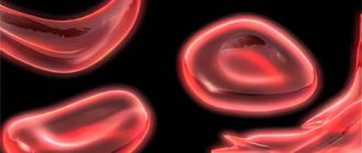
- Benign and malignant tumors: cause blood clots in the blood;
- Tuberculosis: promotes the appearance of necrosis, particles of dead tissue are sometimes released.
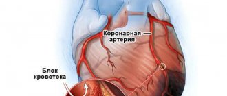
Vascular diseases - infarction or embolism of the kidney, venous thrombosis, dissecting aneurysm of the abdominal aorta, also cause disruption of the outflow of urine, and therefore are conditionally considered a risk factor for the development of renal colic.
Allergic reactions, injuries and diseases of the pelvic organs and retroperitoneal space lead to narrowing of the lumen of the ureters. Developmental anomalies (bends, kinks) of elements of the urinary system lead to disruption of the outflow of urine and can cause renal colic.
The attack begins due to changes in the internal structures of the upper urinary tract due to scarring: normal contraction is impossible due to decreased function of muscle tissue.
An attack sometimes begins in the complete absence of predisposing factors. For example, a person sits calmly in front of the TV or has just climbed from the first floor to the second. However, in most cases, the symptom complex is preceded by severe physical exertion, stress or trauma.
MAKE AN APPOINTMENT WITH A UROLOGIST
Klokov Andrey Nikolaevich
Urologist-andrologist, doctor of the highest category
Make an appointment
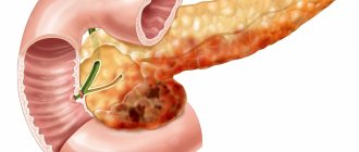
Installation of nephrostomy percutaneous (puncture) nephrostomy
A nephrostomy tube is a thin tube with holes at one end called a pig tail. The opposite end of the nephrostomy is represented by a straight tube and serves to connect to a bag for urine collection.
The role of a nephrostomy is the same as that of a stent (ensures unimpeded outflow of urine from the kidney), but the difference lies in the installation method. In the case of installing a stent, the operation is performed through the natural urinary tract, and when installing a nephrostomy, through a puncture in the lower back in the projection of the affected kidney. This procedure is used in cases where it is not possible to install a stent through the natural urinary tract.
Nephrostomies can be installed for several weeks or months. In the case of renal colic, a nephrostomy is installed for a period of about two weeks. The operation, like ureteral stenting, is performed in an endourological X-ray operating room, where there is the necessary equipment for ultrasound (ultrasound machine with a special adapter for punctures) and X-ray television control (C-arm). In our clinic, such an operating room operates around the clock.
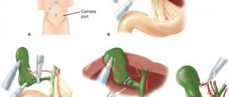
Operation percutaneous nephrostomy.
We will inform you in advance about the stages of the operation and tell you what to expect during the installation of a nephrostomy. The operation is performed under local or intravenous anesthesia, which eliminates or minimizes discomfort. The choice of anesthesia method depends on patient preference and medical indications.
The operation begins with positioning on the operating table. You can lie on your healthy side or on your stomach. It depends on your build and the location of the kidney relative to the costal arch. Visualization of the kidney and monitoring during surgery is carried out, as mentioned above, under ultrasound and X-ray television control. The exception is pregnant women, where x-rays are not used.
Using ultrasound guidance, the doctor finds the puncture site in the lumbar region where the trauma to the kidney will be minimal, and the stent curl will be securely located in the pelvis. The puncture is carried out with a needle, 15-20 cm long and 2 mm in diameter. After the puncture of the pelvis is carried out through the lumen of the needle, a conductor (a very thin string) is installed into the cavity system of the kidney, through which a nephrostomy is carried into the kidney.
As a rule, after installing a nephrostomy, the symptoms of renal colic subside and you can be discharged home after a few days. It is important to drink more fluids than usual for the next few days after the procedure. If you do not have a fever, then calculate 30 ml of drinking water per kilogram of weight per day.
Patients often ask the question: what kind of nephrostomy care is needed and what should I change in my lifestyle in these 2-3 weeks?
- Hygienic measures allow you to take a shower, followed by treating the nephrostomy site with an antiseptic solution and dressing.
- A lifestyle that is not associated with heavy physical activity and heavy lifting (walking, driving, working at a desk) is allowed.
- There are no dietary restrictions unless there are other recommendations from the attending physician.
- The main thing is the functioning of the nephrostomy. The amount of urine excreted, of course, is individual, but it is necessary to ensure that the urine bag is regularly filled.
Pathogenesis
The development of renal colic occurs in patients in approximately the same way:
- Due to the cessation of the normal outflow of urine, newly formed by the kidneys from the blood, pressure increases in the ureters, calyces and pelvis.
- Despite the change in urine output, urine does not stop being produced.
- Due to this problem, the muscles of the ureter begin to contract with an unnatural frequency. Contractions develop into spasms - a sharp and stable shortening of the muscles. Accompanied by severe pain.
- At this time, the kidney tissue swells and becomes inflamed without timely medical intervention: this is how the body reacts and signals a problem.
- Due to the increased parenchyma of the organ, the nerve endings are pinched, causing painful sensations.
Preventive actions
To protect your body and eliminate signs of renal colic in women
, you need to carry out a number of activities:
- do not delay treatment,
- during the treatment of inflammatory processes,
- do not overload your body with physical activity,
- At the first signs of illness, consult a specialist for examination.
Modern treatment methods will help you quickly and effectively cope with the disease. You just need to seek qualified help in time. Specialists will help relieve pain and provide all necessary, timely assistance to restore the outflow of fluid from the kidneys.
Symptoms of renal colic
The most obvious sign of pathology is severe pain in the lumbar region, usually on one side. It does not stop from several minutes to several hours. Almost immediately it moves to other areas: pubis, groin, inner thigh.
The patient becomes restless, irritable, and cannot find a position that helps relieve pain. More often, the sufferer takes a reclining position, tightly pressing the lumbar region - this is where the unpleasant sensations are localized. However, the technique does not help for long; you have to take a different position again. It is on this basis that doctors immediately dismiss other pathologies: acute appendicitis or cholecystitis, for example, in which patients take a lying position and do not strive to change anything.
Severe pain that interferes with life is not the only symptom of problems:
- blood pressure increases;
- nausea and vomiting appear;
- bloating;
- flatulence;
- constipation
Frequent urination may occur, accompanied by additional acute pain in the urethra. There is sometimes blood in the urine. The temperature rarely rises, usually only in case of infection.
Renal colic can subside as quickly as it began. However, more often the symptoms decrease gradually: the pain is replaced by a dull, aching sensation. Only a urologist can determine whether the attacks will continue.
Stent placement (optical internal stenting)
A ureteral stent is a thin tube with many holes that is placed in the ureter to allow urine to drain freely from the kidney. Urine passes inside the lumen of the stent and the stone does not interfere with this. The stent has two fixation curls, one of which is located in the kidney and the other in the bladder. Ureteral stents can be placed for several weeks, months or years. In the case of renal colic, the stent is installed for about two weeks.
Operation optical internal stenting.
Before surgery, we will tell you what to expect during ureteral stent placement. The operation is performed under local or intravenous anesthesia, which makes you feel more comfortable during the procedure. The choice of anesthesia method depends on patient preference and medical indications.
To place a stent, your doctor will first insert a cystoscope through your urethra. A cystoscope is an optical instrument of small diameter that allows you to transmit an image to the monitor of an endoscopic stand. A cystoscope is used to find the ureteral orifice (the opening through which the ureter connects to the bladder). After finding this hole, the doctor will insert a stent into the ureter through the cystoscope. Once the stent is in place, the cystoscope will be removed. The operation in our clinic is carried out under endoscopic, X-ray and ultrasound control.
After the operation, you will be transferred to the recovery room, where medical staff will monitor you. If pain occurs, your doctor may prescribe pain medication to make you feel more comfortable. As a rule, after installation of a ureteral stent, the symptoms of renal colic subside and you can be discharged home after a few days. During the first 48 hours after the procedure, it is important to drink more fluids than usual. Calculate 30 ml of drinking water per kilogram of weight per day. Approximately one day after the procedure, you can return to your normal lifestyle.
Since the stent is a foreign body (even the best quality), you can feel its presence in the form of a change in the act of urination.
You may have:
- Frequent urination, i.e. needing to urinate more often than usual
- A strong, sudden urge to urinate accompanied by bladder discomfort
- Blood in the urine. This may occur as long as the stent is in place. Sometimes this happens after increased activity. If you notice blood in your urine, drink more fluids than usual until the blood goes away
- Pain in the lower back, on the side where the stent is located, at the end of urination
- Pain in the pelvis and lower abdomen
These symptoms usually disappear over time. However, if so-called stent-related symptoms are severe enough to affect your quality of life, your doctor may prescribe medication to relieve symptoms. In rare cases, it is necessary to remove the stent ahead of schedule.
Call your doctor if you have:
- chills - temperature 37.7°C or higher - burning sensation when urinating - cloudy urine - urine has an unpleasant odor - taking painkillers does not reduce pain
Diagnosis of the problem
If urination is disturbed in the pelvicalyceal system, pain can radiate to the navel area and subcostal area. Therefore, the main goal of diagnosis at the initial stage is to differentiate renal colic from attacks of appendicitis and other pathologies of the retroperitoneal space.
Doctors collect anamnesis and conduct specific tests:
- Definition of Pasternatsky's symptom. The urologist taps his fingertips on the back, in the area of the kidneys. The patient feels increased pain. Then the patient submits his urine for analysis - blood agents are determined in the biological fluid.
- Deep palpation of the abdomen. The doctor reveals an enlarged kidney; when pressing on the area where the organ is located, the painful sensations intensify.
- Novocaine blockade according to M.Yu. Lorin-Epstein. At the same time, it confirms or refutes the preliminary diagnosis, reduces pain if the cause is renal colic. If the painful sensations do not go away, in 99% of cases it is not renal colic.
Next, the urologist prescribes laboratory blood tests and a urine test. The level of ESR in the blood increases. During an attack, urine changes consistency, color and other characteristics. Cloudiness, blood clots or threads, and a whitish sediment are observed.
Instrumental diagnostics help determine the cause of the pathology. The formation of stones and narrowing of the lumen of the urinary tract are determined by the following research methods:
- Survey radiography;
- Excretory urography with contrast agent;
- Retrograde pyeloureterography;
- Chromocystoscopy;
- Ultrasound;
- CT and MRI.
The equipment helps to determine the location of stones, the volume of enlarged tumors, and impaired blood supply.
Treatment of renal colic
The primary task is to stop the attack by eliminating spasm of the ureteral muscles. Medicines help to cope with the problem:
- Antispasmodics;
- Analgesics (ganglionic blockers);
- Anti-inflammatory nonsteroidal or steroidal;
- Antidiuretics;
- Antihistamines.
Doctors recommend placing a warm heating pad on the affected area while the ambulance is traveling. Hot baths help reduce symptoms.
Doctors choose different forms of medications. Tablets and capsules help with moderate attack intensity. If the damage is severe, doctors prescribe intravenous or intramuscular injections. Sometimes catheterization of the ureter is required to prevent waste fluid from accumulating.
In difficult situations, when there is a risk of death or disability, doctors resort to radical measures:
- Percutaneous pyelonephrostomy: doctors install a synthetic drainage for normal urine outflow;
- Open (minimally invasive) removal of stones of the pelvis, cup, ureter;
- Kidney decapsulation: doctors excise the kidney capsule, internal pressure decreases;
- Nephrectomy: Doctors choose a method if severe kidney damage, a malignant process or necrosis is detected.
Next, doctors must treat the underlying disease: pyelonephritis, nephroptosis.