Some medications, such as glucocorticoids and immunosuppressants, may suppress the development of vaccine immunity. In such cases, vaccination tactics should be discussed with your doctor.
For vaccines against the new coronavirus infection, the duration of vaccine immunity remains one of the main questions. It is very difficult to predict the duration of protection of a particular vaccine. This is usually determined in practice by recording the frequency of infections in people vaccinated during mass vaccination after different amounts of time, as well as measuring the titer of protective antibodies.
What are antibodies? And how to decipher the results of the analysis?
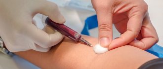
Antibodies are proteins that the immune system produces in response to infection. In laboratory diagnostics, it is antibodies that serve as markers of infection. The general rule for preparing for an antibody test is to donate blood from a vein on an empty stomach (at least four hours must pass after eating). In a modern laboratory, blood serum is examined on an automatic analyzer using appropriate reagents. Sometimes a serological test for antibodies is the only way to diagnose infectious diseases.
Tests for infections can be qualitative (they answer whether there is an infection in the blood) or quantitative (they show the level of antibodies in the blood). The level of antibodies for each infection is different (for some there should be none at all). Reference values (normal values) of antibodies can be obtained with the test result. In the online laboratory Lab4U you can take a set of tests for all TORCH infections at one time and with a 50% discount!
What is the "positivity rate"?
Semi-quantitative tests are used in most cases to determine antibody levels after vaccination. They do not show the exact amount of antibodies formed. But with their help, you can assess the intensity of the immune response - by the brightness of the sample, if the test is carried out using the enzyme-linked immunosorbent assay (ELISA).
Tests from different manufacturers have different scales. In most cases, state laboratories use Vector-best test systems, and their cut-off level is 1. That is, if the indicator is lower, there are no antibodies. From 1 to 1.1 is the so-called gray zone. If the result is higher than 1.1, it means that we have a strong positive reaction of the immune system.
The “positivity rate” shows how many times the level of antibodies in the sample exceeds the threshold level for their recognition. So, for example, the indicator of 15, which the president recently spoke about, is a good result. The majority of those vaccinated with Sputnik V or CoviVac have approximately the same or even higher rate of positive immune response.
At the same time, I would like to note once again that other test manufacturers may have a different scale and, accordingly, different indicators. At the same time, when you receive the test result in the laboratory, you can always see the reference values on the form and compare the result with them.
Various classes of antibodies IgG, IgM, IgA
Enzyme immunoassay determines infectious antibodies belonging to various Ig classes (G, A, M). Antibodies to the virus, in the presence of infection, are detected at a very early stage, which ensures effective diagnosis and control of the disease. The most common methods for diagnosing infections are tests for IgM class antibodies (acute phase of infection) and IgG class antibodies (sustained immunity to infection). These antibodies are detected for most infections.
However, one of the most common tests - hospital screening (tests for HIV, syphilis and hepatitis B and C) does not differentiate the type of antibodies, since the presence of antibodies to the viruses of these infections automatically assumes a chronic course of the disease and is a contraindication, for example, for serious surgical interventions. Therefore, it is important to refute or confirm the diagnosis.
A detailed diagnosis of the type and amount of antibodies for a diagnosed disease can be done by taking an analysis for each specific infection and type of antibodies. Primary infection is detected when a diagnostically significant level of IgM antibodies is detected in a blood sample or a significant increase in the number of IgA or IgG antibodies in paired sera taken at an interval of 1-4 weeks.
Reinfection, or repeated infection, is detected by a rapid rise in the level of IgA or IgG antibodies. IgA antibodies have higher concentrations in older patients and are more accurate in diagnosing ongoing infection in adults.
A past infection in the blood is defined as elevated IgG antibodies without an increase in their concentration in paired samples taken at an interval of 2 weeks. In this case, there are no antibodies of classes IgM and A.
What test can be done before vaccination?
Currently, IgG antibody tests are most often performed by people about to get vaccinated to ensure they have not previously had COVID-19 asymptomatically or have naturally acquired immunity. If the test shows a positive IgG antibody titer, vaccination can be postponed.
What if a person wants to check the level of antibodies after vaccination? Now many people compare their performance.
You need to understand that the tests used in laboratories are different and the scales they use are also different. Therefore, if you try to compare results (although there is not much point in this), then you must at least be sure that you tested with the same test.
IgM antibodies
Their concentration increases soon after the disease. IgM antibodies are detected as early as 5 days after onset and reach a peak between one and four weeks, then decline to diagnostically insignificant levels over several months, even without treatment. However, for a complete diagnosis, determining only class M antibodies is not enough: the absence of this class of antibodies does not indicate the absence of the disease. There is no acute form of the disease, but it may be chronic.
IgM antibodies are of great importance in the diagnosis of hepatitis A and childhood infections (rubella, whooping cough, chickenpox), easily transmitted by airborne droplets, since it is important to identify the disease as early as possible and isolate the sick person.
References
- Encyclopedia of clinical laboratory tests / ed. WELL. Titsa. - M.: Labinform, 1997. - P. 213-215, 223-226.
- JVaillant, A., Jamal, Z., Ramphul, K. Immunoglobulin. In: StatPearls, 2021.
- Sathe, A., Cusick, J. Biochemistry, Immunoglobulin M. StatPearls Publishing, 2021.
- Histology (introduction to pathology) / ed. E.G. Ulumbekova, Yu.A. Chelysheva. - M.: GEOTAR-Media, 1997. - P. 528 – 530.
- Kumar, V., Abbas, A., Fausto, N. et al. Robbins and Cotran Pathologic Basis of Disease, 2014. - 1464 p.
IgG antibodies
The main role of IgG antibodies is the long-term protection of the body from most bacteria and viruses - although their production occurs more slowly, the response to an antigenic stimulus remains more stable than that of IgM class antibodies.
Levels of IgG antibodies rise more slowly (15-20 days after the onset of illness) than IgM antibodies, but remain elevated longer, so they may indicate a long-standing infection in the absence of IgM antibodies. IgG may remain at low levels for many years, but upon repeated exposure to the same antigen, IgG antibody levels rise rapidly.
For a complete diagnostic picture, it is necessary to determine IgA and IgG antibodies simultaneously. If the IgA result is unclear, confirmation is carried out by determining IgM. In case of a positive result and for an accurate diagnosis, a second test, done 8-14 days after the first, should be checked in parallel to determine the increase in IgG concentration. The results of the analysis must be interpreted in conjunction with information obtained in other diagnostic procedures.
IgG antibodies, in particular, are used to diagnose Helicobacter pylori, one of the causes of ulcers and gastritis.
INTERPRETATION OF RESULTS.
In healthy individuals under 60 years of age, negative results for antibodies to nuclear and cytoplasmic components should be expected; in a low titer (weakly positive result), these antibodies are still found in 2-3% (according to some data, up to 10%) of practically healthy people, but it is never possible to predict whether these people will develop an autoimmune disease in the future (so, according to According to some authors, antibodies to DNA begin to be detected long before the onset of clinical manifestations of SLE; the latent period can last for years). After 60 years, the frequency of weakly positive results constantly increases with age. The clinical significance of these results remains unclear; as a rule, they are detected only in the primary (screening) dilution of 1:40. Based on this, if a positive result is detected at a screening titer of 1:40, titration is always necessary, at least to a diagnostic titer of 1:80. In any case, it seems advisable to carry out regular clinical and laboratory follow-up of individuals who have been diagnosed with low-titre ANA. LIMITATIONS OF THE METHOD A positive result suggests the presence of certain diseases (see below), however, like the result of any laboratory tests, it may require confirmation or clarification using other methods, as well as the establishment of clinical and laboratory correlations. It should be borne in mind that various drugs can cause the formation of autoantibodies (hydralazine, procainamide, phenytoin, carbamazepine, isoniazid, chlorpromazine, penicillamine, levodopa, penicillin, phenylbutazone, oral contraceptives, quinidine). The most common targets of these iatrogenic autoantibodies are nuclear histones, which leads to the development of a homogeneous or homogeneous peripheral type of luminescence.
Antibody analysis in the diagnosis of TORCH infections
The abbreviation TORCH appeared in the 70s of the last century, and consists of capital letters of the Latin names of a group of infections, the distinctive feature of which is that, while relatively safe for children and adults, TORCH infections during pregnancy pose an extreme danger.
The blood test for TORCH infection is a comprehensive study, it includes 8 tests:
— Determination of antibodies to herpes simplex virus type 1,2 IgM and IgG
— Determination of antibodies to cytomegalovirus IgM and IgG
— Determination of antibodies to the rubella virus IgM and IgG
— Determination of antibodies to Toxoplasma gondii IgM and IgG
Often, infection of a woman with TORCH complex infections during pregnancy (the presence of only IgM antibodies in the blood) is an indication for termination.
What else is prescribed with this study?
Humoral immunity (immunoglobulins IgA, IgM, IgG, IgE, circulating immunocomplexes, complement components C3, C4)
17.51. Ven. blood 8 days
2,740 ₽ Add to cart
Immune status (screening) (Phagocytic activity of leukocytes, cellular immunity, total immunoglobulin IgE, immunoglobulins IgA, IgM, IgG)
27.960. Ven. blood 3 days
5 880 ₽ Add to cart
Total immunoglobulin IgE
17.2. Ven. blood 1 day
400 ₽ Add to cart
Cellular immunity (T-lymphocytes, T-helpers, T-cytotoxic cells, Immunoregulatory index, B-lymphocytes, NK-T cells, NK cells, Leukocyte formula)
17.50. Ven. blood 3 days
4,170 ₽ Add to cart
Serum paraprotein typing using immunofixation
50.28.2181 Ven. blood 14 days
4,000 ₽ Add to cart
That is, antibodies destroy viruses and bacteria?
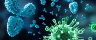
Not really. The purpose of antibodies is to neutralize the “intruder” so that it cannot reproduce, or to mark it for destruction. For example, they stick to viral particles, preventing them from entering the cell. Since the virus is unable to reproduce outside cells, it dies. Antibodies can also clump bacteria together into clumps, which are then devoured by phagocytes, or activate a system of immune proteins that will break through the bacterial membrane and kill it.
An antibody works like a molecular master key: it is born already tailored to a certain structural feature of a foreign body. For example, this could be a site (spike) on the surface of a viral particle through which it binds to a cell. The antibody blocks the spike and the virus cannot infect the cell. Related viruses may have similar structural elements, and then antibodies to one member of the family will be effective against another. For example, some people who have had SARS in the past (caused by the SARS-CoV-1 virus) already have effective antibodies against its relative, SARS-CoV-2, which causes COVID-19 infection.
Normal value for immunoglobulin G
The norm of immunoglobulin G for newborns is 3.97–17.65 g/l. This indicator is due to the fact that the body of a child who has just been born still uses antibodies received from the mother.
In the first year of a child’s life, he begins to produce his own antibodies. At this age, the normal immunoglobulin level for boys is 3.97–17.65 g/l, and for girls – 3.91–17.37 g/l. Norm up to two years: for boys – 4.75–12.1 g/l, for girls – 4.88–12.26 g/l.
Then the amount of immunoglobulins in the blood reaches the same level as in adults. For boys and men, the norm is 5.4-18-22 g/l, for girls and women - 5.52-16.31 g/l.
Immunoglobulin therapy is a natural defense mechanism
Nowadays, immunoglobulins are used to combat various infectious diseases - tick-borne encephalitis, cytomegalovirus, staphylococcus, and also as a therapy for patients with allergies and primary immunodeficiency. In the latter case, treatment using normal human immunoglobulin is indicated. For these types of immunodeficiencies, when the patient’s body is not able to produce antibodies on its own, the normal human immunoglobulin preparation makes up for the lack of antibodies that usually circulate in the blood of a healthy person, providing patients with a high quality of life.
In case of treatment of allergies, an antiallergic immunoglobulin drug is used. Immunoglobulins also help fight autoimmune diseases such as systemic lupus erythematosus, Guillain-Barré syndrome or Kawasaki syndrome.
Immunoglobulin therapy, due to its natural nature, is often prescribed to pregnant women when many antibiotics are contraindicated due to the negative effect on the fetus. At the same time, anti-Rhesus immunoglobulin is the only way to avoid hemolytic disease of the fetus in case of Rh conflict during pregnancy.
Today, the pharmaceutical industry can offer preparations of specific immunoglobulins for various types of microorganisms. But even if the required drug is not available, it can be created quite quickly. This is one of the key advantages of immunoglobulins - the ability to urgently obtain specific antibodies. Thus, immunoglobulin therapy is indispensable when new strains of infection, for example, the influenza virus, appear in epidemics, especially in the absence of a vaccine. It is not surprising that immunoglobulin therapy has become especially relevant during the COVID-19 pandemic.
What can affect immunoglobulin G?
It is important to know that during intense muscle loads, as well as psychological stress, the level of immunoglobulin G can physiologically increase. In addition, immunization in the last 6 months or taking medications may increase the level.
Agents that increase the concentration of immunoglobulin G:
- valproic acid;
- carbamazepine;
- phenytoin;
- chlorpromazine;
- penicillamine;
- dextran;
- oral contraceptives;
- methylprednisolone;
- gold preparations;
- estrogens.
The level of immunoglobulin G can be reduced by:
- burns received;
- radiation exposure;
- taking cytostatics and immunosuppressants;
- intestinal pathologies that are accompanied by protein loss, for example, ulcerative colitis, Crohn's disease;
- renal failure, nephrotic syndrome.
Background: How antibodies turned into a “magic bullet”
Immunoglobulins (antibodies) are special proteins that participate in the immune response. They bind to fragments of dangerous “enemies” of our body - bacteria, viruses, fungi, our own tumor cells, toxins, and trigger a mechanism to destroy the threat. Antibodies can be called an evolutionary invention of humans, like all vertebrates. Our body produces about 2-3 grams of various antibodies per day. In total, a huge number of unique variants of immunoglobulin varieties can be synthesized in the human body throughout life. Thanks to this, our body is able to select a specific antibody to any of the dangerous agents it encounters.
Scientists began studying these processes in the second half of the 19th century. It was then that immunology was born, the first data appeared on the body’s ability to recognize and defeat the causative agent of the disease when encountering it again. Of course, the foundation was laid by the English physician Edward Jenner, who at the end of the 18th century invented the first vaccine against smallpox. Later, scientists found out that such protection is possible thanks to special substances in the liquid part of the blood - serum. This means that the serum can be introduced into the body instead of vaccination and achieve temporary protection or help an already sick person cope with an infection. The first to successfully demonstrate this in practice was the German scientist Emil Behring at the end of the 19th century, when he invented a drug for the treatment of diphtheria based on the blood serum of those who had recovered from the disease.
Emil Bering with assistant
At that time, diphtheria claimed thousands of children's lives around the world - every twentieth child in Europe and the United States fell victim to this insidious disease. Doctors were powerless, so the first success of Behring's anti-diphtheria serum became a real Christmas miracle for some. On the night before Christmas 1891, dying from diphtheria, patients at the Berlin Children's Hospital received an injection of life-saving Behring serum. Then this injection turned out to be the last chance at life for many young patients, but it did not become a salvation for everyone.
His colleague and friend, immunologist Paul Ehrlich, helped Bering refine and perfect the anti-diphtheria serum. His deep knowledge of immunology helped to more accurately calculate the dosage of antitoxin and prepare purified, safer sera. In 1894, the new version was successfully administered to more than 200 young patients.
In 1901, Behring received the first ever Nobel Prize in Physiology or Medicine. In the official formulation, the serum he created was called “a victorious weapon against disease and death.” Seven years later, Paul Ehrlich was also awarded the Nobel Prize - “for the discovery of antibodies and the substantiation of the humoral theory of immunity.”
The term “immunoglobulins”, which is popular today, appeared much later - in 1959 it was proposed by the Belgian immunochemist Joseph Heremans. This name very well reflects the dual nature of antibodies - functionally they are immune factors, and in their structure they belong to the group of blood serum proteins called “globulins”.
In 1972, the chemical structure of antibodies was deciphered; for this, American immunologist Gerald Edelman and English biochemist Rodney Porter also received the Nobel Prize. The next two important discoveries in this area occurred in 1984 - the substantiation of the idiotypic chain theory and the development of a method for producing monoclonal antibodies, which also received the Nobel Prize. From the same period, the development of methods for purifying immunoglobulins began, which became a key condition for the effectiveness of therapy. Today the standard in this field is chromatography.
More than a hundred years of research and four Nobel Prizes - no other discovery in the field of physiology and medicine has received such attention and recognition.