Detailed description of the study
Estriol (E3) is a sex hormone that is one of the main forms of estrogens (female sex hormones) in the body. In addition to estriol, the group of female hormones includes estrone (E1) and beta-estradiol (E2). The main function of estrogen is considered to be the growth and development of the genital organs, as well as tissues that affect reproductive function.
Thus, during puberty, estrogens promote the development of the genital organs - the uterus, fallopian tubes, ovaries and vagina - and the formation of the mammary glands. In addition, female sex hormones perform other important functions:
- Affect the function of the musculoskeletal system: stimulate bone growth and suppress their destruction;
- Participate in fat metabolism: promote their deposition in subcutaneous tissue;
- Regulate water and electrolyte balance: retain sodium in the body, participating in the development of edema.
However, the latter action is of practical importance only in the body of a pregnant woman due to the massive release of estrogens into the blood.
Estriol is a steroid hormone with the weakest estrogenic activity. In numerical comparison, its bioactivity is approximately 12 times inferior to estrone, and 80 times inferior to beta-estradiol. Estriol becomes the main estrogen during pregnancy - up to 90% of all forms of estrogen during this period are made up of it. Normally, its level in the blood increases already at the end of the first trimester, when the placenta, the intermediary organ between the fetus and the mother, begins to form. Estriol is a marker of normal fetal life and uteroplacental circulation.
Assessing estriol concentrations during pregnancy helps identify developmental disorders of the fetus or placenta.
It has been established that a decrease in the level of free estriol in the second trimester of pregnancy is a laboratory sign of Down syndrome and Edwards syndrome. These pathologies relate to chromosomal diseases, that is, they are manifested by a pathological increase in the number of chromosomes in the 21st and 18th pairs.
Low concentrations of free estriol during pregnancy may also reflect the development of the following diseases:
- Smith-Lemli-Opitz syndrome is a congenital disorder of cholesterol metabolism;
- X-linked ichthyosis - excessive keratinization of the skin, formation of scales;
- Primary or secondary adrenal insufficiency (in a child);
- Preeclampsia;
- Trophoblastic disease (hydatidiform mole, choriocarcinoma).
In some cases, there is a decrease in the concentration of free estriol due to intrauterine infections (toxoplasmosis, rubella, cytomegalovirus and herpetic infections). Also, a decreased level of the hormone is recorded in women while taking a number of medications - antibiotics (penicillin, ampicillin), hormonal drugs (glucocorticoids, estrogens), dinoprost trimethamine and others.
Free estriol is used as a laboratory marker for the above diseases. For a comprehensive assessment of the fetal condition, this study is recommended to be carried out in combination with other markers of developmental abnormalities - alpha-fetoprotein (liver) and β-hCG.
One of the early and first signs of carbohydrate metabolism disorders in women of the older age group are complaints of signs of atrophic vulvovaginitis (AV). This is due to the fact that in peri- and postmenopause women develop pathological processes caused by age-related deficiency of sex steroids (mostly estrogens), which under physiological conditions cause an increase in glycogen synthesis in intermediate cells and proliferation of the vaginal epithelium. Under the influence of estrogens, blood supply to the vaginal wall improves, the transudative function of the mucous membrane and its elastic properties are restored. A normal level of estrogen ensures the formation of lactic acid, which suppresses the growth of pathogenic and opportunistic microorganisms, maintains the acidic environment of the vaginal contents and the population of lactobacilli in the vaginal biotope. Estrogens stimulate the secretion of immunoglobulins, which is one of the factors of local immunity that prevents the development of ascending urinary tract infections.
As a rule, the frequency of atrophic vulvovaginitis and the severity of pathological changes in women without diabetes mellitus (DM) depend on the duration of postmenopause [1–3]. Thus, 7-10 years after the cessation of menstruation, vaginitis is observed in almost half of women, and after 10 years and later its frequency increases to 73-75%. Patients with carbohydrate metabolism disorders are characterized by earlier and more severe damage to the vaginal mucosa [1–5].
The main manifestations of atrophic vaginitis are dryness, itching, pain in the vagina, burning, discomfort, discomfort in the urethra and vagina associated with the act of urination, dyspareunia, and the appearance of contact bleeding. Atrophic changes caused by hormone deficiency are associated with a decrease in blood flow and blood supply to the mucous membranes, destruction of elastic and collagen fibers. In addition, a change in the cellular composition occurs in the vaginal epithelium and underlying connective tissue, and in the epithelial cells there is a decrease in the production and content of glycogen, which leads to a weakening of the protective properties and facilitates infection of the mucous membranes. Colonization of the vagina with lactobacilli is reduced, the amount of lactic acid is significantly reduced and the pH of the vaginal contents increases to 5.5-6.8. Changes in the microcenosis of the vagina create conditions for colonization of the vaginal mucosa by exogenous and endogenous flora, which increases the risk of developing bacterial vaginosis, infectious diseases of the vagina and other organs of the genitourinary system. Changes occur in the vagina, leading to a decrease in its depth and lumen due to loss of elasticity and weakness of the pelvic floor muscles or their atrophy, smoothing of vaginal folds, thinning of the vaginal epithelium with subsequent infiltration of lymphocytes. Such atrophic changes lead to the development of urogenital, sexual and trophic disorders, and also serve as a background for the long-term chronic course of inflammatory processes in the lower parts of the genital tract.
In recent years, more and more attention has been paid to finding optimal ways to solve problems associated with the treatment of atrophic vulvovaginitis (AV). The relevance of this direction is determined by the tendency towards generalization and chronicity of inflammatory processes, the development of serious pathophysiological and pathomorphological changes in the affected tissues and organs, and the involvement of the immune, nervous, endocrine, reproductive and other systems of the body in the pathological process. Vaginitis refers to diseases that in themselves do not pose a direct threat to a woman’s health. However, in the lower parts of the reproductive tract, opportunistic microorganisms accumulate and are constantly preserved in extremely high concentrations, which can lead to the manifestation of the inflammatory process.
“Atrophic diabetic vulvovaginitis” (ADV) is commonly understood as acute or chronic inflammation of the mucous membrane of the vagina, vestibule of the vagina, vulva, as well as the external genital organs, associated with decompensation of carbohydrate metabolism and manifested against the background of atrophic changes in the mucous membrane of the urogenital tract associated with prolonged estrogen deficiency (A R. Grigoryan, 2004). It is important to note that estrogen deficiency does not always play a major role. Most often, for the development of this disease, only a sudden or prolonged increase in blood sugar levels is sufficient [1, 2, 5, 6].
ADV can be acute or chronic; uncomplicated and complicated (erosive, ulcerative, ulcerative-necrotic). It is characterized by all 5 classic signs of inflammation: hyperemia, swelling, pain, local increase in temperature of surrounding tissues, dysfunction (dyspareunia, frequent urination, etc.) [6-9].
The main methods of objective diagnosis are: general examination, cytological examination, determination of the pH of vaginal contents, extended colposcopic examination, microbiological examination [10].
Diagnosis of atrophic vulvovaginitis includes:
1) complaints of dryness and itching in the vagina; recurrent discharge, often regarded as a symptom of recurrent colpitis; contact bleeding;
2) objective examination methods:
- extended colposcopy: thinning of the vaginal mucosa, bleeding, petechial hemorrhages, numerous translucent capillaries are determined;
- colpocytological study - determination of the karyopyenotic index (KPI), which decreases to 15-20 with the development of atrophic processes in the vagina, or determination of the maturation index (MI), which is assessed by the shift of the formula: a shift of the formula to the left indicates atrophy of the vaginal epithelium;
- determination of vaginal pH (vaginal pH in untreated postmenopausal women is 5.5-7.0, depending on age and sexual activity; in sexually active women, the pH is slightly lower; the higher the pH, the greater the degree of atrophy of the vaginal epithelium).
The evolution of treatment for AV has undergone significant transformation over the last century, from topical antiseptic douches through systemic antibiotics to hormone replacement therapy (HRT), as well as the topical use of various estrogen-containing drugs. The local route of drug administration makes it possible to reduce the pharmacological load on a woman’s body; its advantages are also simplicity and ease of use, the absence of absolute contraindications (except for individual intolerance to the components of the drug), as well as the possibility of use in patients with extragenital pathology, namely, carbohydrate metabolism disorders.
In this regard, rational and effective treatment of AV, for which the local use of drugs is most preferable, is a difficult, but extremely urgent task.
The purpose of this study is to determine the dependence of the pH level, the maturity of the vaginal epithelium, and the vaginal health index on the duration of diabetes mellitus in peri- and postmenopausal women. To evaluate the clinical effectiveness of the drug estrocad (vaginal suppositories of estriol 0.5 mg) in the treatment of ADV in this category of patients.
Material and methods
For the period 2000-2010. In the clinic of the Federal State Institution Scientific Research Center of the Ministry of Health and Social Sciences of the Russian Federation (director - Academician of the Russian Academy of Sciences and Russian Academy of Medical Sciences I.I. Dedov), 1352 women with carbohydrate metabolism disorders, who were in peri- and postmenopause and agreed to participate in the study, were examined. The age of the patients was 40–65 years (mean age 54.3±7.8 years). 636 (47.1%) patients were in perimenopause (mean age 48.7±4.5 years); in postmenopause - 716 (52.9%) aged 55-58 years (average age 58.2±3.7 years). Type 1 diabetes was present in 451 (33.3%) women: 296 (66%) patients in the perimenopausal phase, 155 (44.0%) in the postmenopausal phase. Type 2 diabetes was verified in 620 (45.8%) women: 274 (44%) in the perimenopausal phase, 346 (66%) in the postmenopausal phase. Impaired glucose tolerance (IGT) was detected in 281 (20.8%) women: 136 (48.3%) in the perimenopausal phase, 145 (51.7%) in the postmenopausal phase.
Vaginal microbiology was studied in all patients with the preliminary exclusion of sexually transmitted infections; Extensive colpocytological studies were carried out - the maturity of the vaginal epithelium was determined, the vaginal health index was calculated, and vaginal microcenosis was studied [10].
15 postmenopausal women with diagnosed ADV (without signs of a fungal infection, according to the results of bacteriological and bacterioscopic studies), in addition to strict compensation of glycemic levels during the day, were offered treatment with the drug estrocad (vaginal suppositories of estriol 0.5 mg), deep intravaginally 2 times a day week for 3 months. For the treatment of ADV, 15 women in the control group were offered only strict compensation for the underlying disease and traditional soap-containing intimate hygiene products.
Research results
When examining 214 patients with type 1 diabetes at the age of 56.3±10.7 years and 356 with type 2 diabetes at the age of 52.6±8.9 years, 209 (209) had complaints of dryness, itching and burning in the vagina 97.6%) and 341 (95.7%) women, respectively. 198 (92.5%) women with type 1 diabetes and 314 (88.2%) women with type 2 diabetes complained of dyspareunia; for recurrent discharge from the external genitalia (not bloody) - 143 (66.8%) and 186 (52.2%) women, respectively. Prolapse of the vaginal walls of 0-2 degrees was detected in 21 (9.8%) women with type 1 diabetes and in 286 (80.3%) women with type 2 diabetes.
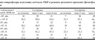
Data on the dependence of the pH level, maturity of the vaginal epithelium, and vaginal health index on the duration of the underlying disease in women with type 1 and type 2 diabetes are presented in Table. 1 and 2.
When analyzing the data obtained, direct correlations were revealed between the duration of diabetes and the pH of the vaginal contents (r=0.45 and r=0.48, respectively, p<0.01), an inverse correlation between the duration of diabetes and the maturity values of the vaginal epithelium, and also the vaginal health index in both groups of women (r=–0.58; p<0.01; r=–0.49; p<0.01). The same trend was revealed in a comparative analysis of the level of compensation for diabetes with the listed indicators. Thus, in women with HbA1c% <7.5%, the pH was 4.1–4.6, and in women with HbA1c% ≥9% it was 7.8–8.9 (p <0.001).
A comparative analysis of pH, maturity of the vaginal epithelium, vaginal health index depending on the level of HbA1c% in the studied groups is presented in table. 3.
When analyzing microbiological diagnostic data in a group of women with clinical manifestations of AV, it was revealed that out of 570 women, only 112 (19.6%) patients with diabetes had “conditionally normocenosis” [10] (for healthy people - 43%); bacterial vaginosis - 62 (10.8%) patients (for healthy ones - 15%); and atrophic colpitis - 228 (40%) women (for healthy ones - 1%).
In connection with complaints of itching and burning in the external genital area in combination with specific clinical manifestations of the urogenital tract, we used the nosological concept of “atrophic diabetic vulvovaginitis,” which had a direct correlation with the level of HbA1c% (Table 4).
Clinically, ADV manifested itself as itching and burning in the external genital area (95% of patients). A gynecological examination revealed hyperemia (93%), swelling (90%), less often - microerosions and cracks (26 and 41%, respectively).
A study of the microflora composition in 298 women with diabetes with clinical manifestations of ADV revealed the absence of lactobacilli in 87.6% of cases; Colonies of opportunistic microorganisms were found, including gram-positive cocci - 68.7%, low titer Staphylococcus epidermidis (8-103 CFU/ml) - 11%. A high pH level (7.8–8.9) was detected in 93% of women, and a low maturity value of the vaginal epithelium (25–30) was detected in 89% of cases. Severe leukocytosis (the number of leukocytes in the vaginal contents is 38.6±10 in the field of view) was determined in 96.7% of patients.
15 women of the main group with verified “acute atrophic diabetic vulvovaginitis” (without signs of a fungal infection according to the results of bacteriological and bacterioscopic studies), in addition to strict compensation of carbohydrate metabolism, used the drug estrocad (vaginal suppositories of estriol 0.5 mg), deep intravaginally 2 times a week within 3 months. The control group consisted of 15 women with verified acute atrophic diabetic vulvovaginitis, who were offered strict compensation of carbohydrate metabolism. The data is presented in table. 5-8.
In all 15 (100%) patients of the main group, after 3 months of using the drug estrocad, the pH level decreased by an average of 30%, the maturity value of the vaginal epithelium increased by 15-25%, and the vaginal health index in the subgroups of women with decompensation of the underlying disease increased by 40 -50%. In the control group, a study of the composition of the microflora before and after the end of the study revealed the absence of lactobacilli in 93% of patients, a high pH level (7.8-8.9) in 87.6% of cases, a low maturity of the vaginal epithelium (25-30) in 89% of women and pronounced leukocytosis of the vaginal contents (38.6±10 leukocytes in the field of view) - in 96.6%. In 98.7% of women with type 1 diabetes and 93.4% with type 2 diabetes, a direct correlation was revealed between the duration of diabetes, the degree of compensation of carbohydrate metabolism and the pH of the vaginal contents.
Thus, specific clinical manifestations of the urogenital tract in women with diabetes have a direct correlation with the degree of compensation of carbohydrate metabolism, and ADV is a consequence and an early marker of decompensation of carbohydrate metabolism. The presented results of therapy with the ADV drug estrocad showed the high effectiveness, safety and acceptability of this drug immediately after treatment. In the complex pathogenetic treatment of both acute and chronic ADV in patients with carbohydrate metabolism disorders in peri- and postmenopause (in addition to strict compensation of carbohydrate metabolism), it is necessary to include the local use of drugs containing estrogens, such as the drug estrocad, to normalize the vaginal health index and increase quality of life.