Anomalies of the uterus and vagina are observed in 4.3-6.7% of all women of childbearing age. They cause infertility in every eighth case, and in 12.6-18.2% they cause recurrent miscarriage, placental abruption, abnormal fetal position and other complications.
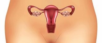
One of the most common anomalies of the uterus is its infantility (also called hypoplasia) - a defect associated with the small size of the main reproductive organ. With a slight decrease in the size of the uterus, the anomaly does not manifest itself with any symptoms, but a significant degree of hypoplasia can be suspected even in adolescence - by the late appearance and extreme pain of menstruation.
Causes of uterine infantilism
A girl’s uterus begins to form during the prenatal period – at the 5th week of its development. By the time of birth, it is fully formed, but still very small. Until the age of 9-10, it grows slowly, spending the first 3 years in the abdominal cavity, and then descending into the pelvis. From 10 to 13-14 years, her growth accelerates greatly, and by the time of puberty she should reach her normal size: 48±1 mm in length, neck length 26±1 mm (that is, a total length of 72-76 mm), 33 mm in thickness, 41 mm in width. If this does not happen, then one of the situations has occurred:
- The uterus either initially failed to develop: it was prevented either by intoxication that affected the mother’s body during the prenatal period, or a disorder occurred at the level of genes or chromosomes, due to which the organ will no longer be able to grow;
- The uterus began to develop, but the girl’s body (mainly her endocrine system) was adversely affected. This could happen as a result of:
- suffered severe influenza: the virus “likes” to affect the main endocrine organs - the hypothalamus and pituitary gland;
- frequent respiratory tract infections: ARVI, chronic tonsillitis;
- nicotine or drug intoxication;
- constant stress (they also affect the hypothalamus), including those caused by significant mental and physical stress at school;
- hypovitaminosis;
- tumors of the pituitary gland or hypothalamus;
- damage to the ovaries by mumps, measles, rubella viruses;
- insufficient nutrition of the girl, including as a result of her following a diet for weight loss;
- surgery on a girl's ovaries.
Underdevelopment of the central nervous system. Brain hypoplasia
Intrauterine underdevelopment of the structures of the central nervous system is one of the most serious defects affecting the functioning of the entire organism.
You can guess about the underdevelopment of the central nervous system structures from the symptoms, which are detected almost from the moment of birth. Thus, with hypoplasia of the cerebellum, which is responsible for the coordination of movements, the child experiences incoordination and deterioration in muscle tone. And when he begins to walk, a balance disorder is revealed. Hypoplasia of the corpus callosum, which is responsible for the coordinated work of both hemispheres, can lead to a variety of consequences. First of all:
- muscle spasms;
- convulsions;
- impaired skin sensitivity and body temperature regulation;
- deterioration of auditory and visual memory.
Brain hypoplasia (or microcephaly) is a proportional underdevelopment of all its structures. In early childhood, such a disorder manifests itself as delayed growth and development, and in later childhood – serious intellectual impairment.
Signs of an infantile uterus
If the teenage uterus does not manifest itself in any way, and a woman can only find out about this anomaly during pregnancy or even after childbirth, then more severe degrees of hypoplasia have their own symptoms:
- late onset of menstruation (after 16 years);
- painful periods;
- irregular periods;
- decreased libido;
- difficulty achieving orgasm;
- infertility or recurrent miscarriage.
Uterine hypoplasia is often combined with other gynecological pathologies: mainly endometriosis, frequent inflammation of the vagina and cervix. They can mask the manifestations of uterine hypoplasia.
Often, girls with uterine infantility have a characteristic appearance: they are short, thin, with very small breasts and a narrow pelvis. Upon examination, the gynecologist sees a small amount of pubic hair, underdeveloped labia and a small vagina.
Classification of the disease
The following types of hypoplasia are distinguished:
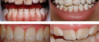
- Systemic enamel hypoplasia (SHE) can affect all primary or permanent teeth or (more often) groups of symmetrically located teeth developing simultaneously.
There are 6 forms of systemic hypoplasia:
- The spotted form is a mild variant of the pathology in which the color of the enamel changes. Smooth, glossy spots of a milky white or brownish color are observed on the teeth. The spots have clearly defined boundaries and are only an aesthetic drawback.
- With the pitted form, pits appear on a group of teeth. They are not only an aesthetic defect, but also a source of pain when the teeth are exposed to thermal irritants.
- With the grooved form, groove-shaped depressions are observed in the enamel. Over time, the cutting edge of the tooth becomes thinner. From the outside it seems that in place of one tooth there are 2: the small one grows from the larger one.
- The cup-shaped form is distinguished by the presence of cup-shaped depressions on a group of teeth. This entails chipping and abrasion of the enamel, increased sensitivity to thermal and chemical irritants.
- The combined form most often develops in children who suffered from both mild and severe pathologies in the first years of life. With this form, cup-shaped and grooved depressions appear on the teeth.
- Enamel aplasia is the absence of enamel - complete or partial. This form of the disease occurs on its own or after other forms of SGE. It represents a significant cosmetic defect. The patient also observes fracture of the cutting edges of the teeth, hypersensitivity, and severe abrasion of the enamel.
- Local enamel hypoplasia (LHE) is characterized by pathology of the enamel of one or two teeth. The cause of its occurrence is an infection developing in the tooth germ, or mechanical trauma to the tooth follicle. As a rule, MGE occurs on permanent teeth. Rarely observed in dairy products, after a jaw fracture or osteomyelitis. Local hypoplasia consists of spots that have unclear boundaries and differ from each other in shape. They are matte, have a yellow or brownish tint, and do not result in thickening of the enamel.
- Focal hypoplasia is a decrease in the size of tooth crowns. They may belong to the same or different periods of development. The teeth are yellowish in color and have a rough surface. Focal hypoplasia is accompanied by increased abrasion of the enamel or the presence of gaps between the teeth. The cause of the development of the disease may be trauma, chronic osteomyelitis, radiation, or delayed teething. The pathology is not only a cosmetic defect, but is also characterized by pain when teeth come into contact with thermal irritants.
There is another special type of disease - tetracycline teeth. This is a pathology in which the color of the teeth changes - they become yellow. The cause of the defect is the use of tetracycline drugs at the moment when the development of the surface layer of enamel and mineralization of dental tissue occurs. Moreover, not the entire tooth changes color, but only the area that was mineralized during the period of medication use. The patient does not feel pain or other discomfort; he only complains about an aesthetic defect.
Infantile uterus and pregnancy
With an embryonic type of uterus, pregnancy can occur only with the use of assisted reproductive technologies, but often it is necessary to resort to surrogacy with the woman’s egg.
The childhood type of hypoplasia makes pregnancy possible, but pregnancy is associated with the risks of premature birth, placental abruption, abnormal position of the fetus in the uterus, and premature rupture of amniotic fluid.
With the teenage type of anomaly in combination with preserved ovarian function, problems with conception and pregnancy usually do not arise. In the early postpartum period, uterine contractions need to be monitored.
Grooved shape
In this case, one or more grooves are formed on the vestibular (front) surface parallel to the cutting edge of the incisors. The depth of damage may vary. As a rule, teeth of the same name are affected symmetrically. As in the case of the erosive form, the anomaly can be seen on the radiograph even before the incisors erupt (horizontal lesions).
Here are some more types of hypoplasia:
- Linear - manifests itself in the form of numerous wavy stripes on the vestibular surface.
- Aplastic is a severe disorder characterized by severe damage to the enamel. Occurs in the case of amelogenesis imperfecta.
- X-ray examination does not reveal the disease.
- Mixed - most often we are talking about a combination of erosive and spotted forms.
Diagnostics and treatment in our clinic
Having established a preliminary diagnosis of uterine hypoplasia and determined its degree by ultrasound, clinic specialists perform hysterosalpingography: this is how they find out the internal structure of the uterine cavity and determine the tortuosity of the fallopian tubes. After this, we need to create a hormonal profile of the woman.
Only on the basis of these studies will we be able to draw up a treatment plan for uterine hypoplasia. The main methods we use for this developmental anomaly are:
- physiotherapeutic methods: magnetic therapy, paraffin, ozokerite applications, endonasal galvanization, mud therapy and other methods;
- vitamin therapy;
- gynecological massage;
- Exercise therapy.
If the degree of uterine infantilism is high, and you want to get pregnant, we will select exactly the assisted reproductive technology that will solve your problem. This could be IVF or fertilization of your egg using the ICSI, PIXI, IMSI method, followed by the introduction of the embryo into a surrogate mother.
Is it worth carrying out the silvering procedure?
Some dental clinics eliminate the initial stages of hypoplasia using the silvering method. The essence of this therapy is that a layer of active metals is applied to the teeth. Upon contact with enamel, crystals of silver phosphate and potassium fluoride are formed, protecting the enamel from destruction.
The Nutcracker Clinic does not use silver plating. We abandoned this method for several reasons:
- Poor efficiency. To remove plaque and restore the nervous structure of the incisors, you need to carry out about 5 procedures over six months.
- The drugs used often cause allergic reactions.
- In case of advanced caries, the procedure is contraindicated.
- After silvering, the enamel darkens, which causes psychological discomfort in the child.
Preventive measures and forecasts
When diagnosing the fertile type of uterine infantility, the possibility of conception is excluded. In this case, pregnancy is possible only with the use of assisted reproductive technologies (ART). If the generative function of the gonads is preserved, in vitro fertilization (IVF) is used using oocytes ready for insemination.
The course of pregnancy in patients with severe endometrial hypoplasia is associated with a high probability of spontaneous abortion and complicated delivery.
In women with miscarriage syndrome, intracytoplasmic sperm injection (ICSI) is performed as part of surrogacy. With minor changes in the structure of the reproductive organ and normal secretion of steroid hormones by the ovaries, the chances of conception and successful pregnancy increase by 45-50%.
Pathogenesis
The pathogenesis of dental enamel hypoplasia is based on a violation of the processes of its formation, which is caused by changes in the structure of the protein matrix of enamel / dentin and disturbances in the process of their mineralization. The pathogenesis of a wedge-shaped tooth defect is based on excessive mechanical load on both the chewing surface of the tooth and the neck of the tooth, as well as stretching of the enamel, which leads to its destruction (cracking) and the formation of a wedge-shaped defect.
The pathogenesis of the wedge-shaped defect is due to the action of force on the enamel in the area of the tooth neck: compression force and tension force. At the same time, the compressive strength of enamel is much stronger than the tensile strength. This contributes to the rupture of intercrystalline bonds of hydroxyapatite and the formation of microdefects in this zone, and subsequently force, mechanical and chemical effects cause an increase in the resulting cracks and the development of a wedge-shaped defect.
What is needed to confirm the diagnosis?
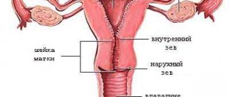
A gynecologist can assume the presence of uterine hypoplasia during examination when there are signs of general infantilism. During a vaginal examination, attention will be drawn to underdevelopment of the vulva, scant hair growth, a short and narrow vagina, and a disproportionate ratio of the uterus to the cervix. The leading criterion is a reduction in the size of the uterus.
Data from laboratory and instrumental studies help determine the diagnosis.
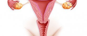
Ultrasound diagnostics provides the most information.
In addition to the reduced size, flattening of the uterine body and a pronounced anterior tilt - hyperanteflexia - will be characteristic. The fallopian tubes are usually long and twisted, which increases the likelihood of an ectopic pregnancy. Often, with general infantilism, ultrasound can detect ovarian hypoplasia. This is associated with a decrease in the formation of sex hormones, so determining the concentration of steroids plays a significant role in the diagnosis of uterine underdevelopment.
Another study that helps confirm uterine hypoplasia is hysterosalpingography (HSG). The uterine cavity and fallopian tubes are filled with a special contrast agent, after which a series of x-rays are taken. The resulting image allows you to see a small uterus with long fallopian tubes, which allows you to confirm the diagnosis.
To determine the hormonal status, the level of hormones in the blood is determined (androgens, estrogens, prolactin, follicle-stimulating and luteinizing hormones, T3, T4 - thyroid hormones).
In doubtful cases, the attending physician has the right to order an X-ray of the skull and an MRI of the brain.
Prevention
To prevent the development of tooth enamel hypoplasia/wedge-shaped defect, it is recommended:
- The correct diet/diet of a woman during pregnancy with a sufficient content of calcium, phosphorus, vitamins B, A, D, E.
- Carefully observe oral hygiene, regularly visit the dentist for preventive purposes and, if necessary, carry out regular enamel remineralization therapy.
- Timely removal/treatment of baby teeth in children with chronic apical inflammatory processes.
- Selecting the right dental/oral care products (pastes, toothbrushes, rinses), using the correct teeth brushing technique, avoiding the use of aggressive drinks (based on chemical composition), such as carbonated drinks.
List of sources
- Groshikov M.I. Non-carious lesions of tooth tissue. ─ M.: Medicine, 1985. - 176 p.
- Dentistry of children and adolescents: Per. from English / Ed. T. Mac Donald, D. R. Avery. - M.: Medical Information Agency, 2003. - 766 p.: ill.
- Mikhalchenko V.F., Aleshina N.F., Radyshevskaya T.N., Petrukhin A.G. Dental diseases of non-carious origin. // Educational and methodological manual, part 1 (non-carious lesions that developed during the formation and mineralization of teeth). - Volgograd, 1998. - 24 p.
- Semchenko I.M. Clinical manifestations of wedge-shaped defects. // Sat. scientific works: Works of young scientists. Anniversary edition. - Minsk, 2001.- p.121-124.
- Lyamzin, S.S. Adequate treatment of wedge-shaped dental defects / S.S. Lyamzin // Dental Market. - 2008.- No. 3. — P.51-55.