Author:
Kuzmina Tatyana Sergeevna dermatologist, Ph.D.
Quick transition Treatment of cutaneous lupus erythematosus
Systemic lupus erythematosus is treated by a rheumatologist. During therapy with antimalarial drugs (hydroxychloroquine, chloroquine), observation by an ophthalmologist is necessary. If you suspect the development of squamous cell skin cancer against the background of discoid lupus erythematosus, consultation with an oncologist is necessary.
Lupus erythematosus rashes on the skin and/or mucous membranes can be an independent disease or a manifestation of systemic lupus erythematosus, a chronic disease associated with the formation of antibodies to components of cell nuclei. Antibodies cause damage and inflammation of various tissues, including the skin.
Forms and complications
Skin manifestations of lupus erythematosus include three groups, each including specific skin manifestations:
- acute cutaneous lupus erythematosus (localized, generalized and toxic epidermal necrolysis-like);
- subacute cutaneous lupus erythematosus (ring-shaped, papulosquamous, drug-induced, erythrodermic, poikilodermic, vesiclobulous, Rowell syndrome);
- chronic cutaneous lupus erythematosus (discoid, edematous, panniculitis, pernioid, lichenoid).
The identification of these forms is due not only to the duration of the skin disease, but also reflects the connection with systemic lupus erythematosus.
Most often, the manifestation of systemic disease is acute forms, while the discoid form is observed only in 5-15% of all cases. The longer skin manifestations exist in isolation, the lower the risk of developing a systemic form of the disease. The risk of systemic disease in the presence of cutaneous manifestations is higher in women and children.
Lupus erythematosus rashes can occur in children (antibodies are passed on to the fetus) from mothers with systemic lupus erythematosus. This form is called neonatal lupus, occurs during the first two months of a child’s life and may be the first sign of systemic lupus erythematosus in the mother.
The systemic lupus erythematosus complex may also include other changes in the skin and its appendages that are not specific exclusively to this disease.
SLE symptoms, complaints and clinical manifestations
Redness (erythema) of the facial skin in the cheeks and nose
This is the most characteristic symptom of systemic lupus, which occurs or worsens when exposed to direct sunlight. Because of its characteristic shape, this erythema became known as “lupus butterfly.” Similar skin rashes may also occur in other areas. The skin of the fingers often changes color under the influence of cold from white to purple-bluish ( Raynaud's phenomenon ). of ulcers on the oral mucosa is possible alopecia ) and brittleness of the nail plates.
Pain and inflammation (swelling) of the joints
They occur frequently, but do not lead to the destruction of joint structures, as happens with rheumatoid arthritis. The small joints of the hands are most often affected, but arthritis of the large joints is also possible. Characteristic signs of inflammatory joint pain are morning stiffness (> 1 hour), increasing pain at rest and weakening with movement.
Pleurisy and pericarditis
A common symptom of lupus is inflammation of the pleural membranes lining the inside of the chest (pleurisy) and the heart sac (pericarditis), which can manifest as pain in the chest during breathing movements.
Kidney damage
When internal organs are involved in the process, kidney damage most often occurs, usually occurring unnoticed in SLE. Edema and a persistent increase in blood pressure are indirect signs of this serious complication.
Inflammatory changes in the nervous system. They can manifest themselves in a wide variety of ways (from headaches and psycho-emotional disorders to convulsive seizures).
Antiphospholipid syndrome. Also, with systemic lupus erythematosus, the formation of “immune agents” (antibodies) to the components of the blood coagulation system is possible. This condition is called antiphospholipid syndrome and is dangerous for the development of various thrombotic complications (vein thrombosis, heart attack, stroke). The presence of these manifestations in a patient at a young age requires the exclusion of SLE and antiphospholipid syndrome.
Nonspecific complaints. Many patients at the onset of the disease notice only nonspecific symptoms of the disease, such as general weakness, increased body temperature, weight loss, and lack of appetite.
Symptoms
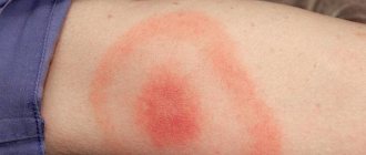
The skin manifestations of lupus erythematosus are diverse. A significant manifestation, almost always associated with a systemic disease, is a rash on the face, shaped like a butterfly: with wings on the cheekbones and cheeks and a body on the bridge of the nose and bridge of the nose. The rash may appear as raised or deep-lying elements of pink and bluish-red color. There is a tendency to form ring-shaped lesions covered with very dense scales that are painful when removed. In some cases, the so-called lupus erythematosus pernio is formed, in which painful bright red bluish nodules appear on the toes, hands, nose, and ears in cold weather. Damage to blood vessels is accompanied by the appearance of rashes resembling bruises; dilated vessels can be visible in the nail beds. Some patients with SLE have symptoms resembling lichen planus.
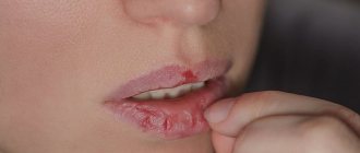
There are also severe lesions with the formation of blisters and peeling of the skin, which is more typical for the onset of a systemic disease. Changes can affect the mucous membranes, especially often the mucous membranes of the lips and oral cavity. Lupus lesions on the scalp may result in hair loss. Diffuse hair loss and hair thinning are also possible.
Diagnosis and treatment of SLE at the Yauza Clinical Hospital
Diagnosis of systemic lupus erythematosus at the initial stage of development makes it possible to achieve rapid and stable remission of the disease and avoid the development of severe complications. In the treatment of lupus, we use drug therapy (steroidal and non-steroidal drugs, biological genetically engineered drugs), methods of extracorporeal hemocorrection.
If symptoms of SLE appear, contact rheumatologists at the Yauza Clinical Hospital. Modern treatment methods, subject to preventive measures, make it possible to maintain a state of remission for years and even decades, give birth to children, and lead an ordinary normal life.
You can see prices for services
Treatment of cutaneous lupus erythematosus
The basis of treatment is corticosteroid hormones. With this disease, they can be applied to the lesions in the form of ointments, or injected directly into the lesion of the affected skin. In severe cases, systemic corticosteroids may be prescribed. As an alternative or to enhance therapy, it is possible to apply calcineurin inhibitors to the lesions.
In cases of severe skin damage and insufficient effect of external treatment, systemic therapy is carried out using antimalarial drugs (chloroquine, hydroxychloroquine), anti-inflammatory drugs (methotrexate, mycophenolate, dapsone, azathioprine, etc.).
Features of the treatment method
The first line of treatment is topical corticosteroids and hydroxychloroquine.
To reduce the total dose and side effects of topical corticosteroids, calcineurin inhibitors are included in treatment.
During treatment with hydroxychloroquine, regular medical supervision is necessary. The condition of the retina must be monitored, since the drug can impair vision with long-term (more than 5 years) use. This side effect is uncommon, but signs of retinal damage may require discontinuation of the drug.
Intralesional administration of drugs (corticosteroid hormones) using multiple superficial injections directly into the lesion of the affected skin allows you to create a high concentration of the drug in the lesion, while reducing the risks of systemic side effects. The medicine is administered by a doctor in a dressing room to a depth of 2-3 mm. The procedure is painful, but the pain is not sharp and largely depends on the sensitivity of the patient’s receptors. To reduce pain, a thin needle is used, and an anesthetic ointment may be used.
All patients must be prescribed skin protection against ultraviolet radiation.
How is lupus erythematosus treated at the Rassvet clinic?
The dermatologist will ask you to talk about the course of the disease and the treatment that was carried out previously. The doctor will examine the skin (including the scalp) and mucous membranes. Dermatoscopy and trichoscopy can be informative. With this disease, to establish a diagnosis in most cases, a skin biopsy will be required with an additional immunofluorescence reaction.
If cutaneous lupus erythematosus is suspected, it will be necessary to discuss the possibility of systemic manifestations and assess the condition of other organs and systems of the body. Additional studies may be required to identify a systemic process (blood tests, urine tests, etc.).
The onset of systemic lupus erythematosus in persons over 50 years of age occurs in 3-18% of cases. Late onset of the disease leads to significant changes in the clinical picture, the course of the pathological process, the therapeutic effect of the treatment, and prognosis [1]. Among elderly patients with lupus erythematosus, the predominance of women is less pronounced than in other age groups [2]. A study from Canada showed that late-onset lupus erythematosus was much more common in people of European descent, while early-onset disease predominated in patients of Asian and African descent [3]. Late diagnosis is often noted. In the older age group, lung damage and serositis are more common than in young patients, while rash in the malar region, photosensitivity, arthritis, and nephropathy are observed less frequently in older patients [1]. In general, younger patients met more American College of Rheumatology diagnostic criteria. For example, despite the increase in antinuclear antibodies, which is similar for young and elderly patients, anti-Sm antibodies, ribonucleoprotein, and hypocomplementemia were more often detected in young individuals. In addition, nephritis, skin lesions, and cytopenia were more often diagnosed in young patients [3]. Severe manifestations of lupus erythematosus in elderly patients are observed less frequently; the cause of death is more often due to infections, cardiovascular diseases, and malignant neoplasms [1–3]. In some cases, intercurrent diseases also take on an unusual course. Thus, the development of pulmonary blastoma in a 62-year-old patient with discoid lupus erythematosus is described. Typically, lung blastoma develops at a younger age, in contrast to non-small cell lung carcinoma [4].
On the other hand, it is noted that, despite the more benign course of lupus erythematosus, the prognosis in elderly patients is always worse, which is due to the high frequency of concomitant diseases, lesions of various organs due to age and longer exposure to classical risk factors for vascular lesions in elderly patients [ 3, 5, 6]. It is also necessary to take into account the decrease in the therapeutic effect of drugs in older age groups. The development of ascites caused by lupus peritonitis in a 77-year-old woman is described. The administration of glucocorticoids was ineffective. Despite some improvement in laboratory parameters such as immunological markers in the blood serum, titers of anti-DNA antibodies, the content of immune complexes in the peritoneal fluid remained elevated. It is assumed that the lack of effect of therapy may be due to impaired vascular circulation against the background of immunological changes [7].
The clinical picture of integumentary forms of lupus erythematosus can also change significantly with age, which determines the special role of differential diagnosis in elderly patients. Improving laboratory methods for diagnosing lupus erythematosus significantly improves its quality, but the greatest difficulties are caused by clinical manifestations [8].
If the patient is photosensitized, a differential diagnosis should be made with solar persistent erythema. The disease is more often observed in women 20-30 years old and affects exposed areas of the skin. On the surface of the lesions, peeling, small papular rashes, hemorrhages and crusts are detected [9].
In rosacea, the leading ones are angioedema disorders. Persistent erythema is found on the skin of the neck, forehead and nose. Depending on the stage of the disease, papules, pustules, telangiectasia, and nodes are detected on an edematous, hyperemic background [10]. Follicular hyperkeratosis and scar atrophy are absent [11].
Skin lesions in various forms of lupus erythematosus have some similarities with Senir-Usher syndrome. Both cases are characterized by localization on the face, butterfly-shaped lesions, and erythematous-squamous rashes. The greatest difficulties can be caused by isolated localization of the lesion on the scalp with the development of alopecia and cicatricial atrophy, but unlike lupus erythematosus, in seborrheic pemphigus the lesions are formed due to the formation of rapidly bursting flabby flat blisters. The course of the disease does not depend on the periods of greatest insolation. In the common form of seborrheic pemphigus, there are no capillaritis. In lupus erythematosus, Nikolsky's symptom is negative [12].
The clinical picture of centrifugal annular erythema of Darier is characterized by the appearance of non-flaky yellowish-pink edematous spots, which quickly turn into raised flat annular elements with a tendency to eccentric growth. Follicular hyperkeratosis and scar atrophy are absent [9].
With nodular chondrodermatitis of the auricle, a dense hemispherical nodule appears, up to 0.5 cm in diameter, the color of healthy skin or slightly bluish-reddish, covered with tightly adjacent scales, after the fusion of which superficial ulceration can be detected. Follicular hyperkeratosis, scar atrophy, and dependence on insolation are absent [9].
Tuberous syphilide begins to develop in the reticular layer of the dermis, without causing noticeable changes on the surface of the skin. Gradually, the tubercle increases in diameter, protrudes above the surface of the skin, and there are no subjective sensations. The formation of a scar or cicatricial atrophy is characteristic. While in lupus erythematosus the primary morphological element is a spot, follicular hyperkeratosis and dependence on insolation are characteristic [9].
With atrophic lichen planus, atrophic changes develop at the site of regressing nodular elements, often ring-shaped. Follicular hyperkeratosis, scar atrophy, and dependence on periods of insolation are absent [8].
Various lesions on the scalp (pyoderma, seborrheic dermatitis, Hoffmann's folliculitis, etc.) are also not dependent on periods of insolation [8].
Hammel's garland-shaped migratory erythema is characterized by multiple garland-shaped lesions accompanied by swelling and itching. Hammel's erythema is a paraneoplastic dermatosis - most often patients are diagnosed with adenocarcinoma of the mammary gland, although cases of combination of Hammel's erythema with gastric cancer, lymphogranulomatosis, Rustitsky-Culler disease, tumors of the brain, genital organs, and respiratory tract have also been described [12, 13]. Cases of the disease have been described in patients without cancer pathology [14, 15].
We present our own observation.
Patient K
., 74 years old, was sent to hospital treatment with a presumptive diagnosis of Hammel's erythema?
Accompanying illnesses
. Cardiac ischemia. Angina pectoris of functional class II. Circulatory failure stage I. Hypertension II degree (AH II, high risk), cerebrovascular disease. Chronic cerebral ischemia. Peptic ulcer, without exacerbation. Gastroesophageal reflux disease. Catarrhal esophagitis. Postcholecystectomy syndrome (cholecystectomy in 2012) Chronic pancreatitis, without exacerbation. Chronic colitis. Diverticulosis of the large intestine. Polyp of the rectosigmoid region. Cysts of both kidneys. Focal formations of both lobes of the thyroid gland. Varicose veins of the lower extremities.
Upon admission, the patient complained of rashes on the skin of the face, neck, torso, upper and lower extremities, accompanied by moderate itching, worsening in the evening hours.
She considers herself sick for 2 years, when for the first time after undergoing cholecystectomy and taking medications for this, she discovered a rash on the skin of her left forearm and back. Subsequently, she received treatment in a hospital setting at the place of residence for a diagnosis of allergic dermatitis of unknown origin (prednisolone solution 30 mg intravenous drip) with significant improvement. Subsequently, she received outpatient treatment, which included antihistamines and external glucocorticoids. She was sent for a consultation to the “Central” branch of the Moscow Scientific and Practical Center of Dermatovenereology and Cosmetology (MNPCDK DZM). Deputy Head of the Moscow Department of Health Prof. N.N. Potekaev recommended an examination to clarify the diagnosis at the Veshnyakovsky branch of the Moscow Research and Clinical Center for Children's Health.
Upon admission, widespread polymorphic rashes of an acute inflammatory nature were visualized on the skin of the face, ears, trunk, upper and lower extremities, which were represented by moderately edematous foci of macular erythema with ring-shaped large scalloped foci, with a scaly peripheral edge, in places of a serpiginous nature, prone to fusion. On the surface of the elements there were point and linear excoriations and erosive defects with a diameter of 0.2–0.3 cm (Fig. 1, 2, 3).
Rice. 1. Lesions on the face, ear, chest.
Fig. 2. Lesions on the chest, abdomen, thighs.
Rice. 3. Lesions on the back.
Indicators of general blood test, urine, biochemical blood test are within normal limits (including glucose level - 6.3 mmol/l).
Glycemic profile: 8.00 - 6.2 mmol/l, 11.30 - 7.1 mmol/l.
Repeated glycemic profile: 8.00 - 6.1 mmol/l, 11.30 - 7.5 mmol/l.
Pathomorphological examination of skin biopsies: histological changes correspond to the diagnosis: subacute lupus erythematosus.
Test for the determination of autoantibodies to nuclear antigens (total antibodies dsDNA, to histones, Sm, RNP/Sm, SS-A (Ro), SS-B (La), Scl-70, Jo-1, U1-NP-B): result 120.3 arb. units - positive.
To nuclear antigens (total antibodies Sm, RNP/Sm, SS-A (Ro), SS-B (La), Scl-70, Jo-1): result 54.8 - positive.
No LE cells were detected.
Data from instrumental research methods
ECG: sinus rhythm, heart rate - 60 beats per minute, horizontal position of the electrical axis of the heart. Moderate changes in the inferior wall of the left ventricle.
EGDS: duodenitis. Mixed gastritis with foci of metaplasia. Duodeno-gastric reflux.
Ultrasound examination of the thyroid gland: echographic signs of diffuse changes in the parenchyma of the thyroid gland, nodular inclusions on the right.
Ultrasound examination of the mammary glands: echographic signs of fibrofatty involution of the mammary glands.
Ultrasound examination of the abdominal cavity and kidneys: echographic signs of steatohepatosis. Diffuse changes in the pancreas. Cysts of both kidneys, microliths of both kidneys.
Consultation with a therapist: coronary heart disease. Angina pectoris of functional class II. Circulatory failure stage I. Hypertension II degree (AH II, high risk), cerebrovascular disease. Chronic cerebral ischemia. Gastric ulcer, without exacerbation. Gastroesophageal reflux disease. Catarrhal esophagitis. Postcholecystectomy syndrome (cholecystectomy in 2012) Chronic pancreatitis, without exacerbation. Chronic colitis. Diverticulosis of the large intestine. Polyp of the rectosigmoid region. Cysts of both kidneys. Focal formations of both lobes of the thyroid gland. Varicose veins of the lower extremities.
Consultation with an endocrinologist: multinodular goiter, euthyroidism. Dysglycemia.
Repeated consultation with an endocrinologist: type 2 diabetes mellitus, newly diagnosed. Multinodular goiter. Euthyroidism.
Thus, in the observation we presented, the debut of lupus erythematosus was noted at 72 years of age. The diagnosis had not been made over the previous 2 years, which could be largely due to the widespread belief that the onset of connective tissue diseases predominates in young people. When examining patients over 60 years of age, a significant part of the research is aimed at searching for cancer, as evidenced by the presumptive diagnosis: Hammel's erythema? Our observation indicates the possibility of the onset of lupus erythematosus in elderly people.
There is no conflict of interest.
Recommendations
- “Cutaneous lupus erythematosus | American Skin Association." www.americanskin.org
. Retrieved 2018-12-11. - ^ a b c
Wollina, Uwe;
Hansel, Gesina; Koch, Andre; Abdel-Nasser, Mohamed Badawi (2006). "Topical pimecrolimus for skin conditions other than atopic dermatitis." Pharmacotherapy expert opinion
.
7
(14): 1967–1975. Doi:10.1517/14656566.7.14.1967. PMID 17020422. - ^ a b c d e f gram h j k l m p o p q r s t you v w X y z aa ab ac announcement ae af ag ah ay hey ak al am an ao ap
James, William
; Berger, Timothy; Alston, Dirk (2005). Andrews Skin Diseases: Clinical Dermatology
. (10th ed.) Saunders. Chapter 8. ISBN 0-7216-2921-0. - ^ a b c d f g gram h i
editor, Fitzpatrick, James E., 1948 - editor.
Morelli, Joseph G. Dermatology Secrets Plus
. OCLC 1010741108.CS1 maint: additional text: authors list (link) - ^ a b c d e f gram h i j k l m p o p q r s t you v w X y z aa ab ac ad ae af ag ah
James J., Jr. (2019)
. Lookingbill and Marks Principles of Dermatology
. ISBN 9780323430425. OCLC 1024315813. - ^ a b
Malagola, R.;
Abicca, I.; Abbouda, A.; Arrico, L. (2015). "Ocular complications of cutaneous lupus erythematosus: a systematic review with meta-analysis of reported cases." Journal of Ophthalmology
.
2015
: 254260. doi:10.1155/2015/254260. PMC 4480931. PMID 26171240. - Kovelas, Dimitrios; Tsellos, Thrasyvoulos George (2008-04-01). "Topical tacrolimus and pimecrolimus in the treatment of cutaneous lupus erythematosus: an evidence-based assessment." European Journal of Clinical Pharmacology
.
64
(4):337–341. Doi:10.1007/s00228-007-0421-2. ISSN 1432-1041. PMID 18157526. - ^ a b
Jessop, Sue;
Whitelaw, David A; Grange, Matthew J; Jayasekera, Prathiva (05/05/2017). "Medicines for discoid lupus erythematosus." Cochrane Database of Systematic Reviews
.
5
:CD002954. Doi:10.1002/14651858.CD002954.pub3. ISSN 1465-1858. PMC 6481466. PMID 28476075. - Erceg, Angelina; De Jong, Elke M.J.G.; Van De Kerkhof, Peter C.M.; Seyger, Marieke M. B. (2013). "Journal of the American Academy of Dermatology". Journal of the American Academy of Dermatology
.
69
(4): 609–615.e8. doi:10.1016/j.jaad.2013.03.029. PMID 23711766. Received 2018-12-14. - Ekelem, Chloe; Pham, Christina; Atanaskova Mesinkovskaya, Natasha (09/05/2018). "A systematic review of the results of hair transplantation for primary cicatricial alopecia." Diseases of the skin appendages
.
5
(2): 65–71. Doi:10.1159/000492539. ISSN 2296-9195. PMC 6388556. PMID 30815438. - Finn, Robin (5 June 1996). “At lunch: print; the gold of glory comes from the crucible of early pain.” New York Times
. - Alyssa Rosenberg (February 2, 2021). Opinion: To understand Michael Jackson and his skin, you need to look beyond race., The Washington Post, May 30, 2021.
- Olivry, Thierry; Linder, Keith E.; Banovich, Fran (2018-04-18). "Cutaneous lupus erythematosus in dogs: a comprehensive review." BMC Veterinary Research
.
14
(1): 132. doi:10.1186/s12917-018-1446-8. ISSN 1746-6148. PMC 5907183. PMID 29669547. - Rosencrantz, Wayne (12/01/2013). "Immune-mediated dermatoses." Veterinary Clinics of North America: Equine Practice
.
29
(3):607–613. doi:10.1016/j.cveq.2013.08.001. ISSN 0749-0739. PMID 24267678.
Mechanism
Most experts consider DLE to be an autoimmune disease because pathologists see antibodies when they biopsy lesions and look at the tissue under a microscope.[5] However, scientists do not understand the connection between these antibodies and the lesions seen in discoid lupus.[5]
It is possible that the light damages skin cells, which then release material from their nuclei.[5] This material penetrates the dermoepidermal junction, where it binds to circulating antibodies, leading to a series of inflammatory reactions by the immune system.[5]
Alternatively, dysfunctional T cells can lead to disease.[5]
