What is Kaposi's sarcoma? Why does this disease occur? What are the symptoms? How is Kaposi's sarcoma diagnosed and treated? How effective can the treatment be?
Kaposi's sarcoma
– malignant vascular tumor of the skin.
Other names for the disease are multiple hemorrhagic sarcomatosis
,
Kaposi's angiosarcoma
.
Kaposi's sarcoma in facts and figures
:
- The disease was first described in 1872 by dermatologist Moritz Kaposi, from whose name it takes its name.
- The causative agent is human herpesvirus type 8
(
HHV-8, HHV-8
). - Kaposi's sarcoma affects about 30% of patients with HIV/AIDS.
- Since around the early 1990s, due to the AIDS epidemic, the prevalence of Kaposi's sarcoma has increased dramatically.
- In general, Kaposi's sarcoma is quite rare among the general population. But it accounts for 40-60% of all tumors in AIDS patients.
General information
Kaposi's sarcoma is a hemorrhagic multiple sarcomatosis , which is caused by multiple malignant neoplasms in the dermis of the skin and soft tissues. The pathology is named after the Hungarian dermatologist Moritz Kaposi, who first described this type of angiosarcoma .
Angiosarcomas are essentially tumors originating from the vascular perithelium and endothelial cells. They are considered extremely malignant and often metastasizing neoplasms. Multiple formations are usually localized on the skin (in 92% of cases), mucous membranes, as well as the larynx, lungs, digestive tract (in 45% of patients) and lymph nodes.
Most often occurs in people 40-50 years old, the ratio of men to women is 3:1. Despite the fact that Kaposi's sarcoma is a rather rare disease, in HIV it is the leader in the frequency of malignant neoplasms and reaches a frequency of 40-60%.
Diagnostics
Diagnosis of Kaposi's sarcomatosis is carried out by an infectious disease specialist, dermatologist and oncologist. First, doctors listen to the patient and collect anamnesis, and then:
- Check for signs of disease.
- They do a biopsy.
- A histological examination is carried out to detect the proliferation of fibroblasts.
- Conduct immunological studies.
- Blood is taken for HIV testing.
The patient is also prescribed additional tests, for example, ultrasound, radiography, gastroscopy, CT scan of the kidneys, MRI of the adrenal glands and others to identify internal lesions.
Pathogenesis
The oncological process leads to the formation of a tumor that has a histological structure with many randomly located thin-walled newly formed vessels with dilations of capillaries and plexuses of bundles of spindle-like cells. Infiltration of macrophages and lymphocytes is observed in the tumor. Due to the vascular nature of the tumor, there is an increased risk of bleeding and hemosiderin .
Tumor multicentric growth from angioblastic mesenchyme begins with activation of herpesvirus 8 in conditions of immunodeficiency caused by HIV infection , old age, drug immunosuppression after transplantation , malaria or other chronic infections and infestations in Africans. As a result of the changes, normal endothelial cells are transformed into malignant ones. Tumor growth is caused by disturbances in the regulation of the cell cycle and is associated with the viral cyclin D , viral IL-6 and others present in herpes viruses. Angiomatous lesions have an inflammatory, granulomatous and tumor stage.
Treatment
The treatment of a patient with sarcomas of the soft tissues of the head and neck requires an integrated approach with the involvement of a number of specialists: a morphologist, a surgeon, a radiation diagnostician, a chemotherapist, and a radiologist.
Surgery
Surgery is the main treatment method for patients with this pathology.
Soft tissue sarcomas grow within the capsule, which subsequently pushes away nearby tissue. This shell is called a pseudocapsule. Surgery involves removing the pseudocapsule en bloc with negative resection margins without damaging it, since violating the integrity of this formation increases the risk of tumor recurrence.
Postoperatively, radiation therapy may be administered to provide local control.
Additional treatments include chemotherapy and radiation therapy.
Radiation therapy
Preoperative radiation therapy provides significant advantages - the operating conditions are improved and the size of the tumor formation is reduced.
The negative aspect of this treatment is the high incidence of postoperative complications of an infectious nature.
Chemoradiation therapy
Treatment that combines chemotherapy with radiation therapy is called chemoradiotherapy.
Chemotherapy can improve the effectiveness of radiation therapy, so they are sometimes used together. This is a combination of systemic and local therapy.
Classification
According to the clinical picture and epidemiology, Kaposi's sarcoma is divided into 4 types:
- Classic - usually found in elderly residents of the Mediterranean, Central Europe and Russia, tumors are localized on the feet, lateral surfaces of the legs and surfaces of the hands, in extremely rare cases - on the mucous membranes and eyelids. Most often it occurs slowly and painlessly over 10-15 years and in a third of cases is associated with other malignant tumors.
- Endemic - typical for the population of Central Africa, Haiti and is diagnosed in children, the peak incidence is the first year of life and young people 25-30 years old, the pathogenesis is characterized by exophytic growth of tumors and damage to internal organs, bones and main lymph nodes with minimal and rare skin lesions.
- Epidemic - associated with AIDS, therefore it is considered a reliable sign of HIV infection, it is usually diagnosed in young people under the age of 37 years, the tumors are brightly colored and have rich contents, and are characterized by an unusual location - on the upper extremities, tip of the nose, mucous membranes and on the hard palate . The course is rapid and involves internal organs and lymph nodes in the pathogenesis.
- Immunosuppressive is an iatrogenic type of sarcoma, typical for recipient patients who have undergone organ transplantation, most often kidney transplantation. The course is chronic and benign, without involvement of internal organs. It develops while taking a special type of immunosuppressive drugs ; after their withdrawal, regression of tumors is observed.
Depending on the location, Kaposi's sarcoma is divided according to ICD-10 code: on the skin - C46.0, in soft tissues - C46.1, on the palate - C46.2, in the lymph nodes - C46.3, other localizations - C46.7, on multiple organs – C46.8, unspecified localization – C46.9.
Complications
The occurrence of complications of Kaposi's sarcoma depends on the stage of development of the disease and the location of the tumors. The following complications may occur:
- lymphatic edema, elephantiasis due to compression of the lymph nodes;
- bacterial infection of damaged tumors;
- limitation of motor activity of the limbs and their deformation;
- bleeding from disintegrating tumors;
- intoxication of the body caused by the breakdown of tumors;
- disruption of the functioning of internal organs when tumors are localized on them.
Some complications lead to life-threatening conditions.
Causes
Human herpes virus type eight (HHV-8) can be detected in 100% of patients. It is associated with Kaposi's sarcoma. It was found that 3-10 years before the development of a tumor, an infection caused by herpesvirus 8 is detected. However, some carriers of the herpesvirus do not develop malignant pigment elements, since the manifestation of sarcoma is influenced by a number of other factors:
- the presence of various substances, for example, cytokines , which are produced by HIV-infected mononuclear cells in response to infection;
- production of interleukins IL-6 and IL-1b, basic fibroblastic growth factor, vascular endothelial growth factor and other tumor growth regulators;
- decreased immune system function;
- a constant source of antigenic stimulus - parasitic or infectious (fungal, protozoan, bacterial, viral) diseases, including cytomegalovirus , Epstein-Barr , herpes , as well as sperm and intestinal injury during homosexual intercourse;
- therapy with glucocorticosteroids and immunosuppressants, for example, taking Cyclosporine ;
- genetic predisposition.
At the same time, the following are at risk:
- HIV-infected males, most often with non-traditional orientation and drug addiction;
- older men of Mediterranean origin;
- people originally from equatorial Africa;
- recipient individuals who have transplanted organs.
Prevention
Primary prevention of Kaposi's sarcoma consists of actively identifying patients and groups at increased risk for developing this disease.
Particular attention should be paid to patients receiving immunosuppressive therapy. In these groups, detection of individuals infected with human herpes virus VIII (HHV-8) is important.
Secondary prevention includes clinical observation of patients in order to prevent relapse (recurrence) of the disease, complications after treatment and rehabilitation of patients.
Symptoms of Kaposi's sarcoma
This type of malignancy is characterized by a slow progression. The first symptoms may take years to appear and may include weight loss, bleeding, fever, and symptoms resembling ulcerative colitis.
Initial stage of Kaposi's sarcoma
Distinctive features tumors or, in more rare cases, located on internal organs:
- have clear boundaries, predominantly purple in color, but can be tinged with red, purple or brown;
- are flat or slightly raised formations above the skin in the form of painless spots or nodules;
- with the classic clinical picture - symmetrical, asymptomatic, but can sometimes cause itching or burning.
In addition, angioreticuloendothelioma can be combined in most cases with damage to the mucous membranes of the palate and lymph nodes.
The clinical picture of the classic type of Kaposi's sarcoma is divided into 3 stages:
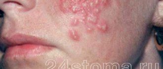
- The first - the initial stage is spotty, that is, at the very early stage, only reddish-bluish and brownish spots of irregular shape with a smooth surface appear, their sizes do not exceed 5 mm (as in the photo of the initial stage of Kaposi's sarcoma).
- The second stage - develops gradually and subsequently the oncoelements become papular - in the form of spherical or hemispherical isolated formations with a dense elastic consistency, their size can increase up to 1 centimeter in diameter. As a result of their fusion, it is possible to form flattened or hemispherical plaques with a smooth and, less commonly, rough surface resembling an orange peel. In addition, lymph node involvement may occur, causing lymphadenopathy .
- The third stage is tumor, in which the neoplasms are nodular, can merge, necrotize, ulcerate and bleed, can be single or multiple, red, bluish or brown in color, with a tightly elastic or soft consistency.
Fungal infections
Candidal stomatitis is diagnosed in the vast majority of AIDS patients (up to 75%) and manifests itself in several clinical forms.
Pseudomembranous candidiasis often begins as acute, but with AIDS it can continue or recur, and therefore is considered as a chronic process. Fungal infection is characterized by the presence of a yellowish coating on the oral mucosa, which may be hyperemic or unchanged in color. Plaque is tightly held on the surface of the epithelium and is difficult to remove. This exposes bleeding areas of the mucosa. The favorite localization of plaque is the cheeks, lips, tongue, hard and soft palate (Fig. 1).
Rice. 1. Pseudomembranous candidiasis. Raid in the sky.
Erythematous, or atrophic, candidiasis develops in the form of bright red spots or diffuse hyperemia, and in AIDS it has a chronic course. The palate is most often affected and acquires an uneven bright red color. The epithelium becomes thinner and erosions may appear. Localization of lesions on the dorsum of the tongue leads to atrophy of the filiform papillae along the midline (in contrast to this picture, age-related changes in the tongue are characterized by diffuse atrophy; in syphilis, atrophy of the filiform papilla takes the form of foci of a mowed meadow) (Fig. 2).
Rice. 2. Erymatous candidal glossitis.
Chronic hyperplastic candidiasis is characterized by the symmetrical arrangement of elements on the mucous membrane of the cheeks in the form of polygonal elevated foci of hyperplasia, covered with a yellow-white, cream, yellowish-brown coating. The hyperplastic form of candidiasis is much less common. Researchers attribute this manifestation to exposure to nicotine from smoking (Fig. 3).
Fig.3. Hyperplastic candidiasis.
Fungal infections of the oral mucosa can be combined with candidiasis of the corners of the mouth - angular cheilitis, which is a sign of generalization of the process.
The diagnosis, which is made on the basis of clinical manifestations, must be confirmed by laboratory tests. Active growth of a large number of colonies (hundreds) on a nutrient medium, detection of mycelium during microscopy of samples indicate the pathogenicity of the Candida fungus. In some cases, a biopsy is necessary.
Treatment of candidiasis can be systemic or local, depending on the extent of the process. Etiotropic effects are mandatory, symptomatic effects depend on clinical manifestations.
Kaposi's sarcoma in women
Kaposi's sarcoma is quite rare in HIV-infected women. This likelihood increases in cases of sexual contact with bisexual men or with those who have been infected with HIV through a blood transfusion.
Epidemic Kaposi's sarcoma, which may be falsely regarded as a cosmetic defect
In the female body, Kaposi's sarcoma is more aggressive. As can be seen in the photo, if it develops against the background of severe immunodeficiency, then the pathology has a widespread, progressive nature, affecting various areas of the skin, especially those that are affected during sexual intercourse - the vagina and anus.
Diet for Kaposi's sarcoma
Diet to boost immunity
- Efficacy: therapeutic effect after 3 weeks
- Terms: 1-3 months or more
- Cost of products: 1600-1800 rubles. in Week
The health of each person depends on a normal, balanced diet, and if the body’s immunity is reduced or an infectious process develops, then it is better to adhere to the following recommendations:
- It’s best to choose lean meats and don’t forget about eggs;
- enrich the diet with fermented milk products and drinks;
- healthy fats should be present every day, these can be nuts, sea fish, seeds, unrefined vegetable oils;
- monitor your water balance - drink at least 8-10 glasses of clean, plain water every day;
- eat more greens and vegetables;
- prepare dishes by boiling, baking or steaming.


