- What is bone cancer?
- Causes and risk factors
- Manifestations and symptoms
- How are bone tumors detected?
- Stages of tumor development
- Treatment methods
- What happens after treatment
- Rehabilitation after surgery
- Prevention
- Survival criteria
Bone cancer is the informal name for a group of malignant tumors that develop in bone or cartilage tissue.
The most common option is secondary bone cancer. These are metastases from other parts and organs of the body that have begun to grow in bone tissue. Features of the course and treatment of secondary bone cancer pathology depend on the type of underlying neoplasia - lung cancer, colorectal cancer, etc.
Primary bone cancer is a rarer disease. This pathology accounts for on average no more than 2% of all cases of bone cancer.
The term primary bone cancer includes various types of sarcomas, as well as chordoma and giant cell bone tumor.
Primary malignant bone tumors are most often a disease of children, adolescents and young adults. In patients over 40 years of age, such cancers are diagnosed extremely rarely.
Types and types
Chondrosarcoma
This is the most common type of primary bone cancer. It begins in the cartilage that lines the joints. Most often found in the upper shoulder or thigh.
There are different subtypes of chondrosarcoma:
- central, primary and secondary chondrosarcoma;
- peripheral chondrosarcoma;
- dedifferentiated chondrosarcoma;
- transparent chondrosarcoma;
- mesenchymal chondrosarcoma.
Osteosarcoma
It is the second most common bone cancer and the first most common in children and young adults. Most often, osteosarcoma occurs in the lower leg and forearm. In older people, it also occurs in the hips and jaw.
There are several different subtypes of osteosarcoma:
- low-grade central osteosarcoma;
- ordinary osteosarcoma (which may be osteoblastic, chondroblastic, or fibroblastic);
- small cell osteosarcoma;
- high-grade superficial osteosarcoma;
- telangiectatic osteosarcoma;
- secondary osteosarcoma (caused by radiation therapy or Paget's disease);
- periosteal osteosarcoma.
Chordoma
Chordoma is a slow-growing cancer that is found at the base of the skull and in the spine. Chordoma is more common in men than in women.
High grade undifferentiated pleiomorphic sarcoma
High-grade undifferentiated pleiomorphic sarcoma is found in the legs, arms, and jaw. It is more common in people over 40 years of age. Previously, this pathology was called malignant fibrous histiocytoma (MFH).
Rare variants of bone cancer pathology
The following primary bone cancers are very rare.
- Ewing's sarcoma . More common in teenagers and young adults. The pelvic bones of the legs, arms, ribs, spine and skull are usually affected.
- Fibrosarcoma . An aggressive type of primary bone cancer that is more common in adults over 40 years of age. It is usually found in the bones of the legs.
- Angiosarcoma . A very aggressive type of primary bone cancer. It develops in the bones of the legs and pelvis, sometimes in several places in one bone.
Prevention
There is no specific prevention. However, you can perform non-specific activities:
- Observation and treatment of precancerous diseases of the skeletal system. Processes that tend to become malignant include chondroma, fibrous degeneration, Paget's disease, and osteochondral exostoses. The malignancy rate of these benign formations reaches 15%. It is important to undergo follow-up examinations annually to detect malignancy at an early stage. Healthy lifestyle. Balanced diet. Avoiding injuries and occupational hazards.
Secondary relapse prevention includes:
- The use of drugs that restore the immune system after chemotherapy and radiation therapy .
- Complete nutrition, rich in vitamins, antioxidants, proteins.
- Rejection of bad habits.
- Prevention of viral infections.
- Preventive examinations with an oncologist. Local relapses can be detected visually. It is important to have examinations during the first 4 years. After this, local relapses are rare.
- Carrying out diagnostic procedures according to the protocol. Metastases develop in 95% of cases within the first 5 years and most often affect the lungs. Bone metastases present with pain, and they appear simultaneously with pulmonary metastases.
Main risk factors and causes of bone cancer
The exact causes of the development of most types of this disease are unknown. But doctors know factors that increase the risk of developing bone sarcomas.
Previous radiation therapy increases the risk of developing bone cancer. The risk is higher for people who receive high doses of radiation therapy at a young age.
Other bone diseases. Some people who have Paget's disease, fibrous dysplasia, or multiple enchondromas have an increased risk of developing bone cancer. Some studies also show that people with soft tissue sarcoma have an increased risk of developing bone sarcoma.
Genetic factors. Some inherited conditions, such as Li-Fraumeni syndrome, increase the risk. People with a strong family history of certain types of cancer are also at risk. Some people develop bone cancer because of genetic changes that occur throughout life rather than because of inheriting a faulty gene. Bone cancer is usually not associated with heredity.
Diet
Dietary table No. 1a
- Efficacy: therapeutic effect after a week
- Terms: 3-10 days
- Cost of food: 900-1100 rubles per week
Diet table No. 5a
- Efficacy: therapeutic effect after 5-7 days
- Time frame: 2-6 weeks
- Cost of products: 1300 - 1400 rubles per week
Diet 11 table
- Efficacy: therapeutic effect after a month
- Terms: 2 months or more
- Cost of products: 1800-1900 rubles. in Week
of diarrhea while undergoing chemotherapy . In this regard, a diet excluding spicy, fatty foods containing fiber (vegetables, nuts, seeds, bran bread) is recommended.
Fried foods and simple carbohydrates, which increase intestinal motility, are also excluded. A balanced diet during this period should contain a sufficient amount of protein (boiled meat, fish, steamed cutlets, meat soufflé). Dishes must be pureed - soups, cereals, meat. During this period, you can use Diet No. 1B and No. 5P . When diarrhea disappears and the patient’s condition improves, the patient is transferred to Table No. 11 . The diet of this diet is suitable for the recovery period. The patient’s diet is complete, containing the necessary vitamins, nutrients and minerals. Daily calorie content is increased (3000 kcal). Food is taken in 5 divided doses. The goal of the diet is to normalize weight while reducing it and improve the function of all body systems.
All foods and cooking methods are allowed, but nevertheless, it is better to adhere to a healthy diet, in which preference is given to stewed, boiled or baked dishes, vegetable and cereal soups that are not fried. To replenish vitamins, the diet should contain fresh seasonal vegetables, fruits and berries. It is important to choose healthy and natural products that do not contain dyes, flavor enhancers or preservatives. The diet must contain dairy products: natural milk, kefir, cottage cheese, cheeses, yogurt. Dairy products are important for replenishing calcium, which is constantly lost from bones. The calorie content of the diet should not be increased by fatty foods, cakes, cream pies, semi-finished products, beef and lamb fat, fatty sausages, ready-made sauces, pastes, mayonnaise, and sweet carbonated water.
Symptoms and signs
Symptoms, especially early ones, are nonspecific in this disease. The most common symptoms of leg bone cancer are severe pain in the affected bone or joint. The pain gradually becomes constant and does not improve with mild painkillers such as paracetamol. The pain is worse at night or during activity.
Other symptoms may include:
- swelling over the affected area;
- abnormal joint stiffness or bone softness;
- problems with mobility, such as unexplained lameness;
- loss of sensation in the affected limb;
- unexplained fracture;
- unexplained weight loss;
- fatigue.
Most people who have these symptoms do not have bone cancer. But the presence of these symptoms for more than two weeks is a reason to consult a doctor.
How to make an appointment with an oncologist
To make an appointment with an oncologist, fill out the online form or call us at the contact number. Our clinic is located in the Central Administrative District, close to the Mayakovskaya, Belorusskaya, Chekhovskaya, Novoslobodskaya and Tverskaya metro stations. We can see you at any convenient time, including weekends and holidays.
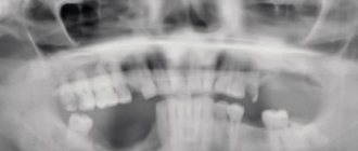
Diagnostics
Bone cancers are difficult to diagnose and the diagnosis process typically involves several different techniques.
- X-rays may reveal damage to the bone or significant abnormalities in its structure.
- Blood tests help check your overall health and find
- CT or MRI are tests that can visualize small bone abnormalities that are not visible with plain x-rays.
- PET scanning and SPECT - these methods allow you to identify very small tumors and find all traces of cancer pathology in the bones.
- A biopsy is the removal of tissue from the outside of a diseased bone for examination under a microscope. A biopsy can be done in one of two ways. In a core biopsy, a local anesthetic is used to numb the area, then a thin needle is inserted into the bone under CT guidance to take a sample. In an open or surgical biopsy, the surgeon cuts through the skin under general anesthesia to remove a piece of bone.
If Ewing's sarcoma is suspected, genetic testing is required before a biopsy to determine a specific marker for this disease. The diagnosis is then confirmed after a biopsy.
Stages
The most common staging system for this pathology is the TNM system. however, the clinic has adopted a more understandable division into stages. There are 4 stages for bone cancer. Typically, stages 1 to 4 are designated by the Roman numerals I, II, III, and IV.
The stages of bone tissue cancer pathology also depend on the degree of malignancy (cell grade), which is higher the more the tumor cells differ from normal ones. This score describes how quickly the abnormal cells grow and divide, and how likely they are to spread.
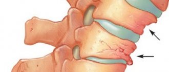
Stages are assigned only to bone tumors that begin in the shoulders, arms, hips and legs (appendicular skeleton), central body (trunk), and skull.
There are no stages for bone cancer pathologies of the spine and pelvis. They are grouped into only one category (category T), depending on the size of the tumor.
When describing the clinical stage, the words localized and metastatic are also used.
Localized cancer means that the growth is only in the bone where it started and has not spread to other parts of the body. Includes stages 1, 2 and 3.
Metastatic cancer means that the cancer has spread to another part of the body, such as the lungs. This is stage 4.
In a simplified version, the system of clinical stages looks like this.
- Stage 1A - Tumor 8 cm or smaller of low grade cells (high differentiation - neoplasia cells look like normal cells).
- Stage 1B - The tumor is larger than 8 cm, or there are tumors in several different parts of the same bone (called discontinuous tumors). Low grade neoplasia cells.
- Stage 2A - Tumor 8 cm or smaller. High grade cells (low differentiation - cells do not look like normal cells).
- Stage 2B - New growth larger than 8 cm in size and of high grade.
- Stage 3 - There are growths in several different parts of the same bone. High grade cells.
- Stage 4 - The cancer has spread to other parts of the body, such as the lungs, brain, other bones, or nearby lymph nodes. This is called metastatic bone cancer. The cell grade can be either low or high.
Recurrent bone cancer
A recurrent bone tumor means that the cancer has returned after treatment. If it returns to the same place where it first appeared, it is called a local recurrence. If it returns to tissue or lymph nodes close to where it first appeared, it is called regional recurrence. It is also possible for the tumor to appear in other parts of the body - this is called distant metastasis or distant recurrence.
Benefits of onco
In our work, we take into account every little detail so that patients’ difficult journey on the road to recovery is as comfortable and safe as possible. Over 30 years of existence in the medical market, I have helped many patients cope with their illness, providing high-quality diagnostics, therapy, and providing support at all stages of treatment. For our patients:
- Modern clinic in the city center. Two large buildings are located within walking distance of several metro stations. There is parking on site with plenty of space for your car. There will also be no problems finding the department or office you need - our employee will take you to where you say and help you find your way around the clinic.
- Favorable environment. All premises of the clinic are decorated in a stylish and beautiful interior. This has allowed us to move away from the depressing hospital environment, so patients feel comfortable. Also, patients and their accompanying persons can visit a cozy cafe and restaurant on the roof with panoramic views.
- High-tech equipment. All departments are equipped with the latest medical technology. The procedures are performed on the latest models of devices.
- High professionalism of doctors. Our employees are leaders of Russian medicine and competent international consultants. Many doctors completed internships in medical institutions in the USA, Europe and Israel. This exchange of experience and knowledge with foreign colleagues allows us to constantly improve the quality of work with patients.
- World standard. We conduct our activities in accordance with domestic and international protocols, which is confirmed by numerous certificates and awards.
Treatment
In Belgian oncology clinics, an interdisciplinary team develops a treatment plan individually for each patient. It is based on the general health condition and diagnostic data in a particular case. When deciding what treatment to offer, your healthcare team will consider:
- type of bone cancer;
- stage and class;
- patient's lifestyle;
- your general health.
Typically, a combination of several treatment methods is used for bone cancer to be most effective.
Surgery
Most people with bone cancer require surgery. The type of surgery depends on where the tumor is, how big it is, what stage of the cancer, and what type of tumor.
In Belgium, all currently known surgical technologies are used. In this case, preference is given to the least disabling techniques.
Limb-sparing surgery involves removing the tumor without removing (amputing) the entire limb . This is the main type of surgery performed in Belgian oncology centers.
After this type of surgery, the limb is reconstructed using bone and skin grafts. It is also possible to install metal endoprostheses of bones and joints. After the operation, the patient retains the mobility of the limb to the fullest extent possible.
Other types of surgery are indicated if bone-sparing surgery is not possible.
A wide resection involves removing the tumor, some normal bone, and soft tissue around the tumor. This type of surgery is also called en bloc resection.
Amputation removes all or part of the arm or leg with the tumor. Most people with amputation use a prosthesis after surgery.
Curettage uses a spoon-shaped instrument with a sharp edge (curette) to remove tumors from bone. As a result, a cavity forms where the tumor was removed. After curettage, the cavity is filled with polymethyl methacrylate, a composite filling material that restores the integrity and strength of the bone. In some cases, curettage is supplemented with cryosurgery to kill any remaining cancer cells.
Radiation therapy
External beam radiation therapy is used before and after surgery. It is also prescribed to treat bone tumors that cannot be removed by surgery or to treat metastases in the lungs.
In Belgium, the most advanced methods of radiation therapy are used - IMRT and Proton therapy.
Intensity modulated radiation therapy (IMRT) is a type of external beam radiation therapy that delivers radiation to the tumor at different angles. It can be used to treat tumors in hard-to-reach areas such as the skull, spine or pelvis.
Proton therapy delivers high doses of radiation directly to the tumor, sparing healthy nearby tissue and vital organs. The Belgian proton therapy centers in Leuven and Charleroi are equipped with some of the most advanced particle accelerators in the world.
Chemotherapy
Chemotherapy is not used for low-grade bone cancer because it usually doesn't work well or worsens the prognosis.
Neoadjuvant chemotherapy is used after surgery to remove high-grade osteosarcoma. Chemotherapy may also be offered for tumors such as:
- chordomas;
- mesenchymal and dedifferentiated chondrosarcoma;
- high grade undifferentiated pleomorphic sarcoma;
- Ewing's sarcoma.
Chemotherapy for bone cancer is usually prescribed as a combination of 2 or 3 different drugs. The medications used will depend on the type of bone cancer.
The most commonly used are carboplatin, doxorubicin and isophosphamide.
Targeted therapy
This method, using the drugs imatinib or sorafenib, has been used relatively recently, but quite successfully, in Belgium for the treatment of inoperable and chemotherapy-resistant chordomas. The drug denosumab also shows good results for giant cell bone tumors.
Other therapies
Various experimental immunotherapy options are available to patients in clinics in Belgium. Clinical trials of methods that have not yet entered into widespread practice can be a salvation in seemingly hopeless cases.
Surgery
The leading role in the treatment of patients with bone tumors belongs to the surgical method.
The principles of surgical treatment of a tumor are as follows:
- radicality – complete removal of the tumor;
- blasticity - removal of the tumor with the capture of healthy tissue to prevent recurrence of the tumor;
- preservation of the limb - without compromising its function.
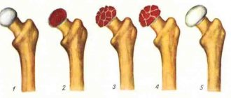
If the neoplasm is benign, then the main method is resection (removal of part of the bone) within healthy tissue.
Resection used for tumors is divided into the following methods:
- marginal - part of the bone structure is removed, there is no complete intersection of the bone, used for tumors of small and medium diameters;
- segmental - removal of the affected area of the bone by a tumor, followed by allo- or autoplasty; autoplasty is understood as replacing the defect with one’s own tissues, like an autograft using parts of one’s own tibia, ilium or fibula; an allograft is the bone elements of the donor skeleton;
resection of a joint or part of it - removal of the affected articular part; after surgery, it may be necessary to compensate for the defect with a prosthesis.
Also, for benign neoplasms, excochleation is used - curettage of the pathological focus with a blunt spoon without damaging the walls of the cavity.
If the tumor is malignant, then most often they use:
- segmental resection;
- resection of the entire joint or part of it;
- amputation;
- disarticulation.
Amputation of a limb involves the removal of its peripheral part. If the removal of part of a limb goes through the joint cavity, the operation is called disarticulation. The advantages of these operations are considered:
- reducing the risk of relapse of the pathological process;
- reducing the risk of infectious complications.
Since the treatment of malignant bone tumors involves the removal of large areas of bone, endoprosthesis replacement is required. There are special endoprostheses that make it possible to replace the bone along with the joint.
Postoperative period and rehabilitation
Recovery from treatment is different for each person and will depend on the type of treatment, your age, your overall health and many other factors. The most important part of rehabilitation is helping a person learn to walk or move again.
There may be several different healthcare professionals on the rehabilitation team.
Physical therapists help you maintain or return to your fitness level through strength and endurance exercises. They teach physical exercises to help muscles become strong again and restore range of motion.
Occupational therapists will take care of your home, work or school. They will recommend changes or tools to help you in your daily life.
Prosthetists will design and manufacture a comfortable and functional prosthesis.
Prognosis and survival
Only a doctor familiar with the specific medical history, type, stage, chosen treatment and other characteristics of cancer can make any reasonable predictions.
The following are prognostic factors for bone cancer.
Spread of cancer
Cancer can spread from where it started to other parts of the body. This spread is called metastasis. Whether the cancer has already spread at diagnosis becomes the most important prognostic factor for bone cancer. Metastasis is associated with a worse prognosis.
The location of cancer spread is also an important prognostic factor. Bone cancer that has spread only to the lungs has a better prognosis than metastasis to other parts of the body.
Tumor location
Tumors found in the legs or arms (distal tumors) have a better prognosis than tumors found in the pelvis, chest, skull, or spine (proximal tumors). This is because tumors on the arms and legs are easier to completely remove with surgery. Tumors in the chest, pelvis or spine are usually discovered later. They are often larger and closer to important organs. These factors make it difficult to completely remove them surgically.
Tumor stage
Tumors in the early stages have a better prognosis than tumors in the later stages.
Tumor size
Tumors smaller than 8 cm have a better prognosis than tumors larger than 8 cm.
Age
People under 40 years of age when diagnosed with bone cancer have a better prognosis than people over 40 years of age.
Clinical picture of osteosarcoma
The disease manifests itself as a progressive increase in the volume of the affected part of the body. The main symptoms of osteogenic sarcoma of the pelvic bones:
- “Deep” increasing pain over several weeks or months.
- The skin over the tumor may become hyperemic and swollen. A pronounced venous pattern is often identified in this area.
- With a large volume of damage, movements in the hip joint may be limited; in some cases, effusion in the joint cavity is detected.
In the area of tumor growth, a painful, dense formation can be felt, which is fused to the bone. A local increase in skin temperature may also be observed.