In gynecology, in recent years, more and more congenital anomalies of the external and external genitalia have been discovered. A defect such as a saddle-shaped uterus is detected, but more often it is bicornuate (more than half of the cases of defects of this organ).
- Norm and deviations
- Classification of pathology
- Causes and pathogenesis
- Signs of pathology
- Saddle uterus during pregnancy
- Saddle uterus and conception
- Diagnostics
- Treatment
Norm and deviations
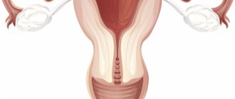
The uterus is shaped like a pear. The upper part is called the bottom, and the one below smoothly extends into the cervix, and then there is the vagina. It is in this way (from the vagina to the uterus) that pathogenic bacteria and sperm enter during unprotected sexual intercourse.
The uterus is usually from 7 to 8 cm in length, and from 4 to 5 cm in width. Its normal weight ranges from 50 to 60 grams. The uterus has tubes, which in medical language are called fallopian. They have fimbriae at the ends (they look like small brushes). The fimbriae can oscillate, which helps the mature egg after ovulation enter the fallopian tube and connect with sperm, if any.
Anomalies of organ development can be of any kind. Moreover, in some cases, deviations in the form and functions of other internal genital organs are noted. The uterus can be one-horned, two-horned, double, etc. One of the forms of a bicornuate uterus is saddle-shaped.
How are female reproductive organs formed?
Before we begin to consider the main topic, we want to explain how the female reproductive organs are formed.
When a girl is in the womb, starting from the 6th week of development, her womb develops in two separate halves, which are called “Müllerian ducts”. Normally, the Müllerian ducts merge with each other into a single structure - the uterus with one cavity, and the septum between the ducts resolves even before the girl is born. Until the 12th week, the uterus acquires the mature morphological shape of a pear. This entire process is completed by the 22nd week of development and leads to the formation of 2 fallopian tubes, one uterus, cervix and vagina.
If the functioning of this system is disrupted, a wide variety of developmental defects can occur. They range from agenesis (absence) of the uterus to more subtle abnormalities, which are classified into specific categories:
- saddle uterus;
- bicornuate uterus;
- uterus with complete and incomplete septum;
- duplication of the uterus;
- unicornuate uterus.
Classification of pathology
The bicornuate uterus can be of three types:
- incomplete
- full
- saddle
Saddle-shaped is a shape of an organ in which there is an expansion in the cross section, and at the bottom there is a depression, which is shaped like a saddle. The two horns of the organ are not very pronounced; they merge, without capturing the bottom of the organ.
The normal shape of the uterus (without pathologies) is designed by nature. It is ideal for carrying a fetus for 9 months. It is worth knowing that some animals, including dogs, have a bicornuate uterus.
What methods exist for diagnosing uterine anomalies (UDA)?
The detection of APM has increased significantly with the advent of 3D ultrasound, which clearly allows one to establish the diagnosis and is also non-invasive compared to X-ray and surgical diagnosis.
Therefore, 3D ultrasound is now considered the gold standard for assessing ARM. For a more accurate diagnosis, we recommend performing a 2D ultrasound in phase II of the menstrual cycle (18-20 days).
3D ultrasound helps: hysteroscopy, laparoscopy, MRI, metrosalpingography (x-ray diagnostics).
Causes and pathogenesis
To date, the exact reasons have not been identified. But the pathogenesis has been sufficiently studied. The uterus begins to form 10-14 weeks after conception. The paramesonephric ducts merge, which makes it possible to form 2 uterovaginal cavities, which are divided by the sagittal septum. By the end of gestation, the septum is eliminated, and the uterus already consists of one cavity (that is, there is one space in it, not divided by anything, no walls).
This means that at the beginning of embryogenesis the uterus is saddle-shaped, and then becomes shaped like a pear. If, during the formation of the organ, harmful factors influenced the body of the expectant mother and the fetus, then the ducts do not merge completely, so the child is born with various defects.
Risk factors for saddle uterus:
- obstetric pathologies that lead to a constant lack of oxygen in the child
- infectious diseases that the mother contracted while pregnant, including influenza and herpes
- heart defects in which the baby inside the mother's belly lacks oxygen
- endocrine disorders in a pregnant woman
- stress in a pregnant woman
- lack of vitamins during gestation
- intoxication of a pregnant woman (smoking, drinking alcohol, etc.)
baby uterus
“Children's uterus” is a pathology in which there is a decrease in the length of the uterine body to 30-70 mm. The small size of the organ often prevents such women from having children.
The prognosis depends on the length of the uterus. The worst prognosis is characterized by an embryonic (rudimentary) uterus, when the length of its body is less than 3 cm.
Reasons for the appearance of a “baby uterus”
A “baby uterus” can be congenital or acquired. The congenital form occurs due to a violation of embryogenesis. The exact reasons for this phenomenon cannot be established. These could be infections, toxins or other unfavorable factors affecting the mother’s body during pregnancy.
The acquired baby uterus is secondary, i.e. occurs against the background of other diseases in adolescence. These may be infections, dishormonal disorders, severe somatic diseases. As a result, the uterus does not fully develop.
Diagnosis of the “baby uterus”
The woman may not feel any symptoms. Sometimes patients complain of irregular cycles, too scanty or, conversely, too heavy menstrual bleeding, and decreased libido.
Upon examination, a woman may have a narrow pelvis and incompletely formed external genitalia.
Diagnosis is carried out using instrumental research methods.
Treatment of the “baby uterus”
Treatment of uterine pathology is aimed at eliminating the causes that caused its underdevelopment. Physiotherapeutic procedures aimed at normalizing blood circulation in the pelvic organs are indicated. It is important to correct hormonal levels and provide the patient with adequate nutrition.
Sometimes infertility develops due to pathology of the uterus due to its small size, as well as concomitant dysfunction of the genital organs. Often pregnancy occurs, but recurrent miscarriage occurs.
If the length of the uterine body is reduced slightly, the pregnancy may end in childbirth. Such women need constant monitoring - they have an increased risk of premature birth.
Saddle uterus during pregnancy
This pathology appears after the child is conceived. There may be a threat of pregnancy. In frequent cases, the doctor speaks of placenta previa or low localization. This means that due to the shape of the uterus, the embryo has attached in the wrong place, and therefore there is a risk of miscarriage.
The higher the term, the greater the risk of the baby’s incorrect position in the mother’s womb. The placenta may prematurely detach, causing external and/or internal bleeding. Early placental abruption threatens not only the life of the unborn child, but also the life of the pregnant woman herself.
Most likely, if the child was carried to term, he will be born prematurely. In the process of childbirth itself, the transmission of nerve impulses during contractions is disrupted. This causes abnormalities in the ancestral forces. In such cases, they speak of incoordination or weakness of labor. But it is impossible to know about such deviations in advance; the doctor can only assume and warn the patient.
Most women with a saddle uterus undergo a cesarean section. There is also a risk of bleeding after childbirth because the uterus does not contract normally. But you shouldn’t think that if you have a saddle uterus, there will definitely be all these complications and consequences. In some cases, gestation and childbirth go well, and the baby is born healthy, and everything is fine with the young mother.
Symptoms
Often, especially with incomplete duplication and saddle uterus, there may be no serious symptoms.
The main pathological conditions are as follows:
- Menstrual irregularities are rare.
- Miscarriage is most often associated with impaired formation of the placenta and, as a consequence, malnutrition of the fetus.
- Infertility is a rare condition, since the duplication of the uterus itself cannot cause infertility. Pregnancy occurs even with complete doubling, and in this case it develops most often in one of the horns of the uterus (less often in both horns). As a rule, the cause of infertility is a combination with other developmental defects.
Saddle uterus and conception
Some experts scare their patients with the fact that the saddle uterus prevents conception. But women with this pathology are not always infertile. This diagnosis can be received by those girls who have a pronounced saddle-shaped organ. Then the egg will not be able to implant normally in the uterus. In some cases, spontaneous abortion is possible. If the saddle pattern is not very pronounced, but the woman still cannot become pregnant, the doctor looks for other causes of this problem. And there can be a great many of them!
As noted above, with a saddle uterus there may also be defects of the urogenital system. A woman may have an imbalance of hormones in her body. Extragenital diseases that occur in a chronic form are also likely.
Many patients ask the question in what position should they conceive if they have a problem such as a saddle uterus. But this is not a correct question, because the likelihood of conception does not depend on the position whether the woman is healthy or not. As you know, sperm are mobile, active, they live quite a long time in the vagina (and move further). So the problem of infertility should be looked for not in the position, but in the state of health (yours and your husband’s).
The saddle shape of the uterus does not prevent sperm from entering the uterine cavity and tubes. Difficulties may arise during the process of attachment of the fused sperm and egg.
General information
Congenital anomalies of the development of the female genital organs, increasingly diagnosed by doctors recently, can become the main cause of the inability to conceive and bear a child.
The saddle shape of the uterus is one of these pathologies, which, according to statistics, is diagnosed in approximately 0.5% of women of childbearing age and can become a serious obstacle to happy motherhood. How to get pregnant with such a diagnosis? Is conception even possible with such a structure of the main female reproductive organ? How to carry a child to term if a saddle uterus is detected already during pregnancy ? We will answer these and other questions on the topic in this material. And to better understand the essence of the problem, we will start with general points regarding the structure of the female genital organs.
Diagnostics
As already noted, it is impossible to make a diagnosis by eye. The following research methods are needed:
- Ultrasound of the uterus and its appendages
Ultrasound does not always detect pathology. Those women who have a strongly pronounced saddle shape of the organ have a greater chance of receiving a correct diagnosis. It is better to do an ultrasound with a vaginal sensor. Visit a specialist in the second half of the cycle, when thickening of the endometrial layer is observed.
- Hysterosalpingography/hysterography
These are methods that use x-rays. Contrast is injected into the uterus, and then pictures are taken with special equipment. The doctor discovers a saddle-shaped depression on them.
- Hysteroscopy
A special optical device is inserted into the uterus, which makes it possible to examine the organ from the inside and understand if there are deviations or not. The method is very accurate in identifying abnormalities of the uterus in girls and women.
- MRI
This method also takes pictures of the uterus at different levels.
Reasons for the formation of abnormalities in the development of the uterus
- infectious diseases suffered by the mother during pregnancy , especially in the first trimester (measles, rubella, influenza, toxoplasmosis, cytomegalovirus infection, syphilis);
- metabolic disorders and endocrine pathology (especially thyroid dysfunction, prolonged fasting, deficiency of vitamins and microelements);
- intoxication (long-term use by a woman before and during pregnancy of alcohol, narcotic substances, and some medications - the most negative effect is in the early stages, when the formation of all organs and systems is formed);
- hereditary factor (chromosomal and gene mutations);
- negative environmental influences (radiation, chemicals, psychological trauma).
Treatment
Therapy is relevant only for those who are faced with the problem of infertility caused by this particular pathology. They are reconstructing the organ. This is a plastic surgery that is done through hysteroscopy. This means that there will be no long anesthesia, and there will also be no large incisions (which will avoid unsightly scars on the body). After a correctly performed operation, the chances of conceiving a child increase many times over.
During gestation, women with a saddle-shaped uterus may experience obstetric complications. Treatment is individual. A woman should observe bed rest, that is, lie down and not lead an active lifestyle. The doctor selects drugs for the patient from a number of tocolytics and antispasmodics. Hormonal therapy is relevant, the drugs of choice are utrozhestan and duphaston.
To ensure that blood circulates normally in the uterus and in the vessels connecting the organ to the placenta, special medications are used. They affect blood clotting and normalize metabolic processes. These are the following medications:
- troxevasin
- Essentiale Forte
- chime
- Actovegin, etc.
Remember that for girls and women of any age, regular examinations by a gynecologist and passing the tests prescribed by him are important. By neglecting your own health today, you can develop a problem such as infertility. Take care of yourself and be healthy!
Removal of the intrauterine septum
Surgical removal of the septum in the uterus is the main treatment method for this pathology offered in our clinic. The hysteroresectoscopy procedure involves dissection and excision of a septum of sufficiently large thickness and density. Today, more modern methods of treating intrauterine septums are also practiced, which include cutting them with a laser.
With a complete septum reaching the internal os of the cervix, its cervical part is usually not affected during surgical treatment. This is done in order to avoid the development of subsequent isthmic-cervical insufficiency.
Surgical removal of the intrauterine septum is a low-traumatic surgical intervention that does not leave scars. This means that after such treatment nothing will interfere with normal conception, pregnancy and natural delivery.
After surgery, the risks of miscarriage and the development of other obstetric complications are significantly reduced. However, pregnancy management in women after removal of the intrauterine septum requires close attention of obstetricians and gynecologists.
Types of adenomyosis
Depending on the distribution of pathological structures in the affected tissues, the following forms of adenomyosis are distinguished:
- Diffuse manifests itself as a uniform distribution of small inclusions of the endometrium throughout the muscular layer of the uterus;
- With the nodular type of disease, endometrioid nodes filled with blood are formed in the muscle layer;
- The focal form of adenomyosis is characterized by the formation of small accumulations of endometrial cells in various areas of the uterus.
Take the first step
make an appointment with a doctor!
There is also a cystic form, which is a further development of nodular or focal adenomyosis and is formed when the nodes hemorrhage followed by the formation of cysts. These types of this pathology can form separately or in combination with each other. The most common form is a diffuse nodular form, in which endometrial cells form a node and areas with their uniform inclusion.
There are also several stages of development of this disease, characterized by the degree of damage to the uterine tissue:
- stage 1 - at this stage, endometrial tissue penetrates only the thinnest layer of the myometrium (muscular wall) of the uterus;
- stage 2 - the pathological process penetrates deeper into the muscle wall, consisting of 3 layers, affecting 2 of them;
- stage 3 - the endometrium extends to all 3 layers of the myometrium up to the serous membrane of the uterus, which borders the surface of the bladder;
- stage 4 - endometrial tissue penetrates the uterine myometrium and affects adjacent organs, causing external gynecological endometriosis.
The signs of uterine adenomyosis and its damage to the body depend on the stage of the disease. Determining the stage of development of pathology plays an important role in its diagnosis and treatment. The earlier it is detected, the more effective and gentle the therapy will be.