Williams syndrome
The diagnosis of Williams syndrome is made based on the patient’s current status, the results of a biochemical blood test, cardiac and molecular genetic studies. Upon examination, a characteristic appearance (“elf face”), disturbances in the formation of teeth and caries, muscle hypotonia with accompanying changes in the musculoskeletal system, signs of mental disability and retardation in physical development are revealed. The picture of a biochemical blood test in Williams syndrome is quite diverse; almost always with this disease, hypercalcemia, an increase in the level of thyroid-stimulating hormone, and a slight decrease in the level of neutrophils are determined. As a rule, studying the patient's hereditary history does not make sense unless one of the parents has Williams syndrome.
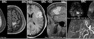
Cardiac studies (EchoCG) can detect congenital heart defects, most often - aortic or mitral valve insufficiency, supravalvular stenosis of the aorta and pulmonary trunk. In older patients with Williams syndrome, thickening of the walls of the aorta and other great vessels and severe arterial hypertension may be detected. Upon examination, indirect signs of the disease may include umbilical and inguinal hernias, convergent strabismus, and a hoarse, low voice. The most reliable diagnostic method for Williams syndrome is molecular genetic analysis performed by a geneticist. To detect the absence of a region of chromosome 7, DNA microarray or fluorescence hybridization (FISH) techniques are used; prenatal diagnosis is possible.
There is no specific treatment for Williams syndrome; palliative measures and psychological correction methods are used. Surgery is sometimes required at an early age to correct congenital heart defects, which can be fatal in severe cases. To reduce the severity of arterial hypertension, traditional drugs are used, mainly from the group of ACE inhibitors. Psychocorrectional work for Williams syndrome allows patients to master speech, and in some cases, reading and writing, and reduces the likelihood of developing anxiety and obsessive-compulsive disorder. A friendly emotional environment in the family plays an important role in reducing the severity of mental retardation.
Forecast and prevention of Williams syndrome
The survival prognosis for those suffering from Williams syndrome is relatively favorable - the greatest threat at an early age is congenital heart defects, but if they are mild or promptly eliminated, patients can live to an old age. Life expectancy is usually slightly lower than that of healthy people, which is due to a persistent increase in blood pressure and disturbances in calcium and carbohydrate metabolism. Although partial social adaptation is possible with Williams syndrome, most patients require careful care and monitoring from relatives. The possibility of any work activity in the presence of this disease is completely excluded. There is no prevention for Williams syndrome, since this disease is usually the result of a spontaneous mutation - only prenatal detection of this pathology is possible.
Acquired talent
Apparently, anyone can obtain one or even several of the listed qualities without much effort on their part, if they are “lucky.” How do you get savant syndrome? It is enough, for example, to get hit on the head with a blunt object in an alley, to dive unsuccessfully into a pool, to suffer a hemorrhagic stroke, or, of course, to be struck by lightning. However, the probability of such “luck” cannot be calculated even by a savant, since thousands are injured every day, and only mattress salesman Jason Padgett, who was severely beaten on a September night in 2002 while leaving a karaoke bar, has shown his ability in mathematics and drawing. In addition to health problems, the former 31-year-old lover of drinking and singing acquired “geometric thinking.” He began to see the world around him in general and mathematical formulas in particular as a collection of lines and geometric shapes. As a result, Padgett took up number theory professionally.
A case is described of a 9-year-old Mexican boy who was wounded by a bullet in the left temporal region during a robbery. In addition to the fact that he suffered right-sided paralysis, he also forgot how to speak, read, write and count. On the plus side: he stopped getting lost in unfamiliar areas (he always knew the way to a new house when his family moved frequently), began to perfectly repair and improve all kinds of mechanical devices and redraw what he saw with photographic accuracy.
Age-related degenerative brain diseases usually put an end to an artist's career. However, the opposite can also happen: in 1998, Bruce Miller and co-authors described in the journal Neurology five cases of a sudden desire and ability to draw in patients with frontotemporal dementia, and the skill developed in parallel with the severity of the disease. When conducting single-photon emission computed tomography in patients, a predominant lesion of the left parts of the brain was revealed.
Treatment methods
Medicine does not have the means to completely cure Williams syndrome, so treatment is based on the correction of physical and mental disorders .
To reduce the severity of physical defects (underdevelopment of internal organs), symptomatic drug therapy , which supports the functioning of the heart, kidneys, and improves the functioning of the stomach and intestines.
Drugs are selected individually depending on the characteristics and severity of pathologies. A number of surgical interventions are often necessary.
Specific drug treatment aimed at correcting mental retardation and mental disorders has not been developed. Experts often prescribe drugs from the following groups:
- Nootropic and neurometabolic medications (Cerebrolysin, Actovegin, Semax). They protect brain cells from hypoxia, improve adaptation and learning, and have a positive effect on cerebral circulation.
- Sedatives (Valerian, decoctions of sedative herbs, Novo-passit). Used if the child shows increased anxiety. With the development of phobias and other mental disorders, work with a psychotherapist is indicated, and the use of stronger means is indicated.
The following correction methods are also used:
- Physiotherapy. Maintains muscle corset tone, preventing the occurrence of curvature of the spinal column, improves motor skills and coordination.
- Massage. Positively affects muscle strength and improves coordination.
- Neuropsychological correction. In the process of special classes, the child learns to take care of himself, talk, read and write, and expands his vocabulary. The best result is obtained if parents actively join in working with the child and surround him with love.
Children are also advised to follow a diet that is specially developed depending on the severity of their deviations.
In the first years, diet therapy is aimed at reducing calcium concentrations. It is recommended to reduce the amount of salt and spices in dishes to reduce the risk of developing arterial hypertension.
Origins of the disease
They started talking about the disorder on a large scale in 1983 thanks to the Swedish scientist Bengt Hagberg. At this time, he and his group studied 35 similar cases in 3 different countries: Portugal, France and Sweden.
However, Hagbert is not the discoverer of the syndrome. It was first discovered by pediatrician Andreas Rhett, whose name the disease bears. He observed two girls who had the same symptoms. He noticed them in the reception line. They sat on their mothers' laps, and they held their hands. The girls swung like pendulums, and then suddenly both began making stereotypical movements with their hands. The children were frozen in one position, detached from the world around them. The gaze was directed to one point. Their synchronicity in movements and behavior was amazing.
The doctor found similar case histories in his written archives, and then went to Europe to find the same patients there. In 1966, he made the first publications of his research, which, however, did not arouse much interest.
Rett called the disease he recorded brain atrophy syndrome. At first it was considered a manifestation of autism or schizophrenia, and only in 1983 was it brought into a separate nosological unit.
Currently, the syndrome is classified as a rather rare genetic disease. It occurs with a incidence of 1 in 15,000. The cause is said to be a mutation in the MECP2 gene. This gene is responsible for the synthesis of a certain protein that affects brain development. Normally this protein is
established some time after birth, must be suppressed by other genes to ensure normal brain development.
If the MECP2 gene is mutated, the protein is not completely inactivated, which causes abnormal brain maturation and provokes the development of Rett syndrome.
Usually the mutating gene is located on the X chromosome, so the disease mainly affects girls.
Positive effects of some deletions on viability
Small deletion changes can have a significant impact on the survival of the organism. For example, the loss of the gene encoding the CCR5-δ32 protein causes immunity to the human immunodeficiency virus. Scientists suggest that this mutation first appeared about 2.5 thousand years ago and over time spread across Europe.
The current distribution is uneven. According to statistics, about 10% of residents of European countries are resistant to HIV. However, in the Scandinavian countries this figure reaches 14-15 percent. Russians and Finns demonstrate a 16 percent level of resistance. For Sardinia, however, the frequency is a modest 4 percent.
A number of scientists have hypothesized that such a spread is determined by the bubonic plague epidemics that took place in the medieval period. It is likely that a mutation in the gene causes increased resistance to this disease. Therefore, in the countries where the Black Death took place, more people with this genotype survived.
Etiology and pathogenesis
Currently, the causes of the disease are not fully understood. Experts have found that the main etiopathogenetic factor of pathology is heredity. The syndrome develops as a result of deletion of chromosome 7 . This rearrangement leads to the loss of a section of the main autoreproducing structure of the nucleus and a disruption in the chromosomal complement of the fetus. A hereditary defect of chromosome 7 is a loss of a long arm containing 25-30 genes. The lost part of the chromosome is not transmitted at the time of conception, and the newborn child develops a characteristic clinical picture. The hereditary form of the disease develops extremely rarely - when the father or mother has Williams syndrome.
There is another theory of the origin of the disease - the spontaneous occurrence of pathology, which was the result of a chromosomal mutation during conception. This assumption allows us to conclude that the disease is congenital and not hereditary. A random chromosomal breakage leads to the birth of a sick child in an absolutely healthy family. The causes of such a genetic anomaly are factors that have a negative impact on the parents’ body. These include: occupational hazards, poor environment, bad habits.
Williams syndrome is characterized by the absence of genes responsible for:
- development and functioning of the brain,
- production of elastin protein,
- carbohydrate metabolism,
- calcium metabolism in the body,
- intellectual development,
- trophic processes in nervous tissue.
Elastin deficiency over time leads to the formation of heart defects and the development of hypertension. Disorders such as calcification of blood vessels and heart valves lead to their thinning, loss of elasticity and rupture under the influence of various factors. Calcification is a very dangerous pathological process, which over time becomes the cause of acute cardiovascular diseases and death of the patient.
The effect of deletions on the ability to fertilize
Deletions that occur on normal chromosomes (autosomes) can in some cases be compensated for by a normal copy of the gene. However, when it comes to sex chromosomes, especially the Y chromosome, the situation changes.
First of all, it should be noted that the genes localized on it do not have a second copy. With a normal number of chromosomes in the set, the Y chromosome turns out to be extremely vulnerable. Combined with a small number of genes, this leads to serious consequences for each change. Of particular interest are mutations affecting the AZF locus and the SRY gene.
Differential diagnosis
Differential diagnosis is carried out with similar diseases, which in their clinical picture have heart defects, developmental delays, and features of the facial skeleton. Such diseases include:
- Noonan syndrome. The disease is genetic and has an autosomal dominant mode of inheritance. The most common heart defect is pulmonary stenosis. The appearance attracts attention with wide-set eyes, drooping upper eyelid, and low-set ears. On examination, a deformation of the chest (keeled breasts), widely spaced nipples are observed;
- Deletion 2 syndrome is transmitted in an autosomal dominant manner. It is distinguished by its characteristics: drooping eyelids, small ears, underdevelopment of the wings of the nose. Upon examination, the child has a decrease in calcium levels and immunodeficiency;
- Smith-Magenis syndrome is not inherited; all cases are De Novo, that is, they occur for the first time and only in one family member. Heart defects are observed in the septum (septum). Appearance is characterized by deep-set eyes, underdeveloped irises, and Mongoloid eye shape. The patient’s behavior also has its own characteristics; self-aggression is observed. When speaking, a hoarse voice is noted;
- Kabuki syndrome. This disease can be inherited either as an autosomal dominant type or as an X-linked one. The most common congenital heart defect detected during examination is coarctation of the aorta. In addition to heart diseases, congenital anomalies of other organs and systems are also observed - malformations of the spine, cleft lip and palate. On the child's face there are inverted lower eyelids and large ears;
- Fetal alcohol syndrome is not inherited and develops when the mother abuses alcohol during pregnancy. A child is diagnosed with septal heart defects. Upon examination, the child is lethargic and disinhibited. The syndrome has its own phenotypic features - short palpebral fissures, thin upper lip, small ears, flat filter.