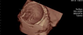
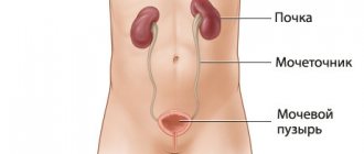
An ovarian dermoid cyst (teratoma) is a neoplasm consisting of tissues that are not typical for this organ. The causes of the pathology are associated with the appearance of anomalies during intrauterine development, which contributed to the formation of teratoma. In most cases, the tumor is benign, but there is a possibility of its malignant degeneration. Treatment of dermoid cysts is successfully performed at the Yusupov Hospital, where experienced gynecologists work who will prescribe the necessary diagnostics and adequate therapy. Appointments with specialists can be made by telephone.
Ovarian dermoid cyst: ICD-10 code
The dermoid type of cyst accounts for 20% of all types of ovarian cysts. The pathology is a congenital anomaly and may not show itself for a long period of time. It can be discovered accidentally during a routine examination or diagnosis of pelvic organ disease.
Unlike ordinary cysts, which are characterized by the accumulation of fluid, cell division occurs in dermoid cysts. Such a cyst is a dense epithelial node with a capsule (usually single-chamber), which contains elements of germinal tissue: hair, teeth, bones, glands, etc. The size of the node can reach 15 cm in diameter, which significantly worsens the woman’s health.
According to ICD-10, a dermoid cyst belongs to the class of benign neoplasms (D10-D37). It has code D27 - benign ovarian neoplasm. In 1-3% of cases, malignant degeneration of the tumor occurs, which requires mandatory consultation with an oncologist.
Immature teratomas, in which active cell division is observed, are prone to malignancy. They can grow into neighboring tissues and metastasize. Moreover, more often the oncological process is detected not in the primary tumor, but in metastases.
Ovarian dermoid cyst: causes
The reasons for the development of ovarian dermoid cysts are not fully understood. Doctors cannot name the exact conditions in which teratoma develops. The most common theory is a failure in the development of identical twins, when one twin absorbs the other. Proof of this is the contents of the dermoid cyst: underdeveloped bone and cartilage tissue, epithelium, hair, elements of glands and other organs that are not related to the ovaries.
A dermoid cyst occurs in utero and develops during human growth. Having reached a certain size, it stops growing. In this case, she is called mature. If the process of cell division continues and it is active, then an immature cyst is implied, the presence of which requires consultation with an oncologist.
Among the reasons for the formation of a dermoid cyst, there are provoking factors that negatively affect the course of pregnancy and can cause various fetal abnormalities:
- Viral and bacterial diseases of the body;
- Infectious diseases of the pelvic organs;
- Presence of bad habits (alcoholism, smoking, drug addiction);
- Food and toxic poisoning;
- Effect of radiation;
- Overdose and uncontrolled use of certain medications.
The impact of a negative factor on the body of a pregnant woman does not necessarily guarantee the appearance of teratoma. However, in this case the woman is at risk of developing fetal pathologies.
Symptoms
This pathology, like other benign formations, may not manifest itself for a long time. A dermoid cyst on the eyelid can be detected upon careful examination as a slightly elongated or rounded elastic “ball” localized under the skin. Symptoms of formation usually appear when it becomes inflamed, suppurates, increases in size, and begins to put pressure on nearby tissues and organs.
A cyst on the eyelid is characterized by the following:
- Usually has a round shape;
- Elastic and dense to the touch;
- Painless on palpation;
- Does not adhere to the skin;
- The skin over the dermoid cyst on the eyelid is of a normal color, without ulcerations, rashes, etc.;
- It does not increase in size for a long time.
The clinical manifestations of a dermoid cyst on the eyelid are usually determined by its size and the age of the patient. Dermoids, which are small in size, do not affect health and do not cause damage to the eye structures. With small sizes, this is just a cosmetic defect. However, as the formation begins to grow, it begins to interfere with clear vision. In addition, the risk of its inflammation and malignancy (the appearance of malignant properties) increases, which becomes the reason for its mandatory removal.
Ovarian dermoid cyst: symptoms
Signs of pathology will depend on the type of neoplasm (mature or immature) and its size. In many cases, the cyst does not manifest itself for a long period of time, so if the disease is asymptomatic, it is difficult to detect.
A mature cyst will have the following symptoms:
- Abdominal pain;
- Unpleasant sensations during sexual intercourse;
- Increased urination;
- Difficulty in defecation.
It should be noted that with a dermoid cyst, the menstrual cycle is not disrupted. The neoplasm does not affect the woman’s hormonal levels and the process of menstruation.
An immature cyst is also characterized by pain in the lower abdomen, as well as general malaise. It is associated with tumor progression. As a result, a woman may suddenly lose weight and experience general symptoms of cancer intoxication.
A dermoid cyst has a stalk that attaches it to the ovary and through which blood channels pass. The presence of a leg is accompanied by the risk of torsion. This is a serious complication that can lead to necrosis of the node and the development of peritonitis. In this case, acute abdominal pain occurs, body temperature rises, and nausea and vomiting may occur.
Prevalence of ovarian cysts
According to various authors, the incidence of ovarian tumors among all gynecological diseases ranges from 8 to 19%.
Foreign researchers claim that more than 80% of women of the reproductive period had a history of an ovarian cyst at least once. However, only 1/4 of them had any clinical manifestations.
In postmenopausal women, the incidence of ovarian tumors ranges from 3 to 18%, but it is at this age that they need to pay special attention due to the high risk of malignancy.
Ovarian dermoid cyst: treatment
The only effective treatment for teratoma is surgical removal. The extent of the operation will depend on the size of the tumor and accompanying circumstances. Most often, treatment is performed laparoscopically.
In case of a benign course of the disease, the cyst is removed along with the ovary and the upper part of the uterus. If it is necessary to preserve reproductive function, the decision to remove the ovary is made individually.
Laparoscopy of ovarian dermoid cyst has positive reviews from both doctors and patients. The operation is quick and the woman can go home the same day. The decision on short-term medical examination is made individually. The operation has a short recovery period and is performed without an abdominal incision. After it is carried out, there are no scars left that would have a negative aesthetic appearance.
In the case of oncological degeneration of the tumor, the appendages are removed along with the uterus. Additionally, a course of chemotherapy is drawn up. Radiation therapy in this case is ineffective.
At the Yusupov Hospital you can undergo a full course of treatment for dermoid cysts, including examinations using the latest equipment. Treatment is prescribed strictly individually based on diagnostic results by experienced gynecologists and oncologists (in case of a malignant process). This approach ensures maximum effectiveness of the prescribed therapeutic measures.
Our doctors who will solve your vision problems:
Fomenko Natalia Ivanovna
Chief physician of the clinic, ophthalmologist of the highest category, ophthalmic surgeon. Surgical treatment of cataracts, glaucoma and other eye diseases.
Yakovleva Yulia Valerievna Refractive surgeon, specialist in laser vision correction (LASIK, Femto-LASIK) for myopia, farsightedness and astigmatism.
You can find out the cost of a particular procedure or make an appointment at the Moscow Eye Clinic by calling in Moscow 8 (499) 322-36-36 (daily from 9:00 to 21:00) or using the ONLINE REGISTRATION FORM.
Prevention and prognosis
Mature teratomas have a favorable prognosis. Due to the presence of a stalk of the tumor, it is recommended to remove it even if it does not cause discomfort. Otherwise, there is a risk of sudden torsion of the leg with subsequent inflammation. In addition, with a teratoma, a woman is at risk of complicated pregnancy.
Immature dermoid cysts have a poor prognosis. They undergo long-term treatment followed by constant monitoring of the condition.
There is no specific prevention that would guarantee the absence of the development of ovarian dermoid cysts. The main actions are aimed at reducing the risk of abnormal pregnancy in order to prevent the development of a pathological process in the fetus. They include a healthy lifestyle before and during pregnancy, a balanced diet, the absence of bad habits, and a responsible attitude towards your health.
At the Yusupov Hospital, you can undergo regular gynecological examinations, which will allow you to suspect pathology in time. Gynecologists at the Yusupov Hospital provide consultations to women planning a pregnancy to eliminate possible risks for the mother and unborn child.
Types of epidermal cysts
Doctors distinguish several types of formations. Classification is carried out according to structure, symptoms and the presence of complications.
The most common diagnosis is atheroma. This formation is localized in the face, arms, neck and scrotum. A single atheroma may appear on the skin or several may appear at once.
This type of epidermal cyst, such as atheroma, has a round shape and soft contents. As a rule, this formation is yellow or red in color.
Atheroma does not bother a person; it can remain the same size or rise slightly above the skin.
When an infection occurs, the formation suppurates and leads to inflammation of neighboring tissues.
Diagnostics
The basis for verifying the diagnosis are:
- data from a visual medical examination or examination by specialized specialists (urologist, neurologist, etc.);
- Ultrasound (detects a cavity formation with smooth contours, clarifies its size);
- CT/MRI (studies are informative for rare localizations of epidermal cysts);
- histological examination (reveals a thinned epidermis without other skin appendages on the inner surface of the cyst and horny scales inside it, which sometimes become calcified).
Treatment methods
Since the formation takes a long time to develop and is benign, it is not necessary to remove it. The decision to undergo surgical intervention is made for cosmetic reasons or in the event of an unsuccessful pathology location or frequent mechanical damage to a neoplasm protruding above the surface of the skin. This increases the risk of bacterial infection with subsequent transition to purulent processes. In this case, radical treatment in the form of removal is the only way to prevent more serious diseases and complications.
Epidermal cysts are treated in several ways:
- If the formation is large, it is removed with a scalpel. A scar remains at the site of the incision.
- Laser removal of atheroma is considered the preferred method of choice due to the minimal list of contraindications and excellent results.
- Burning with liquid nitrogen is affordable, but has its drawbacks - inaccuracy of the effect, prolonged healing of the burn.
Important: Do not try to treat epidermal cysts or other skin formations with folk remedies. This is fraught with serious damage and the appearance of a scar at the site of exposure. In addition, without histology it is impossible to accurately determine the nature of the neoplasm, therefore it is impossible to talk about its benign quality. Any unprofessional interventions can cause serious health problems.
Causes and mechanism of ovarian apoplexy
The causes of ovarian cysts can be different:
- disruptions in the functioning of the endocrine system, abnormalities in the functions of the thyroid gland or adrenal glands;
- unstable hormonal levels, menstrual irregularities;
- infections or inflammations in the ovaries and fallopian tubes;
- abortion or genital surgery;
- side effects from taking certain medications;
- genetic predisposition.
The main causes of ruptured ovarian cysts include:
- excessive increase in intra-abdominal pressure;
- pathological changes in blood vessels in the ovaries, and, as a result, impaired blood flow;
- abdominal injuries;
- inflammatory processes of the ovaries;
- hormonal imbalance;
- uncontrolled use of anticoagulants - blood thinning drugs.
In some situations, a break occurs in the midst of complete health, and even a detailed examination does not allow us to establish the true cause of the disease.
Serous cystadenocarcinoma
Ultrasound reveals a complex cystic-solid formation in the left ovary, and another large complex formation containing both a solid and a cystic component in the right half of the pelvis
A CT scan of the same patient reveals a complex cystic-solid formation with thickened septa accumulating contrast in the right ovary, extremely suspicious for a malignant tumor. There is also bilateral pelvic lymphadenopathy (arrows). Histopathological examination confirmed serous ovarian cystadenocarcinoma (the most common variant)
CT scan and macroscopic photograph of serous ovarian cystadenocarcinoma.
Ultrasound (left) shows a large multilocular cystic formation in the right parametrium; Some of the chambers are anechoic; in others, uniform low-level echogenic inclusions are visualized, caused by protein content (in this case, mucin, but hemorrhages can also look similar). The partitions in the formation are mostly thin. No blood flow was detected in the septa, the solid component was also absent, and no signs of ascites were detected. Despite the absence of blood flow during Doppler ultrasound and a solid component, the size and multilocular structure of this formation allows us to suspect a cystic tumor and recommend other, more accurate diagnostic methods. Contrast-enhanced CT scan (right) shows similar changes. The formation chambers have different densities, corresponding to different protein contents. Histopathological examination confirmed mucinous cystadenocarcinoma with low malignant potential.
Corpus luteum cyst
The corpus luteum can become obliterated and fill with fluid, including blood, resulting in the formation of a corpus luteum cyst.
Ultrasound: corpus luteum cyst. Small complex ovarian cysts are visible with blood flow in the wall, which is detected by Doppler ultrasound. Typical circular blood flow during Doppler examination is called the “ring of fire.” Note the good permeability of the cyst to ultrasound and the absence of internal blood flow, which correlates with the changes characteristic of a partially involuted cyst of the corpus luteum
It should be noted that women taking hormonal oral contraceptives that suppress ovulation usually do not develop a corpus luteum. Conversely, the use of drugs that induce ovulation increases the chance of developing corpus luteum cysts.
Pelvic ultrasound: corpus luteum cyst. On the left, the sonogram shows changes (“ring of fire”), typical of a corpus luteum cyst. On the right, in the photo of the ovarian specimen, a hemorrhagic cyst with collapsed walls is clearly visible.
Corpus luteum cyst on MRI. An axial T2-weighted tomogram reveals a cyst of the involuted corpus luteum (arrow), which is a normal finding. The right ovary is unchanged.
Cystic metastases to the ovaries
Most often, metastases to the ovaries, for example, Krukenberg metastases - screenings of stomach or colon cancer, are soft tissue formations, but often they can also be cystic in nature.
A CT scan reveals cystic formations in both ovaries. You can also notice a narrowing of the rectal lumen caused by a cancerous tumor (blue arrow). Cystic metastases of rectal cancer are clearly visible in the recess of the peritoneum (red arrow), which in general are not a typical finding.
Normal anatomy and physiology of the ovaries during reproductive age
Before considering pathological changes, we will highlight the normal anatomy of the ovary. A woman's ovary at the time of birth contains over two million primary oocytes, about ten of which mature during each menstrual cycle. Despite the fact that about a dozen Graafian follicles reach maturity, only one of them becomes dominant and reaches a size of 18–20 mm by the middle of the cycle, after which it ruptures, releasing the oocyte. The remaining follicles decrease in size and are replaced by fibrous tissue. After the oocyte is released, the dominant follicle collapses, and granulation tissue begins to grow in its internal lining in combination with edema, resulting in the formation of the corpus luteum of menstruation. After 14 days, the corpus luteum undergoes degenerative changes, then a small scar remains in its place - the white body.
Graafian follicles: small cystic formations found in the structure of the ovary normally in all women of reproductive age (premenopausal period). The size of the follicles varies depending on the day of the menstrual cycle: the largest (dominant) usually does not exceed 20 mm in diameter at the time of ovulation (14th day from the start of menstruation), the rest do not exceed 10 mm.
Ultrasound of the ovary is normal. Sonograms show ovaries containing several anechoic simple cysts (Graafian follicles). Follicles should not be confused with pathological cysts.
What do the ovaries look like on an MRI? On T2-weighted MR images, Graafian follicles appear as hyperintense (i.e., bright signal) cysts with thin walls surrounded by ovarian stroma, which gives a less intense signal.
Normally, in some women (depending on the phase of the menstrual cycle), the ovaries can intensively accumulate radiopharmaceuticals (RP) during PET. To distinguish these changes from a tumor process in the ovaries, it is important to correlate them with the patient’s anamnestic data, as well as with the phase of the menstrual cycle (the ovaries intensively accumulate radiopharmaceuticals in the middle). Based on this, it is better for women before menopause to be prescribed PET scans in the first week of the cycle. After menopause, the ovaries practically do not take up radiopharmaceuticals, and any increase in its accumulation is suspicious for a tumor process.
PET-CT of the ovaries: increased accumulation of a radiopharmaceutical (RP) in the ovaries of a woman in the premenstrual period (normal variant).


