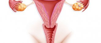
Polyps of the cervical canal are neoplasms in the form of tumors that are benign in nature. The structure of the outgrowths consists of tissue that is necessary for the functioning of the woman’s protective system and her musculoskeletal system.
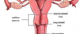
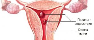
Their location is a sounded channel, to which they are attached using legs of different diameters. Its size determines how far the polyp will extend. If the leg is long enough, diagnosis of the presence of a polyp of the cervical canal is limited to an examination by a gynecologist.
The color of a polyp directly depends on the number of blood vessels in its structure. Burgundy coloring is evidence that the benign growth has a dense structure.
Small formations may have a mild clinical picture. A pathology of impressive size causes significant discomfort to a woman.
Polyp sizes:
- from 2 to 9 mm - small formation;
- from 10 to 30 mm - average growth;
- 40 mm is the maximum size of the tumor.
Despite the benign nature of the disease, a patient with a large number of growths should undergo regular diagnostic examinations. Otherwise, there is a risk of cervical cancer.
Types of polyps by structure:
1. Physical ones. This type of pathology is rarely detected by the woman herself without examination by a gynecologist. It does not produce mucus, and bleeding is recorded in isolated cases. The mild symptoms are due to the fact that the structure of the fibrous formation does not contain glands and is not distinguished by a large number of blood vessels.
2. Ferrous. The voiced pathology is usually small in size, which reduces mucus secretion in a woman. Symptoms of the disease are practically absent.
3. Fibrous - glandular. The mixed type of the two varieties has a clear clinical picture: complaints of pain in the lower abdomen and bleeding of varying intensity depending on the size of the polyp.
4. Atypical. They are dangerous because their growth is uncontrollable. Being a precancerous condition, atypical polyps often require a course of chemotherapy after their removal.
5. Decidual. A similar type is diagnosed in pregnant women. Growths measuring 10+ mm do not have the same surface in different women.
Types of polyps by shape:
- grape-shaped;
- in the form of a tongue;
- round shape;
- oval;
- oddly shaped balls.
There is a similarity in visual characteristics of the pathology with the following diseases:
- endometriotic polyp;
- submucous myoma;
- sarcoma.
Pseudopolyps should be noted separately. Their peculiarity is their occurrence in women during pregnancy. In most cases, doctors recommend removing the growths to minimize the risk of miscarriage.
The causes of the disease do not have a clear genesis, but some factors can be identified - provocateurs:
1. Urogenital infections. After sexual intercourse, which results in damage to the vagina by chlamydia or gonorrhea, the woman’s body’s protective reaction is activated. The epithelium of the cervix begins to grow, creating fertile ground for the emergence of a polyp.
2. Infectious diseases. We are not talking about sexually transmitted diseases, but about endometriosis (proliferation of the inner layer of uterine cells) and vulvovaginitis (inflammation of the vagina).
3. Injury to the cervical canal. Its damage occurs due to an incorrectly placed intrauterine device. He is also injured after an abortion and childbirth with complications.
4. Problems with the cervix. Polyps of the cervical canal can form when diagnosing erosion (pseudo-erosion) and leukoplakia.
5. Hormonal imbalance. In women over 40, pronounced age-related changes are sometimes recorded. When the hormonal balance is disrupted, the cells of the cervical epithelium begin to divide and increase in volume.
6. Stress. With severe mental shock, the immune system bears an additional burden. As a result, its protective block weakens, and the infection easily enters the cervical canal.
Women at risk:
- patients in the age category after 31 years and up to 50 years;
- postpartum period in a woman;
- patients with many gynecological problems.
Diagnosis of pathology:
1. Gynecological examination. During it, the doctor uses special mirrors to analyze the thickening of the cervix and the color of the polyps.
2. Colposcopy. For this procedure, a special optical device (colposcope) is used. Some patients are afraid of experiencing pain during the examination, but it does not require anesthesia.
3. Biopsy. It involves scraping a canal to collect tissue from the cervix. A biopsy is required to make a final diagnosis of the woman. There are several ways to collect material: using a knife, forceps or a surgitron (radio wave scalpel).
4. Ultrasound.
This intravaginal method of examination will help determine the presence and size of polyps in the uterine cavity as a result of their growth. Common myths about cervical polyps:
1. The pathology will disappear on its own. This finding may lead to complications of the disease. The growths cannot disappear even with long-term medication use.
2. Removal of polyps causes heavy bleeding after surgery. It all depends on the method that the surgeons decide to resort to. The low-traumatic method rarely leads to discharge of this kind. In other cases, the woman will feel slight discomfort for 2-3 days.
3. Heavy periods after surgery are normal. Do not be mistaken about this, because their intensity should decrease after the removal of polyps. It should also be remembered that everything depends on the patient’s body, her age and the complexity of the operation.
4. Polyps in the cervical canal are not dangerous. Being a benign formation, they do not threaten a woman’s life. However, in some cases there is a risk of developing atypical cells.
5. You can get pregnant if you have polyps in the cervical canal.
It is possible to conceive a child, but it will be impossible to bear a child if there are large growths. Possible methods for removing polyps from the cervical canal:
1. Cryodestruction. The main condition for this procedure is the use of low temperatures in the form of liquid nitrogen. After freezing the problem area, it is eliminated by cutting off. The result of the operation is the formation of epithelial tissue without pathologies in the area of the cervical canal. The advantages of cryodestruction are the painlessness of the operation and the absence of bleeding after it. Another advantage of freezing surgery is the ability for nulliparous women to become pregnant without problems in the future. You should also not be afraid of the appearance of a scar, which can disperse during childbirth. It simply won't exist. The disadvantage of cryodestruction is the long tissue regeneration after the procedure (1.5 - 2 months) and the impossibility of carrying it out in cities with a small population.
2. Diathermocoagulation. The procedure has several stages: excision of the problem area with an electric knife - cauterization of the growth - burn of the polyp and its death. The advantages of diathermocoagulation are the low probability of infection entering the wound due to the crust formed at the burn site and the affordable price of medical services. The disadvantages of the procedure are still significant: the appearance of a scar in the area of the removed polyp, long recovery after diathermocoagulation, painful surgery.
3. Laser polypectomy. During this procedure, a specialist uses a laser to remove a small growth in the area of the cervical canal. With such a procedure, it is impossible to excise several polyps. Laser polypectomy is an expensive pleasure without any guarantee of the impossibility of a future relapse. Despite all its shortcomings, the procedure does not cause bleeding in the patient and has a slight recovery period after removal of polyps.
4. Amputation of the cervix. A disease with regular relapses can provoke the appearance of apathetic (cancerous) cells. The so-called coning of the cervix is performed so as not to damage the integrity of the uterus itself. The advantage of surgery is the opportunity to get pregnant and give birth after it.
5. Hysteroscopy. A similar method of eliminating polyps in the cervical canal is carried out without inconvenience for the patient. Using a hysteroscope, the doctor examines the problem area. The build-up is then removed by unscrewing it using a loop. If a resectoscope is used, the polyp is cut off at the base. Relapse of the disease occurs in rare cases, because the injured area is cauterized. The operation is carried out within a certain time frame: the end of bleeding is the 10th day of the menstrual cycle. Contraindications to hysteroscopy are the patient’s pregnancy, the presence of an inflammatory process in the intimate area or cancer.
After surgery, you should undergo a course of antibiotic therapy to prevent inflammation of the genitourinary system.
Causes of cervical polyps
Neoplasms on the cervix most often occur due to unstable hormonal levels. Less commonly, the disease is caused by factors such as:
- inflammatory processes developing in the genitourinary system (adnexitis, endometritis);
- the presence of erosive and pseudo-erosive processes, uterine fibroids;
- endocervicitis;
- improper functioning of the ovaries;
- weakened immunity;
- thyroid diseases and sexually transmitted diseases;
- disturbances in the functioning of the nervous system (stress, depression).
At risk are women who start having sex too early or change partners frequently. To protect yourself from the appearance of benign growths in the cervix, you need to use barrier contraceptives during sexual relations. There is a high probability of neoplasms appearing after surgery (curettage, termination of pregnancy).
As soon as hormonal levels are disrupted, the body produces estrogen in large quantities. When there is an excess of this hormone, growths appear on the uterine and cervical walls, which over time turn into benign formations.
Etiology and pathophysiology: some common causes
The content of the article
Cervical polyps occur in 2-5% of women. Neoplasms develop as a result of focal hyperplasia (transformation) of the columnar epithelium (columnar surface cells) of the endocervix. The etiology of the development of the pathology is not completely clear; the proposed reasons are:
- Chronic inflammation of the cervix;
- Abnormal response to the hormone estrogen (cervical polyps associated with endometrial hyperplasia);
- Localization of the cervical vasculature.
The peak incidence occurs between 50 and 60 years of age.
Types of polyps
To correctly determine the method of treatment, you need to know what type of disease the woman is faced with. Polyps appearing in the cervical canal are divided into 4 groups:
- adenomatous;
- glandular-fibrous;
- fibrous;
- mucous membranes.
Adenomatous growths are characterized by large sizes (4 cm or more) and a homogeneous structure. They are the most dangerous and most often develop into cancer. To be on the safe side after removal of the growth, patients are prescribed a course of chemotherapy.
Glandular-fibrous neoplasm is characterized by a heterogeneous structure. It consists of a connective tissue base on which glandular tissue grows. The size of growths of this type on the neck rarely exceeds 2.5 mm.
Fibrous neoplasms most often occur in patients over 40 years of age. They consist of connective tissue cells. In many cases, lack of treatment leads to the appearance of malignant tumors.
Mucous polyps are formed by glandular cellular structures. Most often, such growths appear in patients of reproductive age. Their size rarely exceeds 1.5 cm. This is the safest type of benign formation. After removal, growths rarely appear again. They almost never develop into malignant tumors.
Consequences of the operation
After polyp removal, in most cases the postoperative period proceeds without complications. In order for recovery to occur faster, the following recommendations must be followed:
- Avoid sexual activity for 1 month;
- Do not use tampons as intimate hygiene products;
- Do not take a bath (you can use a warm shower);
- Avoid visiting bathhouses, saunas, and swimming in open water;
- Minimize physical activity.
After removal of the polyp, slight bleeding and mild pain in the lower abdomen may appear. These symptoms do not require contacting a specialist. If the patient's body temperature rises, severe abdominal pain occurs, or heavy bleeding appears, you should immediately consult a doctor.
Symptoms of the formation of polyps on the neck
At the initial stage of development of cervical polyps, the disease is asymptomatic. As the tumors grow, the woman develops the following signs of the disease:
- bleeding that occurs after examinations by a gynecologist or sexual intercourse;
- discharge of leucorrhoea (the presence of an unpleasant odor indicates the presence of an infectious process);
- aching or sharp pain;
- problems when trying to conceive a child.
Aching pain indicates a large size of the polyp. Sharp pain occurs in the event of injury to benign neoplasms. Problems with conceiving a child are explained by the fact that the growth blocks the path of the sperm to the uterus.
The presence of polyposis in women undergoing menopause is indicated by any discharge (scanty and abundant, whitish and brown, with or without odor). Even if the patient is sure that there are no growths, this is an alarming symptom, signaling the presence of problems in the uterus or other reproductive organs.
If the neoplasms in the cervical canal are small in size, then all the signs listed above are absent. The disease can only be determined by examination by a gynecologist. To identify the problem at an early stage, preventive examinations should be performed at least once a year.
What danger does a polyp pose?
Most often, cervical polyps appear against the background of other diseases. They are not dangerous in themselves, but over time they can develop into a malignant tumor. The disease can also cause:
- anemia (due to large blood loss);
- even greater hormonal imbalance;
- miscarriage.
Often, due to the appearance of neoplasms in the cervical canal, it begins to be pinched. In this case, the patient needs to receive urgent surgical care.
If a cervical polyp is detected after pregnancy, there is no need to take urgent measures. During pregnancy, they increase slightly and, as a rule, do not interfere with the birth of the child. Remove growths after childbirth. The gynecologist involved in pregnancy management will monitor the dynamics of the development of the neoplasm and, if necessary, take the necessary measures to prevent premature birth.
Proper nutrition for polyps in the cervical canal
To stabilize the general condition, a woman should follow simple rules:
1. We eat for health. It is necessary to include cereals and legumes in your diet. Among vegetables, you should give preference to carrots, cabbage, zucchini, beets, bell peppers and pumpkin. As for fruits, it wouldn’t hurt to include apples and fenchoya in your diet. Fiber-rich berries include blackberries, strawberries and sea buckthorn. Dairy products can be consumed, but with low fat content.
2. We refuse some products. Margarine will not bring any benefit to a healthy woman, and a patient with cervical canal polyps should absolutely not use it. It is not recommended to get carried away with flour products and sweets. It is better to replace carbonated drinks and coffee with green tea, juices and compotes. Red meat should be excluded from the menu, replacing it with rabbit and poultry meat.
General rules for eating:
- five meals a day in small portions;
- The norm for fat consumption is 100 g per day;
- fluid intake - at least 2 liters per day;
- ban on overeating.
Diagnosis of cervical polyp
Growths are detected during gynecological examinations with speculum. The diagnosis is confirmed after additional ultrasound examination. If necessary, the doctor may prescribe other types of studies (monographic, hysteroscopic). Hysteroscopy is most effective in identifying growths. Based on the results of this study, it is possible not only to confirm the presence of the disease, but also to determine:
- type of polyp;
- number of growths;
- place of localization of neoplasms.
The material taken during the study is additionally sent for histology to exclude oncology.
Treatment of cervical polyps
The need for treatment of polyposis is obvious. If you let the disease take its course, you will have to fight cancer in the future. New growths on the cervix need to be removed. In some cases, the doctor prescribes hormonal drugs or prescribes medications that relieve inflammation.
The growth can be removed by twisting or surgery. The first method is applicable if single tumors are detected. The surgeon, using special sterile equipment, captures and removes the benign growth. When removing the formation, the doctor makes rotational movements. The procedure ends with curettage. Its goal is to eliminate particles of polypous neoplasm that could remain on the cervix.
Multiple growths are removed by surgery. You cannot do without surgery even if a single polyp has a wide base.
Drug therapy relieves symptoms and slows down the growth of tumors. It is not possible to get rid of them with the help of drugs. Doctors usually prescribe:
- hormonal agents;
- antibiotics;
- vitamin complexes;
- anti-inflammatory drugs.
For hormonal therapy, contraceptives and gestagens are prescribed. When hormonal levels normalize, the growth of tumors on the cervix slows down or stops. At the same time, the symptoms are relieved, the cycle is restored, and painful sensations disappear.
Antibiotics are prescribed for polyposis caused by infections or inflammatory processes. Depending on the type of neoplasm on the cervix, drugs of the macrolide, fluoroquinolone, tetracycline or cephalosporin group are used. You cannot self-medicate. An incorrectly selected medical product, even within the same group, can provoke the development of cervical polyps.
Anti-inflammatory drugs are prescribed when there is an inflammatory process in the body. The vitamin complex will help if polyposis occurs due to a weakened immune system.
If a woman stops taking the medications, the growth and development of benign tumors resumes. Over time, you will still have to resort to surgical help. It is better to do this at an early stage of the development of the disease.
Endometrial polyp: how dangerous the disease is and how best to remove it
Gynecologist-surgeon Bulat Ilyasovich Nuriev about hysteroresectoscopy and other methods of surgical treatment of polyps.
Endometrial polyp is one of the most common gynecological pathologies. As soon as the disease becomes known, every woman has many questions. How dangerous is this? Can a polyp turn into cancer? Is it possible to get pregnant with a polyp? Will removing a polyp lead to infertility? The questions are important, and I will try to answer them in this article.
Cancer risk
The endometrium is the mucous tissue that covers the inside of the uterus and serves as the place where the embryo attaches. A polyp is an overgrown area of the endometrium. Most often this is a benign formation. However, there is a risk of malignancy. About 1 in 40 polyps become cancer. The risk increases with the onset of menopause, when 1 polyp in 20 turns into a malignant formation. The only way to avoid this is to remove the polyp.
Danger to pregnancy
Endometrial polyp prevents you from getting pregnant and carrying a pregnancy to term. Even if you succeed in conceiving a child, there is still a possibility of miscarriage with a polyp, since the polyp prevents the embryo from gaining a foothold in the uterine cavity. It is no coincidence that when conception is carried out using IVF, removal of polyps is a mandatory procedure, since embryos are obtained at the cost of great effort and expensive procedures.
Will surgery itself lead to infertility? This depends on the method used for removal, and we will talk about this later.
Ultrasound diagnostics is not 100% accurate
Symptoms of a polyp include: heavy and painful periods, bleeding before and after periods. But this doesn't always happen. In most cases, the disease is asymptomatic and is discovered by chance at a gynecologist's appointment or if a woman encounters difficulties in conceiving.
The main method for diagnosing polyps is ultrasound of the pelvic organs. It is better to do an ultrasound examination in the first half of the cycle, that is, in the first week after menstruation. This is the optimal time when the endometrium is thin and the polyp is clearly visible. But ultrasound diagnostics does not give a 100% guarantee. Therefore, patients are faced with the fact that one doctor found a polyp, but another did not. The reasons for the different results may well be that you were examined on a bad day of your cycle or the location of the polyp is inconvenient for visualization.
No other imaging modalities (MRI, CT, HSG) are advantageous for diagnosing polyps. The only study that will reliably determine whether there is a polyp or not is hysteroscopy.
Why should you refuse hysteroscopy with curettage?
Hysteroscopy is an examination of the uterine cavity using a thin tube with optics that is inserted into the uterus through the vagina. Hysteroscopy can be diagnostic (went in and looked) and operative (went in, looked and removed).
In the case of endometrial polyps, classical diagnostic hysteroscopy is not an effective method. The doctor finds the polyp using a camera. Then he takes out the camera, inserts a curette into the uterine cavity and begins to scrape out the tumor. At the same time, he does not see the polyp itself, but determines its location from memory. This method is very common in domestic gynecology, but has two significant drawbacks:
1. The method is very rough. Scraping with a curette injures the endometrium and uterine cavity, which can lead to infertility. To prevent this from happening, the high skill of a gynecologist-surgeon is required, which no one can guarantee.
2. The method is unreliable. Working blindly, the doctor cannot be 100% sure that he has removed the polyp. In approximately 50% of cases, relapses occur after curettage, since the stalk of the polyp remains intact.
How to remove a polyp without injury and risk of relapse
An alternative to diagnostic hysteroscopy with curettage is hysteroresectoscopy. The difference is that during hysteroresectoscopy there is a special loop on the tube with the camera. Entering the uterine cavity, the doctor, under camera control, immediately cuts off the polyp along with the stalk using this loop.
At the Nuriev Clinic we use a bipolar hysteroresectoscope, which, compared to other models, is more gentle on the endometrium and safe for reproductive health. This instrument cuts away polyps with a thin strip of plasma.
There are other methods:
• The hysteroscope can be equipped with microscissors or microforceps, but this option is only suitable for small polyps. • There is a method of removal using a morcellator: the polyp is sucked into a tube, where it is cut off by a surgical knife. The advantage of this method is that the procedure does not cause heating of the endometrium. However, the method is significantly more expensive than hysteroresectoscopy. • It is also significantly more expensive to remove a polyp using a laser, although this method does not provide any advantages.
Any of these methods can be a good alternative to traumatic and ineffective curettage. The most optimal of them in terms of price and gentle effect on the uterine cavity is hysteroresectoscopy.
Conclusions:
So, what to do if you are diagnosed with an endometrial polyp? There are two most basic tips:
First, talk to your doctor about your pregnancy plans and your risk of developing cancer.
Secondly, if you have been prescribed a hysteroscopy, ask how exactly the procedure will be performed. Often, the term “hysteroscopy” refers to all methods: from primitive curettage to expensive laser.
At the Nuriev Clinic, we prescribe hysteroresectoscopy for most cases.
Removal of polyps surgically
Polypectomy (cleaning the cervix of tumors) is performed using a hysteroscope. The equipment used during removal visualizes the surgeon’s actions, which reduces the risk of error to a minimum. This is a relatively new method of eliminating cervical polyps. Previously, they were gotten rid of by scraping, but this procedure is performed blindly. This increases the likelihood that the surgeon will not completely remove the growth. Remaining particles will quickly provoke a relapse. In addition, curettage is traumatic for the uterus and nearby organs.
You can remove cervical polyps with a laser. This is a minimally invasive method. The growth is removed pointwise. The method is safe and effective. There is no risk of damage to the reproductive organs during the operation. However, the cost of such treatment is more expensive than polypectomy.
Other methods are also used: cryodestruction, radio wave therapy, diathermocoagulation. They are less effective than polypectomy and curettage. Each method has advantages and disadvantages. When choosing, the specialist takes into account the nature and dynamics of the disease, the individual characteristics of the patient.
Prevention of cervical polyps
Treating a disease is more difficult than preventing it. You can minimize the risk of growths by eliminating the reasons for the disease:
- stress, lack of sleep, depression;
- bad habits;
- lack of treatment for infectious and inflammatory diseases;
- unprotected sexual intercourse.
A woman should regularly visit a gynecologist and monitor her sugar levels. Polyps often occur as a result of diabetes. Carcinoma can also provoke the disease. Due to the fact that neoplasms most often appear as a background disease, any infection, inflammation, or pathology must be treated promptly.
The staff of the Health Clinic uses modern equipment of the latest generation during diagnostics and surgical intervention. Patients do not have to wait long for test results, which are obtained in their own laboratory. We have affordable prices. Qualified specialists provide consultations seven days a week.
