Cholestasis syndrome
In the peripheral blood, target-like red blood cells, anemia, and neutrophilic leukocytosis are detected. Within 3 weeks, the content of conjugated bilirubin in the blood serum increases. Biochemical markers of cholestasis are alkaline phosphatase and uglutamyl transpeptidase, leucine aminopeptidase and 5-nucleotidase. In chronic cholestasis, the level of cholesterol lipids, phospholipids, triglycerides, and lipoproteins increases, mainly due to the low-density lipoprotein fraction. At the same time, the concentration of high-density lipoproteins is reduced. The serum contents of chenodeoxycholic, lithocholic and deoxycholic bile acids are increased. The level of albumin and globulin does not change in acute cholestasis. The activity of AST and ALT increases slightly. Bile pigments and urobilin are found in the urine.
Morphologically, the liver with cholestasis is enlarged in size, greenish in color, with a rounded edge. In later stages, nodes are visible on its surface. Light microscopy reveals 6-irubinostasis in hepatocytes, sinusoid cells, and tubules of the third zone of the lobule. “Cirrus” degeneration of the hepatocyte, foam cells surrounded by mononuclear cells, are revealed. Necrosis of hepatocytes, regeneration and nodular hyperplasia in the initial stages of cholestasis are minimally expressed. In the portal tracts (first zone), proliferation of ductules and the presence of bile thrombi are observed; hepatocytes turn into bile duct cells and form the asal membrane. Obstruction of the bile ducts is promoted by the development of fibrosis. Mallory bodies may form in cholestasis. The microvasculature of the liver and its cellular elements undergo reactive changes. Swelling of the cells lining the sinusoids, their dystrophic changes, and the presence of vacuoles containing bile components or their metabolites are observed. By electron microscopy, changes in bile canaliculi are nonspecific and include dilatation, edema, thickening and tortuosity, loss of microvilli, vacuolization of the Golgi apparatus, and hypertrophy of the endoplasmic reticulum. In the liver (hepatocytes, Kupffer cells, bile duct epithelium) there is excessive deposition of copper and metalloproteins, lipofuscin, cholesterol, and other lipids. Changes in liver biopsy in the early stages of cholestasis may be absent.
In the early stages of cholestasis, the liver is not microscopically changed, in the later stages it increases in size and has a greenish color. Microscopic signs of cholestasis in the liver are clumps of bilirubin in the cytoplasm of hepatocytes and lumps of bile (bile thrombi) in the lumens of dilated bile canaliculi. Rupture of the bile canaliculi leads to the release of bile into the intercellular space with the formation of “bile lakes”. Morphological signs of cholestasis are usually more pronounced in the central zones of the hepatic lobule. With long-term disorders of biliary excretion, these changes are visible in the intermediary and then periportal zones. As already noted, there are three forms of cholestasis: intracellular, intratubular and mixed. In the early stages, one or another form of cholestasis is rarely expressed. Intracellular cholestasis is observed with drug (aminosine ) damage, intratubular - with subhepatic jaundice, mixed - with viral liver damage. Coagulation of bile in the interlobular bile ducts is detected only in sectional preparations.
Hydropic and acidophilic dystrophy in the liver is observed already on the 7th day. In rare cases, the cytoplasm of hepatocytes, located around thrombosed bile canaliculi that do not accept dyes well, appears reticulated and contains pigment granules - “pinnate” degeneration of hepatocytes. Progressive dystrophy leads to necrotic changes in the parenchyma.
The following types of necrosis in cholestasis are distinguished:
- focal necrosis of hepatocytes (sensitivity to staining is reduced, the nucleus disappears, hepatocytes are replaced by leukocytes);
- necrobiosis of a group of hepatocytes in a state of “pinnate” degeneration ends in biliary or reticular (mesh) necrosis;
- centrilobular zonal necrosis of hepatocytes (usually in sectional preparations).
Alteration of the parenchyma is caused by the toxic effects of bile components, as well as mechanical pressure from dilated thrombosed bile canaliculi. Stagnation of bile and necrobiosis of hepatocytes are accompanied by inflammatory mesenchymal cell reactions (they occur no earlier than the 10th day of stagnation), then hyperplasia of reticulin fibers in the lobule and proliferation of connective tissue in the portal field occur - the beginning of the formation of biliary cirrhosis. Stagnation of bile is also accompanied by proliferation of cicholangioles. In the liver tissue, the content of glycogen and RNA is reduced, the amount of lipids is increased, there is a positive PAS action of glycoproteins, protein and its active groups, the activity of oxidoreductases is reduced and CP and ALP are increased. The lumen of the tubules is widened from 1 to 8 μm; there are no villi at the biliary pole of the hepatocyte, or they are shortened and take the form of a balloon or bubble. The ectoplasm of the pretubular zone of the hepatocyte is expanded, the Golgi apparatus is increased in size, and hyperplasia of the smooth ER is noted. The number of lysosomes is increased, they are randomly located in hepatocytes (not only in the peribiliary zone, but also at the vascular pole), and also extend into the space of Disse. Mitochondria show signs of dystrophic changes. The junction of cells in the area of the bile canaliculi appears intact. The ultrastructure of the altered liver is identical to intrahepatic and extrahepatic cholestasis. The existing differences are quantitative: with extrahepatic cholestasis they are more pronounced.
Bile thrombi consist of granular components (bile itself) and ring-plate formations of free bilirubin and have a coarse-grained structure localized in mesosomes. Bound bilirubin, having the form of small grains, is located in the vesicles of the EPS, and sometimes lies freely in the cytoplasm.
There are differences in the nature of damage to the intrahepatic bile ducts in various diseases. VH is characterized by the formation of catarrhal and obstructive cholangitis, PBC is characterized by destructive cholangitis, and subhepatic jaundice is characterized by pericholangitis.
Stagnation of bile in the liver is naturally accompanied by proliferation of cholangioles (ductular proliferation). Proliferating bile ducts may be no different from ordinary bile ducts. Sometimes proliferating bile ducts do not have a clear lumen and are formed by two rows of oval cells with an elongated nucleus and basophilic cytoplasm. A significant number of ducts in the portal field indicates their proliferation.
Proliferation of the bile ducts has an adaptive-compensatory significance and is aimed at correcting bile secretion. When the cause of bile stagnation is eliminated, the ductular reaction is reduced, and the portal triad is completely restored.
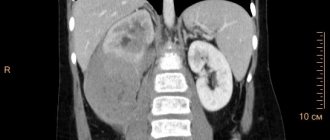
The results of clinical and biochemical studies do not always make it possible to distinguish between intrahepatic and extrahepatic cholestasis. The diagnostic examination algorithm is of great importance. Extrahepatic mechanical obstruction with the development of biliary hypertension is supported by pain in the abdominal cavity (observed when stones are localized in ducts, tumors), palpable gallbladder. Fever and chills may be symptoms of cholangitis. The density and tuberosity of the liver upon palpation reflect advanced changes or tumor damage to the liver. The diagnostic examination algorithm involves, first of all, performing an ultrasound of the abdominal organs, which makes it possible to identify a characteristic sign of a mechanical blockade of the bile ducts - suprastenotic dilatation of the bile ducts (the diameter of the common bile duct is more than 6 mm). When dilatation of the ducts is detected, it is advisable to conduct cholangiography. The procedure of choice is endoscopic retrograde cholangiography (ERCH). If retrograde filling of the bile ducts is impossible, percutaneous transhepatic cholangiography (PTCHG) is used. If there are no signs of extrahepatic obstruction of the bile ducts, a liver biopsy is performed. This procedure can be performed only after excluding obstructive extrahepatic cholestasis (to avoid the development of biliary peritonitis). Cholescintigraphy with technetium-labeled iminodiacetic acid also helps to identify the level of damage (intrahepatic or extrahepatic). The use of magnetic resonance cholangiography is promising.
Description
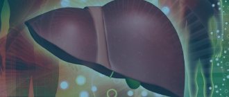
Cholestasis is a condition characterized by a slowdown or complete cessation of bile secretion, which is accompanied by an increase in the content of substances secreted along with bile in the blood.
According to statistics, approximately 10 out of 100 thousand people experience cholestasis. It can also be noted that cholestasis is diagnosed in men somewhat more often than in women. In most cases, this pathology is observed in people over 40 years of age.
The most common causes of cholestasis include:
- damage to the liver parenchyma as a result of prolonged consumption of alcoholic beverages, taking medications that have a hepatotoxic effect;
- liver infection;
- congenital metabolic disorders, for example, galactosemia, cystic fibrosis and others;
- inflammatory damage to the bile ducts;
- cholelithiasis;
- acquired or congenital obstruction of the bile ducts;
- sarcoidosis;
- oncological disease of the hepatocellular system;
- hormonal changes in the body, for example, during pregnancy.
Cholestasis is usually divided into the following types:
- intrahepatic - occurs as a result of a decrease in the production of bile and its release into the bile capillaries;
- extrahepatic - in most cases indicates obstruction of the bile ducts.
According to the mechanism of development, the following types of cholestasis occur:
- partial – characterized by a decrease in the amount of bile produced;
- total - bile does not enter the duodenum;
- dissociative – characterized by a decrease in the body’s amount of substances necessary for the production of bile.
As a rule, the prognosis is quite favorable. In most cases, with cholestasis it is possible to achieve complete recovery or long-term remission. This is achieved through timely diagnosis and specialized treatment. In some cases, complications develop, in particular, liver failure may develop, which threatens to disrupt the normal functioning of other organ systems. It is also important to follow preventive measures, which include excluding spicy, fried foods and alcoholic beverages from the diet. In addition, it is necessary to promptly treat the pathology that causes the development of cholestasis.
Treatment
Photo: img2.vitaportal.ru
To eliminate the phenomenon of cholestasis, the cause of its occurrence should be identified. In some cases, surgical intervention is required to eliminate the factor that caused the development of cholestasis. For example, if there are stones in the gallbladder, a cholecystectomy is performed.
You should also follow a special diet for cholestasis, which consists of limiting the consumption of fried, smoked and spicy foods. In the diet, it is recommended to replace animal fats as much as possible with fats of plant origin. Preference should be given to vegetables and fruits that contain large amounts of vitamins and microelements necessary for the normal functioning of the body. It is important to choose low-fat varieties of meat products. Low-fat dairy products are also allowed. It is strongly recommended not to forget about the existence of various cereals rich in grains. It is strictly forbidden to consume alcoholic beverages, strong coffee and tea. In addition, it is important to give up bad habits such as smoking and drug use.
The following groups of drugs are used as symptomatic treatment:
- hepatoprotectors necessary to protect hepatocytes (liver cells);
- cholesterol, the action of which is aimed at binding bile salts, thereby eliminating skin itching;
- vitamin K. Used for bleeding associated with cholestasis;
- vitamin or vitamin-mineral complexes. Prescribed when the clinical picture of hypovitaminosis develops.
During the rehabilitation period, non-drug treatment methods are used, the use of which is aimed at stimulating the protective mechanisms of the human body. For example, physiotherapy, massage or physical therapy may be prescribed.
Symptoms
Photo: womanadvice.ru
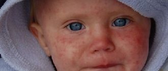
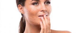
For quite a long time, the only symptom of cholestasis is skin itching, which worries mainly at night and decreases during the day. Severe itching of the skin leads to scratching of the skin, which is dangerous due to secondary infection of these scratches, resulting in pustular rashes. Complaints of pain in the epigastric region or right hypochondrium may also appear. In some cases, there is irradiation of pain to the right shoulder, shoulder blade or right half of the neck. The intensity of pain varies; with the development of biliary colic, the pain becomes unbearable and painful. Dyspeptic disorders manifest themselves in the form of nausea, vomiting, and a feeling of bitterness in the mouth. In addition, as a rule, there is a decrease in appetite, in exceptional cases there is a complete loss of appetite. Decreased appetite over a long period of time leads to weight loss.
Also, the development of cholestasis will be indicated by yellowness of the skin and visible mucous membranes. Most often, the sclera of the eyes turn yellow first. In addition, so-called xanthelasmas, yellowish-brown formations, may appear on the eyelids. As a rule, these formations are small in size and are located symmetrically in most cases. There are also xanthomas, which, unlike previous formations, are located on the chest, back or elbows and have a yellow tint. Since the outflow of bilirubin into the intestines is blocked, as a result of which its removal from the body along with feces is disrupted, discoloration of feces is observed. Also, feces may contain an increased amount of fat, which is associated with impaired digestion of fat supplied with food, resulting from a violation of the flow of bile into the intestines. This phenomenon will be indicated by a “greasy” sheen of feces. In addition, the urine becomes dark in color (the color of beer), which also indicates the possibility of cholestasis.
Since bile is involved in the absorption of vitamins that enter the body with food, with cholestasis a clinical picture of hypovitaminosis is observed. This condition is characterized by the following:
- fatigue, decreased performance and concentration;
- increased drowsiness, especially during the daytime;
- dry skin;
- increased fragility of hair and nails;
- muscle weakness;
- decreased twilight vision;
- increased bleeding of gums.
Folk remedies
Photo: Ponchikov.net
As is known, in some cases surgical intervention is required to eliminate the cause of cholestasis. That is why it is strongly recommended not to self-medicate at home, but to immediately consult a specialist at the first symptoms of cholestasis. It is important to understand that the use of traditional medicine in this case will not only not provide any help, but can also cause harm. However, there are folk remedies that can be used for preventive purposes. We offer to your attention the following:
- take 20 g of pre-prepared rose hips and 10 g of crushed nettle leaves. Mix the listed ingredients and pour two glasses of boiling water over them, then place in a water bath and boil for 15 minutes. After which the resulting decoction should be infused for 1 hour, having previously wrapped the container containing the decoction. The drink becomes ready for use after careful straining. It is recommended to consume 50 ml throughout the day;
- Dry the birch leaves and chop them finely. Take 1 tablespoon of plant material and pour one glass of boiling water, then heat over low heat in a water bath for 30 minutes. Then the resulting decoction must be allowed to brew for half an hour, after which it should be strained. It is recommended to consume 1/3 glass 3 times a day 30 minutes before meals;
- mix chopped chicory root and dried peppermint leaves in equal proportions. To prepare the infusion, you will need 10 g of the resulting plant collection. Pour 10 g of the collection into glasses of boiling water and let it brew for 1 hour. After this, the resulting infusion should be filtered through a strainer. One glass of infusion is drunk during the day. The course of treatment varies from 1 week to 1 month;
- Grind 1 glass of flax seeds and pour them with three glasses of milk. Then place on low heat until approximately 1/3 of the original volume of liquid remains in the container. After this, the solution is carefully filtered through a strainer. It is recommended to drink one glass per day. The duration of use of this recipe is 10 days, after which a break is taken;
- take 3 drops of peppermint oil and one teaspoon of honey. Mix the listed components thoroughly. The resulting mass should be consumed 3 times a day. The duration of the course is 1 month;
- mix red beet juice with black radish juice in equal proportions. It is recommended to consume 1 glass per day, preferably in the morning. The duration of the course is 1 month.
The information is for reference only and is not a guide to action. Do not self-medicate. At the first symptoms of the disease, consult a doctor.