Reasons for appearance
The exact causes of the disease have not yet been established. The main one is regular exposure to ultraviolet radiation. It affects the dermis, epidermal layers, blood vessels, sebaceous glands, and melonocytes.
Gradually, under the influence of sunlight, the disturbances increase, reaching the peak of the disease.
The following factors contribute to the development of pathology:
- genetic predisposition;
- weakened immune system;
- influence on the skin of chemicals (resinous substances, oil, sand, etc.);
- past infections;
- age-related changes (the disease most often affects people over 50 years of age).
Due to weak immunity, AIDS carriers, people with problems with the nervous and endocrine systems, as well as patients who have undergone chemotherapy or complex operations are more prone to the appearance of keratosis.
Some types of keratoses often affect young people. This usually applies to red-haired or fair-haired people with gray, blue or green eyes. Research shows that by the age of 40, 60% of the population has at least one element of keratosis.
Over the age of 80, everyone has some type of this pathology.
Are there any new developments in this direction on the world market?
Well, for example, from 1998 to the present, the Australian biopharmaceutical company Peplin has been studying the topical treatment of actinic keratosis with the drug Ingenol Mebutate, which is the first in a new class of formulations and is derived from milkweed juice. This ingredient has a long history of traditional use for a variety of skin conditions, including the topical treatment of skin cancer and precancerous skin lesions. The company plans to enter the third phase of the trial soon.
Types and signs of keratosis
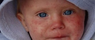
A person may not pay attention to the signs of the initial stage of the disease. It can manifest itself as unnoticeable roughness on any part of the body (cheeks, bridge of the nose, forearms, scalp, ears, etc.). At first the formation is solid, small in size, red or brown in color. The skin around the affected area may peel off, itching begins, and hair loss may also be observed at the site of the keratoma.
There is no unified classification of the varieties of this disease, because it has not been fully studied. According to etiology, the following types of keratosis are distinguished:
- Congenital - appears at birth or at a younger age. Very rare.
- Acquired - appears in adulthood, less often - in adolescence.
- Symptomatic – occurs due to external factors.
According to the affected area, local (single areas of skin are affected) and diffuse (large areas of the skin are affected) keratosis.
There are also several types of clinical manifestations.
Keratosis follicularis
The formation of horny plugs in the hair follicles is observed. These are dead cells that have separated from the skin. By forming nodules, they interfere with hair growth. Most often, keratosis pilaris appears on the abdomen, face, shoulders, buttocks, neck, and armpits. Such manifestations are characteristic of the cold season; closer to summer, symptoms may disappear. This type of pathology is also called pilar keratosis. If the nodule grows more than 3 mm, pain may occur.
Consumption of allergens can cause an exacerbation of the inflammatory process.
Seborrheic keratosis
This type of disease is characterized by plaque-like or nodular formations with a warty surface. The top of the keratomas is covered with a brown or black keratinized substance. As a rule, it occurs over the age of 50 years, therefore it is also called actinic keratosis.
The most common areas for the appearance of formations are the face, chest, neck, etc. It does not occur on the palms and soles. The development of this form of the disease proceeds slowly and usually becomes chronic. Actinic keratosis does not develop into a cancerous tumor, but a malignant tumor can masquerade as keratosis. In case of inflammation of the affected areas, bleeding and rapid growth of the formation, you should immediately consult a doctor.
Actinic keratosis
Appears on exposed areas of the body. At first it looks like uneven, rough skin. Over time, it develops into a scaly, flaky, compacted spot, ranging in color from skin color to brownish. Such formations may look like growths, rising above the skin. Mostly the face, neck, and chest are affected by keratinization. Such keratosis can transform into cancerous tumors, so it is necessary to be regularly monitored by a dermatologist.
Nevi (aka moles)
My first article about moles is on the website of the PLASTES center https://www.plastes.ru/nevus.html Here I want to pay more attention to the issues of additional diagnostics and the safety of removing pigmented skin tumors.
We will immediately remove the questions about why moles appear on the skin and why melanomas arise from them from consideration as uninteresting and, in our case, idle. Let's start the conversation right away from the moment the mole for some reason began to bother me.
Here is a typical example of a letter from my correspondence with patients:
“...I don’t know what to do and what to do? Very scary. I am 33 years old. And all my life I have lived with a mole about 1 cm in diameter. It is swollen and with villi, almost black in color, but with uneven edges. On the thoracic region. Never bothered me. And starting in June of this year, the mole itself began to itch around the mole, as if it was pulling inward. All this is tolerable. I turned to our oncologists, they said that as long as it looks like a benign tumor, it should be removed (literally from the head physician of the N-sky oncology dispensary). The consultation lasted no more than 5 minutes. Visual inspection, without any equipment. I asked to prescribe tumor markers, they refused, they said we didn’t see any point. Nothing was really explained. Very scary. I read about melanoma. What should I do? The general condition is strange: it seems like a cold, but something is wrong. A little later I was told that at the consultation they identified my mole as a congenital pigmented nevus. Are there any guarantees that the outcome of the surgical intervention will be good??? I was really very frightened by the stories of friends that people died after removing a mole...”
Well, what can I say? I perfectly understand the state of mind and fear of this girl. There are several such letters a day, and such patients have to be constantly reassured and explained the same thing.
The most important thing is to understand: until the tumor is removed and subjected to histological examination, there will be no diagnosis!!! And no additional research methods (tumor markers, dermatoscopy, CT, PET, etc.) will ever give us a definite answer to the question of whether it is melanoma or not.
Another question is how to remove the tumor to minimize the risk of adverse consequences. This is where the main “terrible secret” lies - in the method of removal. In order not to let my thoughts wander, I will immediately reveal this secret. If I have even the slightest suspicions and doubts, I insist on COMPLETE SURGICAL EXCITION OF THE NEOPLASM .
Now I will explain why. I completely agree that any intervention in a tumor can lead to rapid uncontrolled progression (growth of the tumor itself and the appearance of metastases). That is why ALL SUSPICIOUS MOLES SHOULD BE REMOVED SURGICALLY, IMMEDIATELY AND COMPLETELY WITH THE HEALTHY SKIN AROUND!!! For exactly the same reason, IF THERE ARE SUSPECTS, YOU CAN NOT DO A PARTIAL BIOPSY, YOU CANNOT DO ELECTROCOAGULATION OR LASER REMOVAL!!!
See how simple everything turns out to be. That's where all these scary stories come from. The doctor simply underestimated the degree of danger, did not attach importance to suspicious symptoms and, instead of complete excision, performed coagulation (with electricity or laser - it makes no difference). The tumor was “disturbed” and began to grow. If the same tumor had been immediately and completely removed, then no further unfavorable sequence of events would have occurred.
However, please note that I am not saying that complete excision is an absolute guarantee of safety. Even after complete surgical excision, the tumor can progress into metastases. But only if it really was melanoma. As you already understand, it is possible to find out whether it was melanoma or not only after a histological examination.
Next, I will present several cases when I insisted on complete surgical excision of pigmented tumors.
| In general, nothing unusual. This is what “calm” moles look like under a microscope. I posted this illustration to show that a dermatoscope is not the most accurate diagnostic device. The indication for excision in this case was a feeling of discomfort and itching in the area of the mole. Histological conclusion - pigmented nevus. |
| A young woman has a pigmented nevus in the décolleté area. Over the past few months, he has noticed a change in color (it has become more intense) and an increase in the size of the mole. Even these two signs are enough to make a diagnosis of “dysplastic nevus” (restless mole) and surgical excision. And, despite the danger of a rough scar, I had to have surgery. Histological conclusion: dysplastic nevus. That is, pre-melanoma. The result of the operation is shown in the image on the far right. There is no more tumor. There are also dangers of degeneration into melanoma. I don’t know what would have happened if the operation had not been performed or the woman had applied later. I also cannot say what would have happened if electrocoagulation had been performed instead of surgery. Perhaps this would become a provoking factor for tumor transformation. |
| This case also illustrates well my approach to choosing a method for removing “moles.” This is the spot that appeared on the young man’s skin on the plantar surface of his foot. It appeared suddenly; there was nothing in this place before. This spot does not cause any discomfort. What to do? If you think that by looking through a deramtoscope, I will immediately make the correct diagnosis - you are mistaken. I have never seen such a picture before. Guided by the principle of cancer vigilance, which says “if the diagnosis is not clear, think about cancer,” I insisted on surgical excision. The histological answer was unusual - pinpoint hemorrhage. Apparently, a microtrauma occurred, which the patient did not even remember. You may think that the operation was performed in vain. To which I will answer your question: what if it turned out to be melanoma? Once again, I will note the main thing - we will never know the exact diagnosis until we remove the tumor. And in such situations it must be removed immediately and completely. |
These are just a few examples from my practice. If I have time, I will add to this gallery. Finally, a few points about moles:
- The majority (99.9%) of moles do not pose any danger to life or health. They can be safely removed in different ways: electro- or laser coagulation, surgery. It all depends on the size and location.
- If a mole bothers you (no matter how), be sure to see an experienced doctor. In such cases, it is preferable to perform complete surgical excision with mandatory histological examination.
- You can ask questions at
Treatment of the disease
Treatment should be carried out by a dermatologist.
You need to visit a specialist after the first appearance of keratomas, since it is important to exclude the malignant nature of the formation. Treatment is long-term and complex, usually including a number of activities.
Conservative therapy
It is carried out with the aim of reducing the number of keratoses before moving on to radical methods of treatment.
Therapeutic agents reduce symptoms and alleviate the course of the disease, but do not cure completely.
Therapeutic agents reduce symptoms and alleviate the course of the disease, but do not cure completely.
To soften keratonic areas, applications using drugs with urea (content - from 12 to 30%) are used: Keratosan, Ureaderm, Ureatop, Akerat..
The following drugs are used in therapeutic treatment: Fluorouracil, Efudex cream, Diclofenac Gel 3%, Imiquimod. Special shampoos are used to treat keratosis of the scalp. Retinoids are taken internally, which help reduce the growth rate of formations, as well as vitamins of groups A, B and C. Additionally, courses of physiotherapy are prescribed.
Radical methods of treating keratosis
Since conservative therapy for keratosis does not guarantee complete cure, it is often necessary to proceed to radical measures - direct removal of the formations. The use of drastic techniques is especially justified if there is a risk of keratosis degenerating into cancer.
The following radical methods of therapy exist:
- Cryodestruction - freezing with liquid nitrogen.
- Radio wave removal. The formation is excised using a radio knife under the influence of radio waves.
- Electrocoagulation - the doctor performs cauterization using high-frequency electric current.
- Laser destruction - a targeted effect of a carbon dioxide laser is applied to the keratoma.
- Photodynamic therapy - methyl aminolevulinate is applied to the affected area with further exposure to a light wave of a certain length. All this leads to necrosis of the affected tissues.
- Surgical removal - the skin is scraped with a curette (special instrument).
- Dermabrasion – removal using an abrasive brush.
Other neoplasms
Very often we have to deal with papillomas . As a rule, these are small (2-3 mm) growths on the skin, with a thin base, soft consistency and pale pink color. They are usually located in the area of skin folds ( axillary and groin areas, submammary folds, neck ). Apparently, the cause of their occurrence is skin irritation in conditions of high humidity. Papillomas are not dangerous in terms of cancer . They can be easily removed using an electrocoagulator . Some comrades manage to “grow” papillomas to absolutely mind-boggling sizes, fearing the adverse consequences of removal. Do not be afraid! Here are some examples of papillomas that could have been removed much earlier.
Inflammatory tumors, the so-called pyogenic granulomas, . They are indeed very ominous. A red tumor appears on the skin , which grows very quickly and bleeds . There is only one solution to the problem in such situations - removal with histological examination.
Something else needs to be said about hemangiomas . These are tumors consisting of altered blood vessels . As a rule, they are congenital and can reach quite large sizes. In adults, hemangiomas often appear on the skin in the form of small red spots. Special treatment for small hemangiomas is not required. Removal is possible for cosmetic reasons.
BLESS YOU!!!
Forecast and prevention of the disease
The prognosis of the disease depends on the time of initiation of treatment. However, in order to avoid relapses, it is necessary to follow certain recommendations.
The following measures can be taken as prevention:
- use moisturizing creams;
- eat well so that food provides the skin with all the necessary “building” elements;
- limit exposure to direct sunlight to reduce exposure to ultraviolet radiation;
- when in the sun, use sunscreens (ointments and creams) with a high SPF level;
- When working with chemicals, you must use appropriate skin protection.
Keratosis is a skin condition that requires constant monitoring. In addition to an aesthetic defect, sometimes formations can develop into malignant tumors. And to prevent this, it is necessary to undergo regular examinations and monitor changes in the skin.
Solar lentigo
Solar lentigo in the photo above.
These are flat, defined pigment spots that appear on sun-exposed areas of the body. Lentigo is characterized by an increased accumulation of melanocytes. There are two types of lentigo:
- Sunlight is a sign of skin damage from ultraviolet radiation
- Senile - usually appears in old age