What is hepatic coma
Hepatic coma is a serious condition of the body that developed as a result of acute inflammatory processes and chronic liver diseases. Despite the fact that many are immediately placed under emergency hospitalization, the survival prognosis for such a diagnosis is disappointing.
The disease is accompanied by loss of consciousness, lack of natural reflexes, and circulatory disorders. Hepatic coma does not occur as a separate disease; it is a complication of long-term disturbances in the functionality of the organ. It is often preceded by chronic failure, in which there was no appropriate treatment and health maintenance.
A serious condition develops when metabolic products (phenols, ammonia, etc.) accumulate in the body, which must be eliminated naturally. Due to their accumulation, a toxic effect occurs that affects the brain. That is why hepatic coma affects the functioning of the central nervous system, namely, the inhibition of its functions.
Description of the syndrome
Hepatic coma is a severe disease caused by extensive damage to the parenchyma (functional tissue) of the liver and brain structures. According to ICD 10, hepatic coma has code K72.
The mechanism of development of the pathological process is associated with damage to brain cells by toxic substances that enter the body from the outside and are also formed in the process of internal metabolism. One of the most dangerous metabolites is ammonia. Phenols and other substances toxic to nervous tissue pose a danger. The liver affected by the pathological process is not able to cope with toxins, so these substances freely enter the arterial bloodstream and are carried to all organs.
Hepatic coma requires emergency care. The condition is difficult to correct and often ends in death even with timely intensive care.
Kinds
Depending on the clinical manifestations, there are several types of hepatic comas:
- Endogenous type. Toxic substances are contained in large quantities in the blood, are produced in it, and are carried through the bloodstream throughout the body. The main cause of endogenous coma is toxic liver damage. They often appear in chronic alcoholism, when metabolic acidosis develops, then alkalosis, and later the effect of poisons affects the brain.
- Exogenous type. Toxic substances enter directly into the blood, leaving the liver itself unaffected. Exogenous coma often develops in liver diseases with pronounced portocaval anastomoses.
- Mixed type. It means a combination of endogenous and exogenous factors. At the same time, the amount of toxic substances in the body increases rapidly, they are in the bloodstream, various organs of the gastrointestinal tract, and brain damage quickly occurs.
There is also a fourth type - false hepatic coma. The main reason for its development is potassium deficiency, which develops against the background of chronic cirrhosis of the liver. A person may take diuretics incorrectly or in excessive amounts, which flush out this substance. Potassium deficiency can also occur against the background of frequent diarrhea and vomiting, when the body is constantly struggling with dehydration. Metabolic alkalosis gradually develops, due to which ammonia cells penetrate into the brain and affect it.
Jaundice
Hepatitis
22144 November 26
IMPORTANT!
The information in this section cannot be used for self-diagnosis and self-treatment.
In case of pain or other exacerbation of the disease, diagnostic tests should be prescribed only by the attending physician. To make a diagnosis and properly prescribe treatment, you should contact your doctor. Jaundice: causes of occurrence, what diseases it occurs with, diagnosis and treatment methods.
Definition
Jaundice is a condition in which the skin and mucous membranes turn various shades of yellow. Moreover, not in all cases this indicates pathological processes in the body.
Types of jaundice
There are false and true jaundice. False jaundice occurs when carotenes accumulate in the skin due to excessive consumption of carrots, beets, oranges, pumpkin, or when certain drugs are ingested (acriquine, picric acid, etc.).
True jaundice is a consequence of an increase in the concentration of total bilirubin in the blood plasma.
Jaundice discoloration of the skin and icterus (yellow pigmentation) of the sclera occur when the concentration of total bilirubin in the blood plasma is more than 35-45 µmol/l. The milk of nursing women, pleural and pericardial effusion, ascitic fluid, and semen may have a yellow tint.
The clinical picture and duration of true jaundice depend on the underlying disease, against the background of which an increase in blood bilirubin developed.
There are:
1. Hemolytic (prehepatic) jaundice:
- jaundice due to genetic diseases (hereditary microspherocytosis, hereditary stomacytosis, etc.);
- autoimmune (acquired) hemolytic anemia.
2. Parenchymal (hepatic) jaundice:
- caused by infectious diseases (viral hepatitis A, B, C, D, E; herpetic hepatitis, cytomegalovirus hepatitis, yellow fever, infectious mononucleosis, relapsing fever, intestinal yersiniosis, psittacosis, leptospirosis, salmonellosis, pseudotuberculosis, etc.);
- caused by non-infectious diseases (acute alcoholic hepatitis, drug-induced hepatitis, liver cirrhosis, poisoning with oxidizing agents based on nitric acid, hydrazine and its derivatives, chloroethane, ethylene glycol).
3. Constitutional jaundice
(Gilbert syndrome, Crigler–Najjar syndrome, Dabin–Johnson syndrome, Rotor syndrome).
4. Mechanical (subhepatic) jaundice
occur against the background of cholelithiasis, cholangitis, cicatricial strictures of the extrahepatic bile ducts, cancer of the head of the pancreas, major duodenal papilla, and gall bladder.
Hemolytic jaundice is characterized by moderate manifestations, lemon staining of the skin, sclera and mucous membranes, a slight enlargement of the liver and often a noticeably enlarged spleen. Anemia is observed.
In acute viral hepatitis, dull aching pain in the right hypochondrium and in the joints is disturbed, the temperature rises, appetite decreases, and the patient experiences weakness. Yellowness of the skin, dark urine and insufficiently colored feces appear. There may be bruises on the skin as a result of hemorrhages. Skin itching is not typical for acute viral hepatitis, since sufficient evacuation of bile through the bile ducts is ensured. When interviewing a patient, it is possible to identify a risk factor for developing the disease (contact with patients with hepatitis, recent surgical operations, blood transfusions, injections and manipulations associated with damage to the skin and mucous membranes).
With obstructive jaundice, the clinical picture may begin acutely or develop gradually.
With the acute onset of the disease (cholelithiasis), colicky pain suddenly appears in the right hypochondrium radiating to the right and to the back, chills, and fever. Then jaundice and skin itching occur. With the gradual development of the disease (cancer of the head of the pancreas), the patient may be bothered by moderate dull or pressing pain under the right costal arch, nausea, belching, and weight loss. Palpation of the abdomen reveals pain in the right hypochondrium. The yellowness of the skin gradually increases, the urine becomes dark in color, the feces become lighter, as less bilirubin is released into the intestines.
Constitutional jaundice is characterized by moderately severe intermittent (periodic) jaundice. Selective staining of the skin of the face, nasolabial triangle, palms, soles and axillary fossae is noted. Jaundice occurs or worsens after physical or mental stress, exacerbation of a concomitant disease, or alcohol intake. The liver and spleen do not increase in size.
As a result of long-term toxic effects of high concentrations of bilirubin on the body, damage to the central nervous system develops, foci of necrosis form in parenchymal organs, the cellular immune response is suppressed, and anemia occurs. Children may experience serious complications: kernicterus, mental retardation, cerebral palsy.
Possible causes of jaundice
True jaundice is the result of an imbalance between the formation and excretion of bilirubin. The cause of this disorder may be increased formation of bilirubin (hemolytic jaundice), a failure in its transportation to liver cells and excretion by these cells, as well as deterioration in the binding processes of free bilirubin (parenchymal jaundice). Obstructive jaundice occurs as a result of deterioration in the secretion of bilirubin through the extrahepatic ducts.
The basis of the mechanism of development of hemolytic jaundice
lies hemolysis (increased destruction of red blood cells), when the life span of red blood cells is significantly shortened, sometimes to several hours. Hemolysis can be intravascular and extravascular. Intravascular hemolysis occurs as a result of mechanical destruction of red blood cells in small blood vessels (marching hemoglobinuria), turbulent blood flow due to dysfunction of prosthetic heart valves, as a result of incompatibility of red blood cells in the AB(0), Rhesus or any other system, due to direct toxic effects ( snake venom), etc.
Extravascular hemolysis occurs in the spleen and liver as a result of the capture and destruction of altered red blood cells. The spleen is capable of capturing and destroying slightly altered red blood cells, the liver – red blood cells with major disturbances.
Hepatic (parenchymal) jaundice
are caused mainly by damage to hepatocytes (liver cells). In some cases, this syndrome is associated with damage to liver cells and stagnation of bile, for example, in acute and chronic viral hepatitis. In others, the release of bilirubin is impaired (with cholestatic jaundice, cholestatic hepatitis, primary biliary cirrhosis).
Obstructive (mechanical) jaundice occurs when there is a disturbance in the outflow of bile through the extrahepatic bile ducts.
.
, enzymopathic or constitutional jaundice occurs
. The most common is Gilbert's syndrome, which develops against the background of impaired uptake of free bilirubin and its binding to glucuronic acid due to deficiency of the enzyme glucuronyltransferase.
In 60-80% of newborns, yellowness of the skin is observed, which appears on the 2-3rd day of life.
Jaundice in newborns can be caused by a high release of red blood cells during childbirth, a high content of fetal hemoglobin, rapid destruction of bilirubin after birth, and a deficiency of conjugating enzymes in the liver. In this case, the rise in bilirubin levels does not reach critical values, and the child’s condition remains satisfactory.
Breast milk jaundice (jaundice from mother's milk) is not considered a pathology and is associated with the baby's body's reaction to the fats contained in breast milk. This condition develops on the 3rd-7th day after birth, when the baby already receives a sufficient amount of mother’s milk. Stopping breastfeeding for 24-48 hours leads to a sharp decrease in bilirubin and a decrease in the severity of jaundice. If the baby continues to receive breast milk, jaundice persists for 4-6 weeks, then gradually decreases - the condition of the skin and mucous membranes returns to normal by the 12-16th week of life.
Hemolytic disease of newborns is caused by incompatibility of the blood of mother and fetus for various antigens, when the body of a pregnant woman produces antibodies that penetrate the placental barrier into the blood of the fetus and cause destruction (hemolysis) of its red blood cells. The intensity of jaundice depends on the level of bilirubin - when critical numbers are reached, this enzyme can affect neurons in the brain, resulting in the development of bilirubin encephalopathy (kernicterus).
Which doctors should I contact?
If jaundice appears, to clarify the diagnosis and determine further treatment tactics, the patient should consult a general practitioner, or if the patient is a child. In the future, a consultation with a hematologist, an infectious disease specialist, may be indicated.
Diagnosis of jaundice
To determine the cause of jaundice, a set of laboratory and instrumental examination methods is performed (according to indications):
- clinical blood test with determination of hemoglobin concentration, number of erythrocytes, leukocytes and platelets, hematocrit and erythrocyte indices (MCV, RDW, MCH, MCHC), leukoformula and ESR (with microscopy of a blood smear in the presence of pathological changes);
Stages and symptoms
The severe condition of the body during hepatic coma develops gradually. The intensity of progression of inflammatory processes depends on the causes that provoke them, the general condition of the human body, the presence of chronic diseases, and other factors.
The following stages of coma are distinguished:
- Prekoma. Symptoms of hepatic coma at this stage affect the mental state of the person. He develops increased anxiety, melancholy, unjustified fears, sleep disturbances, and decreased performance. Confusion and loss of consciousness occur periodically. From the gastrointestinal tract, nausea, vomiting, and frequent abdominal pain are observed.
- Threatening stage (decompensated). It is characterized by severe disturbances of consciousness, in which a person cannot control himself. Fainting, hallucinations, and memory loss often occur. Coordination of movements is seriously impaired, it is difficult for a person to walk in a straight line, he gets tired quickly, his hands tremble all the time. Difficulty in breathing appears - it becomes noisy and intermittent.
- Complete coma. The person completely loses consciousness, reflexes, including pathological ones, are absolutely absent. The skin acquires a pronounced yellow color, which allows doctors to quickly determine the cause of the lack of consciousness. During diagnosis, it is discovered that the size of the liver is much smaller than normal.
In complete coma, various breathing disorders appear; Cheyne-Stokes breathing (with a constantly changing frequency) or Kussmaul breathing (deep, noisy) is more often diagnosed.
Signs of deterioration develop alternately, depending on the stage of the coma.
Symptoms of precoma (harbingers of the syndrome)
Hepatic precoma is a condition that precedes the onset of a deep coma. At this stage the following symptoms appear:
- a sharp decrease in body weight;
- loss of appetite;
- sleep disturbance: drowsiness during the day and insomnia at night;
- constant nausea;
- tachycardia;
- a feeling of bitterness in the mouth;
- unmotivated behavior;
- increased sweating;
- bright yellowness of the skin and mucous membranes;
- darkening of urine.
The precoma stage gradually turns into the stage of threatening hepatic coma and into the stage of the coma itself.
Causes
Hepatic coma is the most severe stage of hepatic encephalopathy, the algorithm for its development is the same in all cases:
Blood test indicators for liver cirrhosis
- metabolic processes are disrupted;
- metabolic products are not eliminated from the body naturally, they accumulate or toxins immediately enter the bloodstream;
- with an increase in their number, the poisons affect the central nervous system;
- When the brain is damaged by toxins, coma occurs.
There are many reasons why these processes occur, often acute or chronic liver diseases. These include hepatitis of various types (alcoholic, viral, toxic), cirrhosis, the appearance and increase of tumors in the liver, and all kinds of diseases associated with general intoxication.
A common cause is complications after portacaval shunt surgery.
There are also predisposing factors that themselves cannot cause such a severe pathological condition, but can contribute to its development against the background of acute or chronic liver diseases. These include:
- bad eating habits, abuse of fatty, fried, and other heavy foods;
- eating too much protein;
- prolonged diarrhea, vomiting, developed against the background of gastrointestinal diseases, which remain without proper treatment and lead to dehydration;
- frequent or uncontrolled use of diuretics;
- uncontrolled use of sedative drugs.
Some weight loss diets suggest taking diuretics daily. If you start taking them without a doctor's prescription, you can cause dehydration and deterioration of the liver, which will lead to serious consequences.
HEPATIC ENCEPHALOPATHY in the practice of an emergency physician
What clinical symptoms allow one to diagnose PE? What are the manifestations of neurological disorders at different stages of development of PE? What are the features of the development of PE in alcoholism? What mistakes should a doctor avoid when treating PE?
Etiology and pathogenesis
Hepatic encephalopathy (HE) is a potentially reversible disorder of nervous and mental activity that occurs in any liver disease that occurs with insufficiency of hepatic cellular function. In most cases, PE complicates the course of the terminal stage of chronic diffuse liver diseases or acute necrotizing (fulminant) hepatitis. It has been established that with liver damage of any etiology, PE can lead to the development of coma and become the direct cause of death of the patient (see Table 1).
| Table 1. Causes of development of PE. | ||
| Encephalopathy variant | Survival | Etiology |
| Acute encephalopathy without liver fibrosis/cirrhosis | 20–45% | Viral hepatitis. Alcoholic hepatitis. Acute poisoning (chemical and biological poisons, drugs). Withdrawal syndrome |
| Acute encephalopathy due to fibrosis/cirrhosis of the liver | 70-80% | Forced diuresis Vomiting, diarrhea Bleeding Gastrointestinal tract Infections Alcohol intake Withdrawal syndrome Constipation Excess protein in the diet Surgical interventions Taking sedatives |
| Chronic portosystemic encephalopathy in end-stage liver disease | 100% | Portosystemic shunting Intestinal contamination Excess protein in the diet |
| Figure 1. Mechanism of development of PE. |
The pathogenesis of PE is not fully established. It is believed that the development of HE is the result of a combined effect on the central nervous system of several mechanisms, the activity of which is initiated and maintained by a pronounced impairment of the hepatic clearance of toxins and metabolites (see Figure 1). The most important factors for the development of PE are considered to be:
- a rapidly increasing increase in plasma ammonia concentration;
- an increase in plasma concentration and an imbalance between the synthesis and catabolism of neurotransmitters and their precursors in the central nervous system.
Clinical symptoms and diagnosis of PE. The type and amount of excess, “toxic” metabolites circulating in the plasma and central nervous system, to varying degrees, correlate with various symptoms of PE, which include:
- disturbance of consciousness;
- personality change;
- intellectual disorder;
- actual neurological disorders (see Table 2).
| Table 2. Glasgow Consciousness Scale (GCS). | ||
| Symptom | Symptom severity | Point |
| Consciousness | oriented | 5 |
| confusion/lethargy | 4 | |
| Verbal reaction | the answer is out of place | 3 |
| slurred sounds | 2 | |
| No answer | 1 | |
| executes commands | 6 | |
| purposeful response to pain | 5 | |
| undirected response to pain | 4 | |
| Motor reaction | flexion response to pain | 3 |
| extensor response to pain | 2 | |
| no reaction | 1 | |
| spontaneous | 4 | |
| Eye reaction | per voice | 3 |
| for pain | 2 | |
| No | 1 | |
| Total indicator (10–15 points): stupor, 5–10 points: precoma, 0–5 points: coma. | ||
Mechanism of development of PE
Early signs of disturbances of consciousness (US) in PE include a decrease in spontaneous movements, a fixed gaze, lethargy, and apathy. Impaired consciousness in PE is, in principle, characterized by drowsiness and inversion of the normal rhythm of sleep and wakefulness. In this case, deterioration of the condition and transition from drowsiness to coma can occur within a very short time. To determine the degree of NS, the Glasgow scale is often used (see Table 2). However, in clinical practice, to assess the depth of NS specifically in PE, a simpler qualitative scale is used, according to which:
- Stage I of NS is characterized by weakened concentration, euphoria and anxiety;
- stage II is characterized by the appearance of drowsiness, disorientation, personality changes and inappropriate behavior;
- at stage III of NS, stupor, hypersomnia and confusion develop, but the patient can follow simple commands and pronounce words clearly;
- with IV degree NS, the patient falls into a coma and contact with him is impossible.
Neurological disorders in PE are generally not specific and can also develop with uremia, severe respiratory and heart failure. However, one of the most pathognomonic neurological symptoms of PE is considered to be the development of “flapping” tremor (asterixis), the distinctive feature of which is the patient’s inability to maintain a fixed position. The greatest severity of hyperkinesis of the muscles of the limbs when maintaining a constant posture and its decrease during movement make it possible to differentiate asterixis from tremor in alcoholic delirium and neuroencephalopathy.
The most reliable way to determine the degree of intellectual impairment in PE is to conduct a number-binding test (Reitan test). Changes in personality and intelligence in PE are the most difficult to diagnose, since they are always superimposed on constitutional, previously acquired disorders and always require dynamic assessment. The greatest difficulties in clinical practice are encountered in the differential diagnosis of PE and personality disorders that develop with chronic alcohol abuse (toxic or alcoholic encephalopathy, the presence of which does not directly affect the outcome of acute PE, but determines the risk of developing delirium against the background of alcohol withdrawal syndrome). It is important that in alcoholism, acute PE can develop with any of the clinical and morphological forms of alcoholic liver disease (see Figure 2).
| Figure 2. Natural history of alcoholic liver disease in chronic alcohol abuse. |
PE in patients with alcoholism generally has the same characteristic signs as in other cases, however, they often have muscle rigidity, hyperreflexia, and foot clonus. Alcoholic delirium differs from “pure” PE by prolonged motor agitation, increased activity of the autonomic nervous system, insomnia, frightening hallucinations and rapid small tremor (see Table 3). Severe anorexia is often observed, accompanied by nausea and vomiting.
| Table 3. Characteristic features of HE and delirium in alcohol withdrawal syndrome. | |
| Hepatic encephalopathy | Alcohol delirium |
|
|
A characteristic feature of PE is the variability of the clinical picture. It is easy to diagnose PE, for example, in a patient with liver cirrhosis,
with massive gastrointestinal bleeding or sepsis, the examination of which reveals confusion and flapping tremor. In cases where obvious causes of deterioration cannot be identified and there are no signs of liver cirrhosis, it is impossible to recognize the onset of PE unless due importance is given to the subtle signs of the syndrome. Anamnesis data obtained from family members who have noticed changes in the patient’s condition or behavior can be of great importance.
In patients with acute PE without signs of chronic portal hypertension (varicose veins of the anterior abdominal wall, edematous-ascitic syndrome, splenomegaly, liver disease or a history of alcoholism), prehospital diagnosis is especially difficult. In these cases, it should be based, firstly, on a thorough examination of the anamnesis, if possible; secondly, to analyze the effectiveness of standard nonspecific therapy for coma. Differential diagnosis in patients without cirrhosis of the liver must be carried out with a number of diseases leading to a sudden and severe impairment of consciousness (see Table 4).
| Table 4. Causes of acute disturbances of consciousness in patients without signs of portosystemic shunting. | ||
| Disease | Characteristic symptoms and anamnestic data | Emergency treatment |
| Hypoglycemia | History of hypotension, bradycardia, diabetes mellitus or hypoglycemic therapy | Administration of concentrated dextrose |
| Acute alcohol poisoning | Smell of alcohol, hypertension, shortness of breath, facial flushing, tachycardia, indications of alcohol intake | Massive infusion and maintenance therapy |
| Alcohol withdrawal syndrome with delirium | Chronic alcohol abuse, last alcohol intake no more than three to five days ago, acute psychosis with agitation and hallucinations | Administration of mannitol, concentrated glucose and diazepam, maintenance therapy |
| Acute diazepine poisoning | Profound disturbance of consciousness, indication of psycho-emotional problems, suicide attempts | Administration of flumazenil, maintenance therapy |
| Acute paracetamol poisoning | Indication of recent inflammatory disease, psycho-emotional problems, suicide attempts | Administration of acetylcysteine, maintenance therapy |
| Acute opiate poisoning | Indication of drug addiction, symptoms of opiate intoxication | Administration of naloxone, maintenance therapy |
| Wernicke's encephalopathy | Nystagmus, bilateral paresis of the abductor muscles, indications of fasting, history of alcoholism | Administration of thiamine |
| Intracranial pathological processes (trauma, heart attack, aneurysm, meningitis, encephalitis) | Anamnestic indications, characteristic symptoms, lack of effect from emergency treatment of coma | Maintenance therapy, emergency hospitalization |
| Endogenous intoxications and hypoxic conditions (uremia, ARDS, acute infectious diseases) | Anamnestic indications, characteristic symptoms, lack of effect from emergency treatment of coma | Maintenance therapy, emergency hospitalization |
In patients with liver cirrhosis and active portosystemic shunting, the most important diagnostic issue is also to determine the causes that led to the development of PE (see Table 5).
| Table 5. Factors contributing to the development of acute HE in liver cirrhosis. |
Metabolic:
|
Bleeding and blood loss:
|
Influence of chemical and pharmacological factors:
|
Infectious diseases:
|
| Constipation |
| High Protein Diet |
In most cases, any of the above factors either directly suppresses the functions of the central nervous system or disrupts them indirectly - inhibiting liver function, increasing the concentration of nitrogen-containing products in the intestine and the amount of blood flowing bypassing the liver through portocaval anastomoses.
Laboratory data occupy a rather modest place in the diagnosis of PE. It is known that neither liver function tests nor elevated plasma ammonia concentrations directly correlate with the severity of disturbances of consciousness.
Treatment of PE
Treatment of PE has three basic goals:
- identification and immediate elimination of factors provoking PE;
- reducing the amount of ammonia and other toxins formed in the intestines during digestion and the activity of microbial flora;
- normalization of neurotransmitter metabolism.
The treatment of PE (stages III and IV) is schematically presented in Table 6.
| Table 6. Treatment of hepatic encephalopathy. |
|
The scope of therapeutic measures for PE is one of the standard emergency care algorithms verified by clinical practice, since the effectiveness of treatment directly depends on the earliest possible start of therapy. In this sense, pharmacotherapy for PE directly relates to the presumption of emergency medical care. However, in practice it turns out that patients with PE, both at the prehospital stage and in the hospital, often do not receive adequate treatment. Moreover, in many cases, patients are prescribed pharmacotherapy, which can provoke a deterioration in the state of consciousness and disruption of autonomic functions. The most common treatment errors include:
- insufficient infusion of concentrated glucose solutions, which, in addition to energy-carrying activity, also have a strong osmotic effect, which is important for threatening cerebral edema;
- unreasonable and uncontrolled administration of isotonic and other non-concentrated polyionic solutions, which entails a direct threat of the development of cerebral and pulmonary edema;
- underestimation of the importance of the planned administration of osmotic laxatives and emergency bowel cleansing with high osmotic enemas (effectively reduce the absorption of ammonia in the intestine and its bacterial contamination, help reduce portal pressure);
- underestimation of the danger of diuretic therapy, massive laparocentesis and the prescription of sedatives in relation to the deepening of the stage of PE.
One of the most significant reasons for inadequate treatment is often the often unsatisfactory diagnosis of PE. Paradoxically, today in all levels of healthcare, against the backdrop of actually existing documents regulating medical activities, the identification and precise qualification of PE as a life-threatening complication of the underlying liver disease has largely lost its practical meaning. For example, in alcoholic liver disease, both in emergency medical services and in the hospital, PE is often not diagnosed at all, since it is included in the rarely used categories of “acute or subacute liver failure”, “acute hepatitis”, “acute alcohol poisoning” or “withdrawal condition”. In the overwhelming majority of cases, when alcoholic liver disease with impaired consciousness is detected, in practice, diagnoses are made that correspond to the headings K.70.0 - K.70.2, which in the case of PE does not at all reflect the real state of affairs (see Table 7). In addition, the term “PE” during diagnosis is often replaced by the term “toxic (alcoholic) encephalopathy,” which entails an underestimation of the severity of the patient’s condition and the risk of death and a late start of specific treatment.
| Table 7. Definition of alcoholic liver and brain disease according to ICD-10. |
Alcoholic liver disease
Alcohol diseases NS
Mental disorders
|
If PE is detected at the prehospital stage, patients with stages III and IV of impaired consciousness should be immediately hospitalized in the intensive care unit.
PE prognosis
The prognosis of PE depends entirely on the severity of hepatic cellular failure and the time of initiation of treatment. In liver cirrhosis, patients with relatively preserved parenchymal function and intense collateral circulation have a better prognosis, while patients with acute hepatitis have a worse prognosis. The prognosis improves significantly if it is possible to quickly eliminate the factors leading to worsening HE: infection, diuretic overdose or blood loss. However, the most effective way to improve the prognosis is to diagnose PE as accurately as possible and start treatment early.
Literature
- Mayer K.-P. Hepatitis and consequences of hepatitis. - M.: GEOTAR - MED., 2001.
- Sherlock S., Dooley J.. Liver diseases. - M.: GEOTAR - MED., 1999.
- Bailey B., Amre D. K, Gaudreault P. Crit Care Med 2003 Jan; 31(1):299-305.
- Carthy MM, Wilkinson ML BMJ 1999 May; 318:1256-9.
- Fedosiewicz-Wasiluk M. Pol Merkuriusz Lek 2002 Aug; 13(74):151-3.
- DW World J Gastroenterol 2002 Dec;8(6):961-5.
- Helewski KJ, Kowalczyk-Ziomek GI, Konecki JJ Wiad Lek 2002;55(5-6):301-9.
- McGuire BM Semin Gastrointest Dis 2003 Jan;14(1):39-42.
- Kelly DA Postgrad Med J 2002 Nov;78(925):660-7.
- Samuel DJ Gastroenterol Hepatol 2002 Dec;17 Suppl 3:S274-S279.
E. I. Vovk MGMSU, NNPOSMP, Moscow
Diagnostics
The sooner the diagnosis is made and the causes of a person’s serious condition are established, the greater the likelihood of a successful recovery. During precoma, that is, the first stage, patients often go to the clinic themselves for help; during the second stage, it is difficult for them to even get to the hospital. Having noticed the first signs of precoma in yourself or your loved ones, it is important to react immediately, before consciousness is lost and other complications develop.
Changes in mental state (depression, apathy, fears, etc.) are often a cause for concern; a person is encouraged to go to the hospital by relatives
Diagnostics includes the following laboratory and instrumental studies:
- Psychometric testing. With the help of tests, an experienced doctor will immediately detect changes in the activity of the central nervous system and assess the degree of their development.
- Blood analysis. Due to metabolic alkalosis, the level of ammonia in the blood increases, which can be determined by passing a clinical test or biochemistry.
- Cerebrospinal fluid analysis. It increases the protein level and some others.
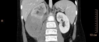
- MRI. Allows you to assess the condition of the brain, the degree of its damage, and morphological changes in its tissues.
The patient is examined urgently, regardless of the stage of development of the coma. The duration of each of them may be short and the person’s condition will deteriorate sharply.
Urgent Care
Treatment of hepatic coma at home is impossible; qualified medical care is required. The first thing to do to help a person is to call an ambulance. If the patient is conscious, he needs to be provided with peace and immobility; if the psycho-emotional state is severe, he must be reassured.
If upon arrival the ambulance crew assesses the person’s condition as critical, assistance is provided before he is taken to the hospital. It includes:
- oxygen therapy with humidified oxygen;
- administration of isotonic saline solutions to eliminate signs of shock and, if possible, maintain consciousness;
- administration of a fast-acting glucocorticoid intravenously;
- if necessary, the introduction of an antibiotic, its choice is considered on an individual basis.
Emergency assistance can only be provided by a doctor; it is prohibited to try to alleviate the patient’s condition on your own, including by giving painkillers.
What are the signs of coma?
Signs of hepatic coma in the initial stage are expressed in the following symptoms:
- dilated pupils, lack of pronounced reaction to light;
- unconscious state;
- the occurrence of minor movements (for example, chewing);
- convulsions;
- inflexibility of the limbs in the joints;
- presence of a reaction to severe pain;
- preservation of corneal and pharyngeal reflexes;
- uncontrolled urination and defecation.
In the deep stage of hepatic coma, the following symptoms are observed:
- absolute lack of reactions to stimuli of any kind;
- muscle cramps;
- lack of reflexes of any kind.
Listed below are the signs that are characteristic of hepatic coma at any stage:
- increased body temperature;
- a specific sweet smell (“liver”) emanating from the oral cavity;
- decreased blood pressure;
- nosebleeds, gastrointestinal or uterine bleeding;
- acceleration of heartbeat.
Clinical manifestations of hepatic coma
One of the dangerous symptoms of hepatic coma, which leads to death, is respiratory arrest.
Treatment
Treatment is carried out in the intensive care unit after hospitalization. Therapy is prescribed on an individual basis, taking into account the degree of coma and other characteristics of the patient. Typically treatment includes:
- administration of glucocorteroids (if they have already been administered by the ambulance team - after 8 hours);
- the use of antibiotics is often prescribed Ceftriaxone, administered intravenously for greater effectiveness;
- antiviral drugs;
- Arninine or glutamic acid to neutralize the effects of ammonia;
- Dopamine or its analogues against arterial hypertension;
- detoxification therapy for general intoxication.
Previously, blood transfusions were widely used in our country for hepatic coma, but today more and more doctors prefer bloodless methods of therapy, given the risks
In severe cases, doctors immediately recommend emergency liver transplantation. Unfortunately, in our country its implementation is complicated by a lack of donors and documentary issues. Therefore, many people prefer to go to foreign clinics. In case of successful recovery, the doctor gives clinical recommendations, which include following a diet and taking medications for life.
Sometimes hepatorenal coma develops or hepatic coma becomes a complication of renal coma. In such cases, hemodialysis is performed, but the likelihood of death is high.
Clinical treatment guidelines
Clinical recommendations include the following activities:
- intestinal sanitation (administration of enemas with electrolytes, administration of oral laxatives through a nasogastric tube);
- detoxification by extracorporeal methods (hemodialysis, hemosorption, plasmapheresis);
- taking medications to suppress factors that provoke the development of coma (hepatoprotectors, antibiotics, glucocorticoids);
- relief of hemorrhagic syndrome (infusion of fresh frozen donor plasma).
Treatment of hepatic coma with worsening neurological symptoms includes artificial ventilation and normalization of body temperature. The only effective method at the last stage of hepatic coma is transplantation of the affected organ.
What is the prognosis and how long do they live after hepatic coma?
How long patients live after hepatic coma depends on the timeliness of treatment and other factors. The survival rate is about 20%; people often die because the disease was diagnosed on time, but it had already progressed, and they never got their turn for a transplant.
Some live for several months, others up to 10 years, doctors explain this by saying that coma is a complication. Much depends on how long the disease that caused the coma lasts, what kind of lifestyle the person led, and what other chronic diseases they have.
Hepatic coma is the final stage of chronic or acute diseases of the organ; its occurrence indicates a person’s inattention to health and neglect of timely treatment. Knowing the pathogenesis, stage, and characteristics of its course, doctors select treatment, but the prognosis for recovery is often unfavorable.