- home
- general surgery
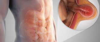
- Gastroesophageal reflux disease
- Reflux esophagitis
As a result of regularly repeated spontaneous reflux of gastric and/or intestinal contents into the esophagus, which have an irritating effect, an inflammatory process develops in the wall of the lower esophagus - reflux esophagitis. This chronic relapsing disease can be considered the most common among adults. Reflux esophagitis is diagnosed in 30% of adult patients, although the figures are very approximate, since many simply do not go to the doctor.
Causes of reflux esophagitis
The disease develops due to contact of the contents of the stomach and duodenum with the mucous membrane of the esophagus, which occurs when the lower esophageal sphincter, located at the border of the stomach and esophagus, is incompetent. As a result of impaired motility of the organs of the gastroesophageal zone, acidic gastric contents, being in the esophagus for a certain time, damage the cells of the mucosa.
Factors contributing to the development of reflux esophagitis are hiatal hernia - hiatal hernia, excess weight, pregnancy, leading to increased intra-abdominal pressure, taking certain medications, and smoking.
You can ask questions and sign up for a consultation by phone: +7 (495)222-10-87
or fill out the form below
Thank you, your question has been sent successfully, we will contact you soon!
Ask a new question
Information about the disease
According to epidemiological studies, about 23.6% of the adult population suffers from gastroesophageal reflux disease in Moscow. The peculiarity is the chronic and recurrent nature of the disease. The reflux of gastric contents into the lower parts of the esophagus occurs spontaneously. At the point of contact with the mucous membrane, an inflammatory process develops, which becomes the cause of discomfort and poor health. In medical practice, the full name of the disease is gastroesophageal reflux disease (GERD).
Symptoms
The clinical picture depends on the severity of the inflammatory process and the condition of the esophageal sphincter; reflux esophagitis is accompanied by a complex of dyspeptic, pulmonary and cardiac disorders. The main symptoms of reflux esophagitis are:
- Heartburn, manifested as a burning sensation behind the sternum, in the area of the xiphoid process, is the most characteristic symptom, occurring in more than ¾ of patients. Unpleasant and painful sensations can appear after eating, during physical activity, bending the body, and even in a horizontal position; wearing a tight belt can provoke heartburn.
- Belching that occurs after eating or carbonated drinks is the second most common symptom of reflux esophagitis. It can appear even in a horizontal position of the body, but more often during physical activity.
- Dysphagia is difficulty passing food that occurs when the motility of the upper parts of the digestive tract is impaired or when stricture is a narrowing of the lumen of the esophagus.
- Odynophagia is a retrosternal or interscapular pain that occurs when food passes through the esophagus. Painful sensations can radiate to the intercostal space and even resemble an angina attack.
- Regurgitation is regurgitation in which the contents of the stomach enter the oral cavity. Appears regardless of body position and physical activity of a person.
There are also extraesophageal manifestations involving other organs, e.g.
- bronchopulmonary: cough, attacks of respiratory discomfort or suffocation at night; the cause is the entry of small particles of stomach contents into the bronchi;
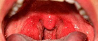
- otolaryngological: hoarseness, signs of rhinitis, pharyngitis due to refluxant entering the larynx;
- dental: thinning of tooth enamel, caries as a result of the aggressive action of acid.
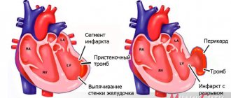
If left untreated, reflux esophagitis can cause severe complications. If the wall of the esophagus is damaged down to the submucosal layer, there is a risk of ulcer formation, which can cause bleeding. Scars formed after healing can lead to stricture, a narrowing of the esophagus.
The most severe complication is Barrett's esophagus syndrome - the squamous epithelium characteristic of the esophagus is replaced by a cylindrical epithelium characteristic of the gastric mucosa. With metaplasia, the likelihood of developing cancer increases by more than 30 times.
Presentation of symptoms and signs
Reflux esophagitis manifests symptoms and signs with varying degrees of severity, which depends on the stage of the disease:
- heartburn (the most common manifestation);
- pain in the chest near the heart;
- lump in throat and difficulty swallowing;
- frequent cough and inflammation of the respiratory tract;
- weight loss;
- hoarse voice;
- poor sleep;
- nausea and belching;
- bloating;
- vomit.
Are you experiencing symptoms of reflux esophagitis?
Only a doctor can accurately diagnose the disease. Don't delay your consultation - call
Classification
To assess the patient’s condition and unify data, a classification is used; depending on the degree of damage to the esophagus, several stages of reflux esophagitis are distinguished.
- I - rounded and longitudinal delimited, non-merging foci of inflammation, spreading to the mucous membrane of the esophagus from the Z-line - the border of the transition of the multilayered squamous epithelium of the esophageal mucosa into the columnar epithelium of the gastric mucosa;
- II - lesions in the Z-line zone merge, but do not cover the entire circumference of the esophagus;
- III - merging lesions covering the entire surface of the mucosa;
- IV - chronic damage to the esophagus, in which fibrous stenosis, shortening of the esophagus, peptic ulcers, and Barrett's esophagus develop.
To determine the stage of reflux esophagitis, it is necessary to undergo an examination, based on the results of which treatment is prescribed.
Types of GERD, manifestations of the disease and complications
In the medical classification, two types of the disease are distinguished - acute and chronic esophagitis. It differs in the type of inflammatory process. In the acute phase, the walls of the esophagus are exposed, and in the chronic phase, the mucous membrane is affected, with the duration of the disease lasting more than 6 months. Reflux esophagitis begins to develop due to improper nutrition, exposure to chemicals, and extensive infections. In the acute phase, there may be an increase in temperature, general malaise, and discomfort as food moves through the esophagus. Patients experience drooling during attacks, belching, and pain. With alcohol abuse, spicy or rough foods, the inflammatory process progresses. The disease enters the chronic phase. If left untreated, the esophagus changes and scars form.
Diagnostics
There are various diagnostic methods that can accurately determine the degree of damage to the esophagus. This is evidenced by the presence of changes due to the inflammatory process, erosions, ulcers, strictures, and metaplasia. The main diagnostic methods include:
- Fibrogastroscopy is an examination of the upper parts of the gastrointestinal tract using endoscopic equipment; during the procedure, it is possible to identify abnormalities in the wall of the esophagus, assess the condition of the mucous membrane, and exclude or confirm diseases of the stomach and duodenum. During the examination, a biopsy can be performed - taking tissue particles from visible pathological areas for the purpose of further histological examination.
- X-ray examination allows you to detect a stricture of the esophagus, ulcerative lesions, hiatal hernia, its size, it is also possible to assess the motility of the esophagus and stomach, the presence of reflux during examination using a barium suspension.
- Daily pH-metry - determination of the level of gastric secretion and the presence of reflux; it is possible to estimate the duration of reflux episodes, which allows you to select therapy and monitor the effectiveness of the drugs used.
- Esophageal manometry is an examination that allows you to determine the tone of the esophageal sphincters and their patency based on measuring pressure in different parts.
- pH impedansometry is a study aimed at assessing esophageal peristalsis and differentiation of gastroesophageal reflux, which is important for determining the nature of the disease and its cause.
Depending on the presence of certain symptoms, other examinations may be prescribed: ultrasound, ECG, consultation with an otolaryngologist, etc. In our clinic, patients have access to all the necessary diagnostic procedures, the examination takes a minimum of time.
How often does laryngopharyngeal reflux occur?
Typical GERD has been on the rise in recent decades, especially in the United States and Western Europe. Its frequency is estimated at 10-20% of the general population.
Calculating the frequency of LPR is not so easy - the symptoms are usually masked and can be explained by many other diseases.
There is a large study looking at the incidence of laryngopharyngeal reflux among people with heartburn. The average frequency was 32.8%, and in people with erosive lesions of the esophagus the frequency was higher, up to 34.9%. With non-erosive GERD it is slightly less - 30.5%.
The actual incidence of laryngopharyngeal reflux without the typical manifestations of GERD is not known, although scientists suspect that it is much more common than we think.
Treatment
Conservative treatment is indicated for patients with mild manifestations. Drug therapy can be aimed at neutralizing the contents thrown from the stomach into the esophagus, reducing gastric acidity, and protecting the esophageal mucosa.
There are various drugs: antisecretory agents, prokinetics and antacids, but they can only eliminate the symptoms. At the end of the course of treatment, signs of the disease return again. It is also worth considering that long-term use of medications that suppress the production of gastric juice can lead to poor digestion of food, which will lead to a number of disorders. When using medications that affect acidity, the risk of malignancy increases.
Let's summarize:
Diagnosis of laryngopharyngeal reflux is not easy; it cannot be solved 100% by the methods familiar to a gastroenterologist; the efforts of other specialists are required to exclude other conditions with similar complaints.
Each case is discussed individually with patients or their families.
Often the solution may be to evaluate the effect of a trial treatment (more on therapy later...)
Treatment of laryngopharyngeal reflux
25, total, today
Surgery
If conservative treatment is ineffective, if complications arise, if there is an esophageal hernia, as well as if there is pain behind the sternum or if there are extra-esophageal signs (cough, hoarseness, etc.), surgical treatment is recommended - fundoplication. The purpose of surgery for reflux esophagitis is to restore normal anatomical relationships in the area of the esophagus and stomach, creating a mechanism that will prevent the reflux of gastric contents into the esophagus.
Most surgeons in domestic clinics perform circular Nissen fundoplication; during the operation, a cuff is created - the fundus of the stomach is wrapped around the esophagus 360°, which prevents reflux in the future. But a valve formed in this way leads to the loss of natural protective mechanisms: the ability to regurgitate and vomit. Large portions of food and drinking carbonated drinks lead to the fact that, if necessary, their removal through the cardia is impossible, discomfort and pain appear. The formed cuff often slips off after 1-2 years, which leads to relapse of the disease.
Conservative therapy
For conservative therapy, the following groups of drugs are used:
- antacids and alginates
- proton pump inhibitors
- prokinetics
Antacids and alginates
Antacids and alginates, topical preparations most often containing aluminum and magnesium salts, these drugs act locally, are not absorbed into the blood and do not have a systemic effect. Their advantage is safety of use, but there is also a disadvantage, a short duration of action. Antacids are not used as an independent method of treatment, but are used as first aid for heartburn, and as an intensification of therapy for the speedy healing of erosions and ulcers of the esophagus. Preparations in the form of gels with alginic acid have proven themselves to be the best, which, when interacting with acid in the lumen of the stomach, form foam, thereby increasing the duration of action of the drug and its effectiveness. Antacids are most often prescribed 40 minutes after meals and at night, or on demand in case of heartburn.
Proton pump inhibitors (PPIs)
Today they are the main drugs in the treatment of reflux esophagitis, these include the well-known omeprazole, lansoprazole, rabeprazole, etc. These are systemic drugs, they block the transport of hydrogen molecules into the parietal cells of the stomach, responsible for the secretion of hydrochloric acid, and how do we We remember from the school chemistry course, this acid consists of a molecule of hydrogen and chlorine, no hydrogen, no acid. These drugs have revolutionized the treatment of reflux esophagitis, significantly improving the outcome and reducing the number of complications. Previously, H2 histamine receptor blockers were used, which were much less effective and had more side effects. It must be said that these drugs are sometimes used now in combination with PPIs to enhance the effect. The main disadvantage of proton pump inhibitors (PPIs) is their fairly rapid removal from the blood, which requires repeated use throughout the day, and even with double use, so-called acid breakthroughs are described, when, more often at night, the acidity of gastric juice sharply increases. Most often, PPIs are prescribed 20 mg twice a day 20 minutes before meals, morning and evening for 6 to 8 weeks. Then maintenance therapy is prescribed at 10 mg per day or 10 mg twice a day.
Prokinetics
Prokinetics are drugs that improve and normalize peristalsis of the gastrointestinal organs, including the esophagus. The most commonly prescribed drug is itopride, which is considered the most effective and safe drug. Motilium is also prescribed. The previously popular cerucal has recently been deprecated due to its central effect on the brain. Prokinetics are prescribed 1 tablet three times a day 20 minutes before meals. A course of 2 weeks is recommended.
Problems of conservative therapy
The vast majority of patients with reflux esophagitis are treated conservatively, achieving good functional results, but unfortunately, therapy is not always effective. As we wrote above. The cause of reflux esophagitis is the reflux of gastric contents into the esophagus. So the first problem with conservative therapy is that it does not eliminate the cause, but only relieves the symptoms. With conservative therapy, reflux remains, but is no longer acidic, and therefore patients do not feel any complaints. Although some researchers believe that due to the neutralization of acid, bile begins to flow into the esophagus, since it ceases to be inactivated in the stomach, and bile is an even more aggressive environment for the development of complications of esophagitis. Although it should be noted that this is still a theory that has not received sufficient confirmation. But at the same time, a number of scientists associate the growth of esophageal cancer with this hypothesis. Since, despite the widespread use of PPI drugs, the incidence of esophageal cancer is steadily increasing.
Taking medications in itself can cause complications. Evidence has been obtained that proton pump inhibitors increase the risk of stroke, osteoporosis and heart attack, and although the risk is not quite high, about 0.4 - 0.6% per year, it is still there.
Well, perhaps the main thing is that the effect of conservative therapy is temporary and is not always sufficiently effective. So, 6 months after stopping therapy, complaints return in 40-50% of patients, and after 12 months in 80-90%, that is, most patients are forced to take medications daily for many years. Considering the sufficient safety of the drugs, this may not be scary, although it certainly reduces the quality of life. But the problem lies in the fact that with long-term therapy, after three or five years, the effectiveness of the drugs decreases and in 20 - 30% of patients complaints reappear and complications of esophagitis develop, despite the constant use of the prescribed drugs.
My approach to treatment
When surgically treating patients with reflux esophagitis, I, like most specialists in European clinics, use a more effective technique that does not have the above-mentioned disadvantages - partial Toupet fundoplication with a cuff rotation of 270°. The technique I improved has a number of advantages:
- the physiological functioning of the esophageal sphincter is restored;
- maintaining a functional esophagogastric valve allows the patient to do without medications throughout his life;
- the ability to belch and vomit—the body’s natural defense reactions—is preserved;
- no pain after overeating or carbonated drinks;
- the number of relapses does not exceed 2% within a year after surgery, after 5 years this figure is about 4%.
A Russian Federation patent has been issued for an improved technique - Toupet 270° fundoplication.
Reflux esophagitis
Renev E.N. Modern treatment of gastroesophageal reflux disease.
Modern treatment of gastroesophageal reflux disease
E.N.
Renev We would like to preface the discussion of therapeutic options for
gastroesophageal reflux disease
(
GERD
) with brief information on the mechanisms of development and diagnosis of this pathology. The possibilities of surgical treatment of GERD will not be discussed in this article.
Definition
So, A.S.
Trukhmanov defines GERD
as the occurrence of characteristic symptoms and (or) inflammatory damage to the distal parts of the esophagus due to repeated reflux of gastric contents into the esophagus [1].
According to the definition of the International Working Group, the term " gastroesophageal reflux disease"
» should be applied to all individuals at risk of physical complications of gastroesophageal reflux, or experiencing a significant deterioration in health-related well-being (quality of life), as a result of reflux symptoms, after adequate reassurance of the benign nature of the symptoms [6].
The term “endoscopically negative reflux disease” should be used in individuals who meet the definition of gastroesophageal reflux disease, but who do not have either Barrett's esophagus or visible mucosal defects (erosions or ulcers) on endoscopic examination [6].
Development mechanisms
Without dwelling in detail on the pathogenetic mechanisms of development of this disease, we will only say that it is based on the effect of acid and pepsin on the esophageal mucosa due to the combination (in varying proportions) of pathological reflux of gastric contents into the esophagus with a violation of its clearance. Pathological reflux of contents, in turn, is caused by dysfunction of the lower esophageal sphincter (either as a result of a decrease in its tone or an increase in the frequency of spontaneous relaxation, or due to its anatomical defect, for example, with a hernia of the esophagus). Impaired esophageal clearance may be caused by decreased saliva production or impaired esophageal motility. As a result of all of the above, there is an imbalance between aggressive factors and protective factors, which leads, but not necessarily, to the occurrence of reflux esophagitis.
Epidemiology
According to S.I.
Pimanova, symptoms of GERD
observed in half of the adult population, and the endoscopic picture of esophagitis is observed in 2-10% of examined people [1]. It must be remembered that GERD is not always accompanied by esophagitis. Up to 50-70% of patients with heartburn at the time of seeking medical help have endoscopically negative GERD [8]. The attitude of a number of practitioners towards endoscopically negative GERD as the mildest degree of this disease that does not require intensive drug therapy is fundamentally incorrect. A number of studies have demonstrated that the quality of life in patients with endoscopically positive and negative GERD is impaired to almost the same extent [29]. Studies have shown that endoscopically negative GERD very rarely develops into reflux esophagitis, which in turn rarely progresses to more severe forms over time [7,8].
Diagnostics
Since
the diagnosis of GERD
is widely described in many manuals, we will dwell only on some of its points.
The main symptom of GERD, observed in at least 75% of patients, is heartburn
[6].
There may also be pain or a burning sensation behind the sternum, belching, etc. Most often, GERD symptoms
occur after eating.
Diagnosis of erosive esophagitis is based on endoscopic examination. Barium radiography has a fairly high sensitivity for severe (98.7%) and moderate (81.6%) esophagitis, but low sensitivity (24.6%) for mild esophagitis [12, 22, 23]. Endoscopy with biopsy is the only reliable method for diagnosing Barrett's esophagus. The severity of erosive reflux esophagitis on the endoscopic picture is divided into 4 degrees A, B, C and D (according to the Los Angeles classification).
pH monitoring
is a sensitive and specific diagnostic test and is especially important for identifying
endoscopically negative GERD
. More than 50 episodes of decrease below 4 are considered as a diagnostic criterion for GERD [1]. In a number of patients, a less significant decrease in the pH of the esophagus occurs, but when most episodes of such a decrease coincide with the onset of symptoms, it allows us to speak of a “hypersensitive esophagus.”
Among provocative tests, the Bernstein test plays a certain role (the appearance of typical symptoms after the introduction of a weak solution of hydrochloric acid into the esophagus and their disappearance after the introduction of saline). Determining the pressure of the lower esophageal sphincter is useful when deciding on surgical treatment.
Treatment
Before moving on to consideration of individual aspects
of the treatment of GERD,
it is necessary to emphasize the fact that its main goal is to quickly relieve patients from the symptoms that bother them. The disappearance of symptoms usually correlates well with the healing of mucosal defects in erosive esophagitis [6].
Lifestyle change
Although according to the GERD Working Group, lifestyle factors do not play a determining role in the development of GERD [6], recommendations aimed at eliminating factors that promote reflux or impair esophageal clearance should be made.
Diet.
It is necessary to stop taking reflux-inducing foods (fatty foods, chocolate and excessive amounts of alcohol, onions and garlic, coffee, carbonated drinks, especially various types of colas) and drugs with low pH (orange and pineapple juices, red wine). However, an attempt to sharply limit a patient’s diet (especially a young one) is rarely possible in practice; your recommendations simply will not be followed. It makes more sense to identify which products cause the appearance or exacerbation of symptoms in a given patient and try to at least give them up. The patient should be informed that overeating should be avoided. After eating, it is advisable not to take a horizontal position or work in an inclined position. The last meal should be 3 hours before bedtime.
Weight control.
Losing weight does not always resolve symptoms, but losing weight may reduce the risk of developing a hiatal hernia. However, giving advice to lose weight is much easier than implementing it. Overweight people sometimes try to hide their lack of a waist by over-tightening the waist belt, which leads to increased intra-abdominal pressure and the development of reflux (as does wearing clothes that are too tight).
Smoking
is a contributing factor to GERD as a result of both sphincter relaxation and decreased salivation and should be discontinued accordingly [7]. Although, according to some researchers, smoking cessation has a minimal positive effect on GERD [6].
Raising the head of the bed is important for patients with nocturnal or laryngeal symptoms (which constitute a small proportion of patients with GERD), but its necessity in other cases is questionable.
A number of medications such as antispasmodics, beta blockers, hypnotics and sedatives, nitrates and calcium antagonists can contribute to the development of reflux.
Antacids
Discussing the use of antacids, of which there are a great many in our time (almagel, phosphalugel, maalox, rutacid, etc.), I would like to emphasize that, in our opinion, antacids do not play an independent role in the treatment of GERD and can only be used as a short-term remedy symptom control. The low effectiveness of antacids is based on the short duration of pH control achieved by their use. Data from many authors confirm the minimal effect of antacids (even in combination with lifestyle changes) in reflux esophagitis, although it is superior to the placebo effect [27]. We suggest that patients (being treated for GERD) use antacids as a method of quickly controlling symptoms that occur, usually after a violation of diet or exercise, and in those with rare (no more than 4 per month) episodes of heartburn without endoscopic signs of esophagitis.
Antisecretory drugs
The most effective way to treat GERD is to reduce acid production in the stomach using H2 blockers or proton pump inhibitors. The goal of this therapy is to increase the pH of gastric juice to 4 and during the period of greatest likelihood of reflux occurring, i.e. not the prevention of reflux as such, but the elimination of the pathological effects of gastric juice components on the esophagus. H2 blockers. Before the advent of proton pump inhibitors, H2 blockers were the drug of choice for the treatment of GERD. There are currently 4 H2-histamine receptor blockers used in practice (cimetidine, ranitidine, famotidine and nizatidine). The mechanism of action of the drugs is to block gastric secretion stimulated by histamine. However, two other stimulation pathways, acetylcholine and gastrin, remain open. It is this fact that is associated with a lower degree of suppression of secretion than with proton pump inhibitors (PPI) and a gradual decrease in the degree of inhibition of gastric secretion with long-term use of H2 blockers, when stimulation of acid production begins to increasingly occur through other mediators (mainly gastrin).
Cimetidine
(First generation H2 blocker). Use 200 mg 3-4 times a day and 400 mg at night. The maximum daily dose is 12 grams.
Ranitidine
(second generation) is used in a dosage of 150 mg 2 times a day, which can, if necessary, reach 300 mg 2 times a day (maximum dose 9 grams per day).
For nighttime symptoms - 150-300 mg at night. Maintenance therapy - 150 mg at night. Famotidine
(third generation) is used at a dose of 20 mg twice daily, with a maximum daily dose of 480 mg. For nocturnal symptoms, 20-40 mg at night, maintenance therapy 20 mg at night.
Nizatiditis
(fourth generation) take 150 mg twice a day or 300 mg at bedtime.
Due to a very wide range of side effects (from androgenic effects to blockade of respiratory enzymes) and inconvenient dosage, cimetidine is not currently used in practice. Of all the other H2 blockers, we prefer famotidine (as the drug with the least common side effects). It must be remembered that all H2 blockers are discontinued gradually in order to prevent “recoil” syndrome - a sharp increase in acidity after stopping treatment.
Based on 33 randomized trials (involving 3000 people), the following data were obtained: the use of placebo led to relief of GERD symptoms in 27% of patients, H2-blockers in 60% and PPI in 83% [7]. Esophagitis was relieved in 24%, 50% and 78% of cases, respectively. These figures suggest the effectiveness of H2 blockers in the treatment of GERD, which, however, is significantly inferior to that of PPI. H2 blockers retain a certain role in the treatment of GERD. They are effective as a treatment for nocturnal reflux [21], even with continued PPI use [25], and as an on-demand therapy.
Proton pump blockers
Their action is based on blocking the ATPase of the proton pump (due to the formation of an irreversible bond with the cystine residue of the enzyme). It must be remembered that PPI only blocks the currently active proton pump. Drugs of this group are absorbed in the form of inactive compounds, turning into the active substance directly in the tubular systems of secretory cells. All PPIs, except esomeprazole, have a short half-life (30-120 min). PPI destruction occurs in the liver, and there are two ways of their destruction - fast and slow. The destruction process is stereodependent. The dextrorotatory isomer decays along the fast path, and the left-handed isomer decays along the slow path. All PPIs, again except esomeprazole (only the levorotatory isomer), are represented by right and left-handed isomers. This fact explains the longer retention of the minimum therapeutic concentration by esomeprazole compared to other PPIs.
PPIs are prescribed before meals (usually 30 minutes before breakfast, with a single dose), so that the effect occurs when the maximum number of active proton pumps is present - 70-80% of their total number. The next dose of PPI again blocks 70-80% of the receptors (remaining and regenerated), so the peak of the antisecretory effect occurs on days 2-3 (slightly faster when using esomeprazole). PPIs are practically ineffective as an on-demand therapy (the onset of symptoms - heartburn, indicates an acid surge that has already occurred, followed, as a rule, by a decrease in the number of active pumps and, therefore, the absence of a target for PPI action).
When analyzing the comparative effectiveness of various PPIs, it can be concluded that there are no significant advantages between omeprazole, rabeprazole, lansoprazole and pantoprazole. The effectiveness of esomeprazole (Nexium) is slightly higher. When comparing the duration of maintaining intragastric pH>4 using different PPIs, data were obtained about better control of gastric secretion when using Nexium (Fig. 1).
Although it should be noted that when using 40 mg of omeprazole, the difference is not so noticeable. The benefits of Nexium are more pronounced in severe forms of esophagitis (grade D) [4]. Omeprazole is used in a dose of 20-40 mg per day (either once in the morning or twice a day). In severe cases, the dose can reach 60 mg per day. Lansoprazole is used at 30 mg/day, pantoprazole at 40 mg/day, rabeprazole at 20 mg/day and Nexium at 40 mg/day. Discontinuation of the drug should also be gradual.
Prokinetic drugs
Prokinetic drugs (domperidone, metoclopramide, and cisapride) may increase lower esophageal sphincter pressure, improve esophageal clearance, and accelerate gastric emptying. Cisapride is only available for limited use in the US due to concerns regarding cardiac arrhythmias (see below). Metoclopamide causes weakness, anxiety, tremor, parkinsonism or tardive dyskinesia in 20-50% of cases. Use 10 mg 3-4 times a day. The maximum single dose is 20 mg, daily dose is 60 mg.
Cisapride.
Although cisapride was generally considered virtually safe, its recent widespread use in the United States has been associated with the occurrence of cardiac arrhythmias. Most often they developed when taking cisapride in combination with drugs that inhibit cytochrome P-450 and increase the level of cisapride. As a result, the manufacturer has partially restricted the use of this drug in the United States. Studies comparing the effectiveness of cisapride 910 mg four times a day with H2-receptor antagonists (ranitidine 150 mg twice a day) and cimetidine (400 mg four times a day) demonstrated their superiority over placebo and similar effectiveness in relieving the symptoms of GERD and cure of esophagitis [9, 18, 24]. The combination of H2 blockers with cisapride gives a better effect than either drug alone, but is inferior to omeprazole [31].
Domperidone
(Motilium) is similar in mechanism of action to metoclopramide, but does not penetrate the blood-brain barrier and, therefore, does not cause central side effects, but increases the level of prolactin in the blood. Use 10 mg 3-4 times a day. None of the drugs gave a good therapeutic effect in severe degrees of esophagitis.
Role of HP infection
current role of HP infection in GERD
remains debatable. Although, according to the Maastrik Agreements, GERD is an indication for eradication therapy, not all authors agree with this. A number of studies have shown that Hp eradication does not cure reflux esophagitis, nor does it have a preventive role in terms of its relapse [32, 14]. The fact that Hp infection can cause either an increase or decrease in gastric secretory function makes its role in the development of GERD even more controversial. Data from some authors even indicate the protective role of HP infection in GERD [10, 28, 30], due to the alkalizing effect, and in the further development of mucosal atrophy.
Almost the only factor justifying eradication therapy for GERD is that chronic use of PPI, against the background of existing HP infection, promotes the development of atrophic gastritis and metaplasia [3, 13, 16]. According to Kuipers EJ [13], who compared the likelihood of developing atrophic gastritis in groups of patients with GERD and HP infection who received omeprazole or underwent fundoplication, it developed in 31% and 5% of patients, respectively. Although another study did not find such a pattern [20]. In turn, eradication therapy does not cause exacerbation or worsening of GERD [15].
In our practice, we test for the presence of HP and perform eradication in patients with GERD only if they have a concomitant disease of the upper gastrointestinal tract whose connection with HP infection has been established (for example, peptic ulcer) or when planning chronic (more than a year) constant use of proton pump inhibitors.
New directions of pharmacotherapy
According to Ciccaglione et al, a drug that reduces the number of spontaneous relaxations of the lower esophageal sphincter - baclofen at a dosage of 10 mg 3 times a day for a month showed significant superiority over placebo, improved pH monitoring of the esophagus and reduced the severity of GERD symptoms [5]. It was also noted to be well tolerated. The drug inhibits 34-60% of spontaneous relaxation of the lower esophageal sphincter and increases its basal pressure [11]. However, there is still insufficient data to justify the widespread use of baclofen in the treatment of GERD.
Treatment regimens
Currently, there are two main tactical approaches to the treatment of GERD, the so-called step-up and step-down. The first is the use of the weakest measures (lifestyle modification, antacids) as the first stage of treatment with the gradual use of increasingly powerful drugs if ineffective (H2-blockers, then their combination with prokinetics and only then PPI). The second treatment option involves prescribing the most effective treatment (PPI), which allows you to quickly relieve symptoms, and then reduce the dose of medications and possibly switch to weaker drugs.
In our practice, we adhere only to step-down therapy because... We believe that the patient comes to us for the fastest relief of the symptoms that bother him, which should be achieved by prescribing a group of drugs from which the best effect can be expected. You should not forget about the advice on lifestyle changes, but in combination with the administration of a standard dose of PPI. As for starting treatment with H2 blockers, and then switching, if necessary, to PPI - you won’t be judged for this, but does it make sense? H2 blockers have no less potential side effects, and their price is not significantly lower. We'll leave them for on-demand therapy and nocturnal reflux episodes. However, there is a very small group of patients with reflux esophagitis refractory to proton pump inhibitor therapy in whom sufficient pH control can be achieved using high doses of H2 blockers [17].
What to do with endoscopically negative GERD? Yes, exactly the same. As mentioned above, the degree of morphological changes in the esophagus does not correlate well with the severity of symptoms [6]. Moreover, in this group of patients there is often a less pronounced effect of antisecretory therapy with longer persistence of symptoms [8]. It must also be remembered that the effectiveness of H2 blockers for endoscopically negative GERD does not exceed that for erosive reflux esophagitis [6].
In severe reflux esophagitis (C, D), therapy with the most powerful PPI (Nexium) or the maximum dose of other proton pump inhibitors is rational.
For nocturnal episodes of heartburn, despite the use of PPI, it is rational to add a single evening dose of an H2 blocker. Antacids can be used as patient-controlled, on-demand therapy.
So, we follow a knowledgeable management strategy when a new patient with GERD appears.
- Proton pump inhibitors in a standard dose (for 2-4 weeks for endoscopically negative reflux esophagitis and erosive esophagitis grades A, B and for 8 weeks for its more severe forms).
- In case of ineffectiveness (determined by the persistence of symptoms after 7-10 days of treatment or the persistence of the endoscopic picture of esophagitis), increase the dose of PPI to the maximum or switch to a potentially more effective PPI - Nexium.
- If ineffective, pH monitoring is required during treatment. Trying to switch to high doses of H2-blockers in combination with prokinetics? Antireflux surgery?
- If effective, gradually reduce the dosage until the drug is discontinued. If symptoms recur, take the minimum effective dose of the drug (every other day therapy or weekend therapy is possible), discuss the possibility of antireflux surgery.
Maintenance therapy
Based on the chronic nature of GERD, there is a need for maintenance therapy. Reducing the dose of medication or attempting maintenance therapy with a drug less potent than the drug used for treatment often leads to a high relapse rate. Only in approximately 20% of patients after a course of treatment, lifestyle changes and periodic use of antacids are sufficient to maintain remission. H2 blockers and prokinetics are ineffective in maintaining remission in patients in whom it was achieved using PPI [2]. Low dose PPI therapy is most effective. The effectiveness of weekend therapy and every other day is controversial.
Conclusion
Drug therapy remains the mainstay of treatment for GERD. PPIs are the drugs of choice for treatment and long-term maintenance therapy. The role of HP infection in the development and natural history of GERD, as well as its effect on the outcome of treatment, are not completely clear. The development of new drugs and comparison of the effectiveness of various schemes for their use is a promising direction for further improving the quality of treatment of this pathology.
Literature
1. Pimanov I.S. Esophagitis, gastritis and peptic ulcer. N. Novgorod. - 2000. 2. Antonson CW, Robinson MG, Hawkins TM, et al. High doses of histamine antagonists do not prevent relapses of peptic esophagitis following therapy with a proton pump inhibitor. Gastroenterology 1990;98:A16. 3. Berstad AE, Hatlebakk FG, Maartmann-Moe H, et al. Helicobacter pylori, gastritis and epithelial cell proliferation in patients with reflux oesophagitis after treatment with omeprazole. Gut 1997;41:735-739 4. Castell DO, Kahrilas PJ, Richter JE, Vakil NB, Johnson DA, Zuckerman S et al. Esomeprazole (40 mg) compared with lansoprazole (30 mg) on the treatment of erosive esophagitis. Am J Gastroenterol 2002;97:575-83. 5. Ciccaglione AF, Bartolacci S, Marzio L. Effects of one month treatment with GABA agonist baclofen on gastro-esophageal reflux and symptoms in patients with gastro-esophageal reflux disease. Gastroenterology. 2002;122:A-196. 6. Dent J, et al. An evidence-based appraisal of reflux disease management the Genval Workshop Report. Gut 1998;44(Suppl 2):S1-S16 (April). 7. DeVault K, Castell D and The Practice Parameters Committee of the American College of Gastroenterology. Updated Guidelines for the Diagnosis and Treatment of Gastroesophageal Reflux Disease. Am J Gastroenterol 1999;94:1434-1442. 8. Fass R. Epidemiology and pathophysiology of symptomatic gastroesophageal reflux disease. Am J Gastroenterol. 9. Galmiche JP, Fraitag B, Filoche B, et al. Double-blind comparison of cisapride and cimetidine in treatment of reflux esophagitis. Dig Dis Sci 1990;35:649-55. 10. Haruma K, Mihara M, Kawaguchi H, et al. Low prevalence of Helicobacter pylori infection in patients with reflux oesophagitis [abstract]. Gastroenterology 1996;110:A130. 11. Holloway RH. GABA-B receptors and control of gastrointestinal motility. In: AGA Research Symposium: GABA-B receptor agonists as novel treatments of reflux disorders. Program and abstracts of Digestive Disease Week 2002; May 19-22, 2002; San Francisco, California. 12. Koehler RE, Weymean PJ, Oakley HF. Single- and double-contrast techniques in esophagitis. AJR 1980;135:15-9. 13. Kuipers EJ, Lundell L, Klinkenberg-Knoll EC, et al. Atrophic gastritis and Helicobacter pylori infection in patients with reflux oesophagitis treated with omeprazole or fundoplication. N Engl J Med 1996;334:1018-1022. 14. Labenz J, Malfertheiner P. Helicobacter pylori in gastro-oesophageal reflux disease: causal agent, independent or protective factor? Gut 1997;41:277-280. 15. Laine L; Sugg J Effect of Helicobacter pylori eradication on development of erosive esophagitis and gastroesophageal reflux disease symptoms: a post hoc analysis of eight double blind prospective studies. Am J Gastroenterol 2002 Dec;97(12):2992-7 (ISSN: 0002-9270). 16. Lamberts R, Creutzfeldt W, Struber HG, et al. Long term omeprazole therapy in peptic ulcer disease: gastrin, endocrine cell growth and gastritis. Gastroenterology 1993;104:1356-1370. 17. Leite LP, Just RJ, Castell DO, et al. Control of gastric acid with high dose H2-receptor antagonists after omeprazole failure: Report of two cases. Am J Gastroenterol 195;90:1874-7. 18. Lepoutre L, Van der Spek P, Vanderlinden I, et al. Healing of grade-II and III oesophagitis through motility stimulation with cisapride. Digestion 1990;45:109-14. 19. Lind T, Rydberg L, Kylebäck A, Jonsson A, Andersson T, Hasselgren G et al. Esomeprazole provides improved acid control vs. omeprazole in patients with symptoms of gastro-oesophageal reflux disease. Aliment Pharmacol Ther 2000;14:861-7. 20. Lundell L, Havu N, Andersson A, et al. Gastric atrophy development and acid suppression therapy revisited. Result of a randomized clinical study with long term follow up [abstract]. Gastroenterology 1997;112:A28. 21. Mann SG, Murakami A, McCarroll K, et al. Low dose famotidine in the prevention of sleep disturbance caused by heartburn after an evening meal. Aliment Pharmacol Ther 1995;9:395-401. 22. Ott DJ, Wu WC, Gelfand DW. Reflux esophagitis revisited: Prospective analysis of radiological accuracy. Gastrointest Radiol 1981;6:1-7. Koehler RE, Weymean PJ, Oakley HF. Single- and double-contrast techniques in esophagitis. AJR 1980;135:15-9. 23. Ott DJ, Chen YM, Gelfand DW, et al. Analysis of a multiphasic radiographic examination for detecting reflux esophagitis. Gastrointest Radiol 1986;11:1-6. 24. Richter JE, Long JF. Cisapride for gastroesophageal reflux disease: A placebo-controlled, double-blind study. Am J Gastroenterol 1995;90:423-30. 25. Robinson M, Yale J Biol Med 1999 Mar-Jun;72(2-3):169-72 (ISSN: 0044-0086). 26. Rohss K, Hasselgren G, Hedenström H. Effect of esomeprazole 40 mg vs omeprazole 40 mg on 24-hour intragastric pH in patients with symptoms of gastroesophageal reflux disease. Dig Dis Sci 2002;47:954-8. 27. Saco LS, Orlando RC, Levinson SL, et al. Double-blind controlled trial of bethanecol and antacid versus placebo and antacid in the treatment of erosive esophagitis. Gastroenterology 1982;82:1369-73. 28. Sehiguchi T, Shirota T, Horikoshi T, et al. Helicobacter pylori infection and severity of reflux oesophagitis [abstract]. Gastroenterology 1996;110:A755. 29. Tew S, Jamieson GG, Pilowski I, et al. The illness behavior of patients with gastroesophageal reflux disease with and without endoscopic esophagitis. Dis Esophagus 1997;10:9-15. 30. Vicari JJ, Peek RM, Falk GW, et al. The seroprevalence of cag A-positive Helicobacter pylori strains in the spectrum of gastro-oesophageal reflux disease. Gastroenterology 1998;115:50-57. 31. Vigneri S, Termini R, Leandro G, et al. A comparison of five maintenance therapies for reflux esophagitis. N Engl J Med 1995;333:1106-10. 32. Werdmuller BFM, Loffeld RJLF. Helicobacter pylori infection has no role in the pathogenesis of reflux esophagitis. Dig Dis Sci 1997;42:103-105. 135. 33. Wilder-Smith C, Röhss K, Lundin C, Rydholm H. Esomeprazole 40 mg provides more effective acid control than pantoprazole 40 mg. J Gastroenterol Hepatol 2002;17 Suppl:A784. 34. Wilder-Smith C, Rohss K, Claar-Nilsson C, Lundin C. Esomeprazole 40 mg provides faster and more effective acid control than lansoprazole 30 mg in patients with symptoms of gastroesophageal reflux disease. Gastroenterology 2002;122 4 Suppl 1:A200. 35. Wilder-Smith C, Claar-Nilsson C, Hasselgren G, Rohss K. Esomeprazole 40 mg provides faster and more effective acid control than rabeprazole 20 mg in patients with symptoms of GERD. J Gastroenterol Hepatol 2002;17 Suppl:A612.
How is the operation performed using a modified technique?
During the operation, a symmetrical cuff is formed from the upper part of the stomach, covering the esophagus at 270°. The anterior right side of the esophagus remains free; The cuff created in this way strengthens the lower esophageal sphincter and prevents the backflow of stomach contents. In the presence of a hiatal hernia, the lower part of the esophagus and the upper third of the stomach are first separated from the adhesions and returned to their natural position. The diaphragmatic hole is sutured to normal size. To prevent relapse and perform diaphragmocruroplasty, I use mesh implants, which reduces the risk of recurrence of the disease from 65% to 1%.
Surgery for reflux esophagitis is performed through laparoscopic access: all manipulations are performed through several small punctures on the anterior abdominal wall. In the future, the puncture marks become almost invisible. The operation is performed using endoscopic equipment equipped with a miniature video camera, which allows all manipulations to be carried out under visual control as accurately as possible. Since important structures (vagus nerve, blood vessels) are located in the surgical area, there is virtually no risk of damage to them.
During the operation, when isolating the esophagus and stomach, I use a dosed tissue ligation device LigaSure (USA), which is capable of “sealing” vessels without the risk of damage to surrounding tissues, as well as the development of bleeding. At the same time, the duration of the operation is reduced. The use of modern suture material promotes rapid recovery.
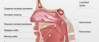
Typical manifestations:
- the desire to constantly clear your throat, especially in the morning or after eating
- persistent dry (no phlegm) cough
- sore throat outside of acute respiratory infections
- hoarseness
- feeling of a foreign body in the throat.
A little more about the main manifestations of laryngopharyngeal reflux.
Laryngitis is one of the common manifestations of LPR.
A condition well described by otolaryngologists.
Although laryngopharyngeal reflux is quite common, it is not the only cause of laryngitis.
Otolaryngologists always rule out postnasal drip, allergies, and irritants from inhaled air (including tobacco smoke from active or passive smoking).
Reflux and chronic cough.
Probable reasons:
- The esophagus and bronchial tree have a common embryonic origin and innervation. Irritation of neuroreceptors in the esophagus and throat can activate the reflex arc and provoke bronchospasm. Moreover, to trigger the reflex, throwing it into the lower part of the esophagus, right next to the stomach, may be enough.
- Direct reflux into the respiratory tract. The mechanism is controversial, since studies have been conducted on the frequency of detection of pepsin (an enzyme in gastric juice) and bile acids in bronchoalveolar fluid, and no difference was found in people with and without a cough.
The connection between reflux and long-term nasal congestion and chronic postnasal drip , an unpleasant condition in which there is a constant sensation of drainage down the back of the throat, is being actively studied.
There is a possible connection between reflux and halitosis - bad breath, disturbances in the perception of tastes and smells.
Getting stomach contents into the ear can cause otitis media, ringing or noise in the ears, and dizziness.
Advantages of laparoscopic surgery
- Less traumatic and no pain in the postoperative period;
- Short period of hospitalization - no more than three days;
- Rapid recovery - after two weeks, patients return to their normal lifestyle.
Since patients often have other diseases that require surgical treatment, so-called simultaneous operations can be performed using laparoscopic access. I have been performing similar interventions for more than two decades. When performing a simultaneous operation during one anesthesia, you can immediately get rid of 2-5 surgical pathologies (for example, calculous cholecystitis, cholelithiasis, tumors, cysts, etc.).
Many, even experiencing painful attacks of heartburn, are in no hurry to see a doctor, trying to alleviate the condition in various ways, including with the help of medications. Is it necessary to treat reflux esophagitis if the symptoms are eliminated with the help of drugs that can be bought at any pharmacy? Of course, since there is a risk of developing severe complications. In addition, in the absence of adequate treatment, you will have to take medications throughout your life and adhere to strict dietary restrictions. At the same time, the effect of the drugs is very short-lived, and any physical activity immediately causes unpleasant and even painful sensations. To determine the extent of the disease and choose the most appropriate treatment tactics, just contact me by email or by scheduling a consultation.
Carrying out surgical interventions for diseases of the esophagus and stomach requires excellent command of surgical techniques, including endoscopic suture, which is impossible without appropriate experience. Over more than 25 years of experience, I have performed more than 2000 surgical interventions for reflux esophagitis, GERD and hiatal hernia. I am the author of monographs and more than 50 scientific papers devoted to these problems. I also regularly conduct seminars and master classes on these diseases, which are attended by specialists from various clinics and centers.
Reflux without pathology
The problem of reflux is familiar to every pregnant woman in the later stages of pregnancy. It is not associated with either infection or overeating. The fetus provokes the release of acid from the esophagus. The growing uterus with the unborn child inside puts pressure on organs, including the stomach, not allowing it to expand and accommodate food.
Forced to be in a constrained state, the stomach is unable to hold incoming food. As a result, acid and fragments of liquid food and drinks easily escape into the esophagus, especially when a pregnant woman is in a horizontal position.
Taking medications even from the antacid group is undesirable during pregnancy, as it can affect the course of pregnancy. Therefore, doctors recommend that pregnant women in their final stages eat frequently, but in small portions. After eating, sit or walk for 40-60 minutes. Do not drink a lot of liquid at one time, as it is easier for the drink to enter the esophagus when the size of the stomach is limited.
If split meals do not help get rid of the problem, you need to consult with your local doctor about choosing a safe option for neutralizing the acid.