Among all diseases of the esophagus, one that occurs most often stands out. This is esophagitis - an inflammatory disease of the esophagus. Frequent symptoms: burning in the chest, discomfort when swallowing, heartburn, belching. Esophagitis is dangerous because, in the absence of proper treatment, it significantly reduces the quality of life and in some cases can develop into a complicated form. In particularly advanced cases, stenosis of the esophagus or perforation of the wall is possible, as well as the development of esophageal cancer - these are life-threatening conditions. To avoid complications, timely diagnosis and treatment are required.
To make a diagnosis, you will need to do several studies: endoscopy and radiography, as well as a biopsy of the esophageal mucosa if indicated. Treatment depends on the etiology and includes both drug therapy and recommendations for lifestyle changes. If pathological changes in the walls of the esophagus are irreversible, then surgical intervention will be required.
At CELT you can get advice from a gastroenterologist.
- Initial consultation – 4,200
- Repeated consultation – 3,000
Make an appointment
Causes
There can be many causes of esophagitis: from mechanical injuries and chemical damage to the mucous membrane to infectious diseases. There are forms of the disease associated with gastritis. In this case, the development of esophagitis is often provoked by the reflux of acid from the stomach. This pathology is specifically called gastroesophageal reflux disease (GERD).
The most common reasons include:
- Mechanical damage. This category includes thermal burns of the mucous membrane, cuts with hard or sharp objects, and ingestion of foreign bodies.
- Damage from chemicals.
- The development of an allergic reaction to food affecting the esophageal mucosa.
Most often, the most severe consequences are observed after burns (thermal and chemical). Infectious esophagitis, as a rule, develops against the background of a general decrease in immunity. In healthy people, viral and bacterial agents do not linger in the esophagus.
Infectious esophagitis can be a complication of influenza, diphtheria and other viral and bacterial diseases. Separately, candidal esophagitis is distinguished, which occurs when the mucous membrane is damaged by fungal flora.
Chronic esophagitis
A separate list identifies the causes of chronic esophagitis:
- Habit of consuming very hot food and drinks.
- Addiction to alcohol.
- Reflux esophagitis, caused by irritation of the mucous membrane with acidic or alkaline reflux from the stomach.
- Work that causes inhalation of caustic fumes.
- Metabolic disorders: vitamin deficiency, intoxication.
- Idiopathic esophagitis - etiology is unclear.
Each form has its own treatment, so at the diagnostic stage it is important to find out the type and causes of the disease.
Variants of the course of reflux disease
The classic version of the disease is manifested by a combination of heartburn, the frequency and intensity of which coincides with the intensity of inflammation and regurgitation. More often, exacerbations occur 1-2 times a year, but a rarely recurrent variant of the course of the disease with exacerbations once every 2-3 years can also occur.
Complications of GERD include the erosive-ulcerative variant of the disease, because it causes bleeding and perforation of the esophagus. Narrowing of the esophagus is also a serious complication of GERD.
Features of GERD in young people
In young people (under 30 years of age), the clinical picture is dominated by pain and dysphagia. Moreover, the development of the disease is often combined with dysfunction of the autonomic nervous system.
Features of GERD in older people
In older people, on the contrary, an asymptomatic or low-symptomatic course of the disease often occurs. Among the complaints, heartburn predominates, which, as the disease progresses, is joined by pain when food passes through the esophagus. Sometimes such chest pain resembles pain due to coronary heart disease.
Features of GERD in obese people
For most people with increased body weight, complaints of dysphagia come to the fore; only a third have chest pain and less than a quarter experience heartburn. This is due to the fact that against the background of obesity, the motor-evacuation function of the stomach is disrupted and gastric secretion decreases.
Features of GERD in obese people
GERD and bronchial asthma have a pronounced mutually aggravating effect. Irritation of the esophageal mucosa by acidic gastric contents causes bronchospasm, and microaspiration of hydrochloric acid into the respiratory tract can further worsen the situation. The presence of bronchial asthma, in turn, leads to a decrease in the tone of the esophagus and a more frequent occurrence of regurgitation and heartburn. Therefore, such mutual aggravating influence requires more intensive treatment of both diseases.
Features of GERD in obese people
The identification of this combination of diseases as a separate variant is due to the fact that there is drug interaction between gastric secretion blockers and antiplatelet/anticoagulant drugs. As a result of this interaction, the drug effect may be reduced, which immediately worsens the course of coronary artery disease.
Symptoms
Manifestations differ for acute and chronic forms. Acute esophagitis is characterized by increasing symptoms. The severity of symptoms depends on the degree of damage to the mucous membrane. The catarrhal form is often asymptomatic.
Main symptoms of esophagitis:
- Pain (sharp or burning). Localized in the chest, it can radiate to the back and neck.
- Dysphagia is a difficulty swallowing solid and sometimes liquid food.
- Heartburn.
- Belching, regurgitation.
- Increased salivation.
- Vomiting with blood (if complications develop).
Symptoms of acute esophagitis disappear after 7-10 days, after which a period of rest may begin. If risk factors persist and there is no treatment, symptoms recur after some time and the disease progresses.
Without treatment, complications of esophagitis develop:
- erosions and ulcers – deep reversible defects of the mucous membrane;
- stenosis is a condition in which the lumen of the esophagus narrows;
- perforation (rupture of the wall of the esophagus) is a life-threatening condition that requires emergency medical attention;
- development of paraesophageal phlegmon and abscess, most often occurring against the background of esophagitis caused by mechanical damage to the wall of the esophagus.
With a long course of the chronic form, degeneration of mucosal cells is possible with the development of a precancerous condition and esophageal cancer.
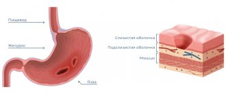
1.Structure of the esophagus
Esophagus
- a muscular tube connecting the throat (pharynx) to the stomach. The length of the esophagus is about 20 centimeters, and its inner surface is a pink mucous membrane. There is a part of the esophagus called the upper esophageal sphincter (UES). This is a group of muscles at the top of the esophagus that work during breathing, eating, belching and vomiting. A person can consciously control this muscle group. The upper esophageal sphincter muscle also holds food and secretions that may come from the windpipe.
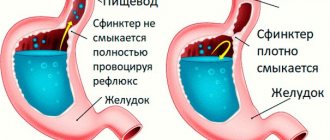
The lower esophageal sphincter (LES) is a group of muscles at the lower end of the esophagus where it connects to the stomach. When the lower esophageal sphincter is closed, it prevents acid and stomach contents from moving back out of the stomach. The muscles of the LES are not subject to conscious control.
Diseases of the esophagus
, in fact, not so little. Some of them are quite serious, some do not require complex and long-term treatment. Depending on the severity of the disease, it may be treated by gastroenterologists or thoracic surgeons, who perform operations on the esophagus when necessary for treatment.
A must read! Help with treatment and hospitalization!
Diagnostics
Diagnosis is carried out in order to differentiate esophagitis from diseases with similar symptoms, as well as to identify the cause of the development of the disease. The easiest way to make a diagnosis is for an acute disease: patients complain of specific pain. A burning sensation behind the sternum (heartburn) is a typical symptom of inflammation of the esophagus, which forces the patient to consult a gastroenterologist. The doctor conducts a survey, finding out the possible causes of the disease, examination, and then prescribes additional examinations.
As part of the diagnosis of esophagitis, the following methods are used:
- Endoscopic examination of the esophagus (esophagoscopy). The study makes it possible to determine the degree of damage to the mucosa and the location of defects.
- Biopsy. Biological material is collected during endoscopy. Histological examination allows us to determine the nature of changes in the mucosa, identify neoplastic processes, and eosinophilic esophagitis.
- To diagnose disorders of the motor function of the esophagus, esophagomanometry is prescribed.
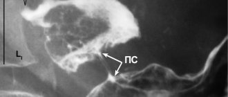
- X-ray examination of the esophagus. The study is used to identify stenosis, diverticula, and hiatal hernia.
- Daily pH-metry to identify reflux disease as a cause of esophagitis.
Based on the examination results, the attending physician chooses treatment tactics.
Pathophysiology
The content of the article
Under normal conditions (in a healthy person), the contents of the gastric juice with high acidity periodically enter the esophagus, but internal protective mechanisms either reduce the amount of acid secreted to a minimum or remove the acid, which is quickly removed by “cleansing” the esophagus. Therefore, symptoms of acid irritation of the esophagus are not felt or are minimal.
Mechanisms that protect the esophagus from stomach acid include the lower esophageal sphincter (sphincter) and normal esophageal motility (motility). When these mechanisms are disrupted, reflux occurs and symptoms of GERD occur.
Gastroesophageal reflux
Treatment
Treatment for esophagitis is determined by its cause and severity. If esophagitis is caused by a chemical or thermal burn, then hospitalization is required. The acute form of the disease is treated with medications, refusal of food for 1-2 days, and diet.
If a patient develops esophagitis of an infectious nature, then therapy aimed at destroying infectious agents is required: antibacterial, antifungal or antiviral drugs. For reflux esophagitis, antisecretory, antacid, and prokinetic drugs are prescribed.
In case of severe pain, painkillers are prescribed. If phlegmon or abscess develops, surgical treatment is required.
Therapy for chronic esophagitis involves eliminating the cause of its occurrence. The rest of the prescriptions are identical: diet, medication, giving up bad habits. Physiotherapeutic procedures are indicated for some patients.
The prognosis is favorable for mild to moderate esophagitis. The success of therapy largely depends on compliance with dietary and lifestyle recommendations. There is no specific prevention of the disease. Prevention of relapse after successful treatment means following the basic recommendations that the doctor gives after recovery.
In the multidisciplinary CELT clinic, you can undergo diagnostics and select effective treatment. Individual approach, the best gastroenterologists and modern treatment methods. Being healthy is easy under the supervision of qualified specialists at the CELT clinic.
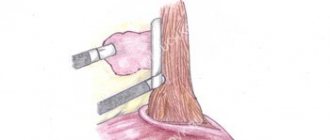
Author's technique of modified fundoplication
During the operation, I use a Toupet partial fundoplication of 270 degrees, in which a cuff is created from the stomach wall to prevent the reflux of stomach contents into the esophagus. Thanks to the improved technique, for which a patent has been received, the functioning of the sphincter between the stomach and esophagus remains undisturbed, while the gag reflex and belching - protective functions - are also preserved.
The surgical intervention is performed using laparoscopy, using high-quality endoscopic equipment, which allows all manipulations to be performed with maximum precision, without the risk of damaging the structures located in this area: vessels, fascial spaces, vagus nerve. During laparoscopic surgery, I create a functional valve between the stomach and esophagus, which allows the patient to do without medications in the future. Due to the fact that all manipulations are performed through 3-4 small incisions, an excellent cosmetic result is guaranteed, since after healing, the marks from the incisions will become almost invisible.
When isolating the stomach and esophagus, I use the LigaSure device for dosed electrothermal tissue ligation, thanks to which the vessels can be “sealed” without damaging the surrounding tissues. The use of the latest generation of absorbable suture material and anti-adhesion barriers allows the operation to be performed quickly and efficiently.
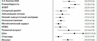
If there are several diseases that require surgical treatment, in our clinic the patient can count on a simultaneous operation. Instead of several interventions, the interval between which should be 5-6 weeks, the person being operated on during one anesthesia can immediately get rid of a number of diseases.
Patients are discharged, as a rule, on days 1-3, and after 2-3 weeks they return to their usual activities. For the first two months you will have to adhere to a strict diet; over the next six months, the diet will gradually expand. In the future, a person who has undergone treatment in our clinic will be able to do without medications and without following a strict diet. We managed to reduce the number of relapses during the first year to 2%; five years after the operation, this figure does not exceed 4%.
I have performed more than 2000 operations on the stomach and esophagus, the accumulated experience is summarized in three monographs and more than 50 scientific publications, which can be found in professional peer-reviewed publications published in our country and abroad.
Our services
The administration of CELT JSC regularly updates the price list posted on the clinic’s website. However, in order to avoid possible misunderstandings, we ask you to clarify the cost of services by phone: +7
| Service name | Price in rubles |
| Gastroscopy (videoesophagogastroduodenoscopy) | 6 000 |
| Colonoscopy (video colonoscopy) | 7 000 |
| Ultrasound of the abdominal organs (liver, gall bladder, pancreas, spleen) | 3 800 |
| Fluoroscopy and radiography of the stomach | 4 800 |
All services
Make an appointment through the application or by calling +7 +7 We work every day:
- Monday—Friday: 8.00—20.00
- Saturday: 8.00–18.00
- Sunday is a day off
The nearest metro and MCC stations to the clinic:
- Highway of Enthusiasts or Perovo
- Partisan
- Enthusiast Highway
Driving directions
Help with cramps
- To quickly alleviate a person’s condition if he has an attack, you need to put a nitroglycerin tablet under his tongue until it is completely absorbed.
- Try drinking a few sips of warm milk or chamomile infusion.
- Inject the drug atropine intramuscularly or intravenously, which also relieves spasms.
- Do a set of breathing exercises - inhale air, hold your breath, count to 5 and exhale slowly.
Clinical picture
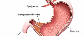
There are physiological narrowings in the esophagus, where malignant growth primarily begins. These narrowings are caused by passing near other anatomical formations - the aorta and the forks of the trachea into the bronchi; at the junction of the pharynx into the esophagus and the esophagus into the stomach there is also a slight narrowing. It is believed that the mucous membrane here is more susceptible to minor injuries from rough food, which means inflammation occurs more often. However, in the cervical region the incidence of cancer is 10%, in the lower third of the esophagus - 30%, and 60% of cancers are formed in the middle segment.
Malignant cells not only grow into the thickness of the organ, as happens with most cancers, they also migrate through small lymphatic vessels. The vessels form a complete lymphatic network inside the esophageal wall, spreading the tumor inside, so the length of the tumor can be 5, 10, or 15 centimeters.
In the advanced stage, localization determines the symptoms, and the first signs are considered to be the feeling of food sticking to the same place or scratching the mucous membrane with a piece of food. As it progresses, it becomes difficult to pass first solid pieces, then porridge, then liquid. All this is called dysphagia. First, the patient washes down pieces of food with water, pushing them through, but after that this no longer helps, nutrition is disrupted, and the person loses weight. Contact of the tumor with food leads to inflammation, an unpleasant putrefactive odor appears, and with regular trauma, the loose mucous membrane of the tumor begins to bleed, and life-threatening bleeding can develop.
Pain occurs as the esophagus contracts with peristaltic waves, the pain is spastic in nature. The growth of the tumor through the entire thickness of the esophageal wall makes the pain constant; it is localized between the shoulder blades. Infiltration of the mediastinal tissue by the tumor involves the recurrent nerve, which is responsible for the movement of the vocal cords, in the process, hoarseness and choking when drinking appear. The nerve can be compressed by lymph nodes enlarged by metastases and the sonority of the voice will disappear.
As with achalasia, a tumor forms an expansion above the narrowing of the esophagus, where food accumulates. Nighttime reflux of accumulated food masses into the windpipe can also lead to pneumonia. And during the day I am worried about severe weakness and fever. If the respiratory tract is involved in the process, an anastomosis may form between the esophagus and the trachea or large bronchi - a fistula, through which food crumbs will enter the breathing tube, causing coughing and pneumonia. If such a fistula opens from the esophagus into the mediastinum, its inflammation will lead to death.
Get a treatment program
Prokinetics
Prokinetics are only effective in treating mild forms of GERD. If GERD is more severe, in addition to prokinetics, drugs that inhibit the secretion of gastric juice are usually prescribed.
Of the prokinetic agents, metoclopramide (10 mg/day orally) is prescribed in European clinics - this is the most commonly prescribed regimen in adults with GERD. Long-term treatment with prokinetic drugs can be dangerous, with serious, even fatal, complications.
ONLINE REGISTRATION at the DIANA clinic
You can sign up by calling the toll-free phone number 8-800-707-15-60 or filling out the contact form. In this case, we will contact you ourselves.
Traditional medicine recipes
As an alternative, you can use unconventional means that can help solve the problem:
- A simple recipe is to take a teaspoon of flaxseed oil on an empty stomach. The procedures are carried out daily, the oil is heated to room temperature. It is better to drink it 20-30 minutes before meals.
- Prepare a decoction of motherwort and valerian root, mix the ingredients in equal proportions, pour boiling water and leave for at least 2 hours. You can supplement the decoction with hops. This medicine will have a good sedative effect.
- Mix flax and anise seeds in equal proportions, grind them in a coffee grinder or use a blender. Pour the mixture with honey, let it stand for 2 days, stirring occasionally. The product can be consumed daily, 1 teaspoon.
- Herbal baths have good results. The procedures are carried out at home, using various herbs: mint, lemon balm, fir needles, hops. Such baths have a sedative effect and are taken before bedtime. You can replace herbal decoctions with essential oils: lavender, ylang-ylang.
Antacids
Until the 1980s, antacids were the standard treatment for mild forms of GERD. They are still used to reduce symptoms of mild reflux. Antacids are taken after each meal and before bed.
Taking antacids before bed
Antacids are also useful for side effects: they relieve constipation (aluminum antacids: ALternaGEL, Amphojel), they can increase loose stools (magnesium antacids: Phillips Milk of Magnesia). Aluminum hydroxide increases the pH of the stomach contents to >4 and inhibits the proteolytic activity of pepsin, reducing the symptoms of indigestion. Antacids do not reduce the frequency of reflux, but they do reduce the acidity of the fluid.
Magnesium hydroxide suppresses the symptoms of acidosis and improves digestion. Magnesium hydroxide antacids osmotically retain fluids in the intestine, which distends the intestinal wall, stimulates intestinal motility, and softens stools (laxative effect). When interacting with hydrochloric acid of gastric juice, magnesium hydroxide is converted into magnesium chloride.
Prevention
You can prevent esophageal diseases if:
- maintain a proper diet;
- avoid overeating and periods of hunger;
- follow safety rules when working with toxic substances;
- expectant mothers should think more about the health of their offspring.
Signs of damage to the esophagus begin with minor symptoms, which later develop into severe forms of disorders. Therefore, you should not try to treat yourself. Early visit to the doctor helps to undergo a timely examination, avoid problems, and stop the disease with the help of diet.