Myopia is a visual impairment in which a person sees well up close, but at a distance is “blurred” or unclear. It is characterized by refractive error in one eye or both.
Today it is considered a disease of civilization. Which is not at all surprising. The rapid growth of computerization of life and technology increases exponentially the growth in the number of cases. It is noteworthy that it occurs most often between the ages of 7 and 18 years. At an older age, it either remains in place and rarely progresses.
The physical basis of the disease is as follows: the image of distant objects in myopic people is focused not on the retina, as in healthy people, but in front of it; a defocused image falls on the retina, so at a distance a person sees objects unclearly and blurry.
Myopia in children
Cases of congenital refractive error of the eye are quite rare and are caused by abnormalities in the development of the organ of vision. This is due to hereditary predisposition if both parents have the same diagnosis.
In children under one year of age, pathology rarely occurs. More often, deviations from the norm of a weak degree in both eyes are formed in school-age children due to increased strain on the eyes and non-compliance with the basic rules of visual hygiene. Long-term entertainment on the computer, smartphone, and watching television has a negative impact on the eyes.
A small child cannot complain about his eyes, since the anomaly does not cause pain. Therefore, parents should be attentive to the behavior of their baby, pay attention to the manifestation of symptoms: low head tilt when reading and drawing; squinting; inability to distinguish distant objects.
What causes myopia?
Myopia occurs when the eyeball is too long compared to the focusing power of the cornea and lens of the eye. This causes light rays to focus at a point in front of the retina, rather than directly on its surface.
Myopia can also be caused by the cornea and/or lens being too curved for the length of the eyeball. In some cases, myopia occurs due to a combination of these factors.
Myopia usually begins in childhood, and you may be at higher risk if your parents are nearsighted. In most cases, myopia stabilizes in early adulthood, but sometimes it continues to progress with age.
Disease “Myopia of both eyes”
Some people with myopia experience worsening twilight vision. In this case, myopia occurs in both eyes. A person has to constantly strain his eyes, which leads to muscular asthenopia - visual fatigue.
In this case, these symptoms are supplemented by the occurrence of pain in the eyes, the appearance of “ache” in the eye sockets, and severe headaches. Progressive myopia requires frequent replacement of glasses and contact lenses, since the previous ones no longer correspond to the degree of the disease and no longer correct vision.
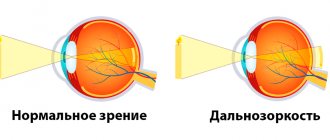
Differences between myopia and farsightedness
Given the above, it is easy to imagine the anatomical differences between two well-known refractive errors: nearsightedness (myopia) and farsightedness (hyperopia). In the first case, the focal point of the clearest image is in front of the retina, in the second - behind it, which causes blurred near vision (farsightedness).
In other words, in a farsighted eye, the distance between the cornea and the retina is not greater (as in myopia), but less than necessary. However, there is a second, not so common cause of hypermetropia: too weak, insufficient refractive characteristics of the cornea itself.
Reasons for the development of myopia
Among the reasons leading to the development of pathology, ophthalmologists identify the following:
- Heredity. If one or both parents have this diagnosis, then the likelihood of the disease manifesting itself in children is extremely high.
- Lack of correct writing and reading skills: long work without breaks, reducing the distance from the eyes to a book or notebook if the student is seated incorrectly, reading in a moving vehicle, lying down, illiterate lighting arrangement. Therefore, school myopia quickly develops and progresses.
- Complements the above unbalanced diet. A lack of vitamins such as copper, manganese, zinc, magnesium can lead to deviations from the norm.
- Lack of physical activity, sedentary lifestyle, prolonged sitting.
- Myopia can also be combined with other diseases of the visual organs, such as astigmatism, strabismus, and keratoconus.
- Hormonal imbalances.
- False myopia, caused by excess accommodation and muscle overload, gradually leads to the development of true myopia.
- Sometimes patients have a history of traumatic brain injury before the onset of myopia.
- Lack of competent diagnosis and correction, non-compliance with the rules of wearing lenses and glasses.
Children of myopic parents should be monitored by an ophthalmologist from birth. All other factors are provoking causes.
Laser vision correction
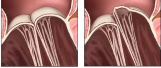
The surgical technology of photorefractive keratectomy (PRK) was the first method of laser correction of myopic vision. A layer of epithelium was removed from the cornea with a scalpel, then the necessary volumes of tissue were evaporated with a laser beam to give the cornea a more correct geometry. The technique involved continued wearing of contact lenses and was associated with a certain risk of complications - in particular, postoperative corneal astigmatism.
The laser epithelial keratomileusis technique (more often called by the English abbreviation LASEK) differs in that the epithelium is not completely removed; After ultra-precise laser action and giving the cornea an anatomically correct shape, the epithelial layer returns to its place. This technique results in faster healing and reduces pain or discomfort.
The modern methodological modification of LASIK (laser in situ keratomileusis) is that the epithelial layer remains in place - it is only incised with a microkeratome and folded back into a flap. After laser ablation (evaporation), the flap takes exactly the same position. No stitches are required, the incision is quickly sealed naturally, and pain is minimized. Wearing contact lenses is also not necessary. However, there are some contraindications to the use of LASIK, for example, an unacceptably thin cornea.
Finally, today’s newest and record-breaking fast method of laser vision correction is Femto- or IntraLASIK. The ablation effect is carried out within a vanishingly short time: the prefix “femto-” means 10-15 seconds. This technique is certainly the least traumatic and the least risky in terms of possible complications or side effects.
It should be noted here that the rate of complications is quite low with any method of laser vision correction.
The most well-known and studied side effects include:
- psychovisual discomfort caused by the “unusuality” of normal vision;
- excessive or insufficient correction, due, for example, to an error in precision calculations;
- corneal astigmatism;
- dryness of the outer membranes of the eye (observed in approximately 20% of cases);
- inflammatory processes as a consequence of an infection or exacerbation of a chronic infection (keratitis, conjunctivitis).
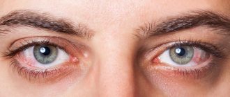
Symptoms of myopia
Most people do not immediately notice worsening vision. Therefore, they visit the ophthalmologist untimely and the disease is not detected at the very beginning. It is often discovered during preventive medical examinations. In most cases, visual acuity decreases gradually, and the person “gets used” to squinting.
The main symptom of the disease is difficulty in viewing distant objects. Over time, the following symptoms appear: constant squinting; headache; pain in the back of the head, temples, eyes; there is a desire to rub your eyes to see objects better; rapid eye fatigue; decreased twilight vision.
The appearance of any symptom requires immediate contact with a qualified specialist who will conduct a competent diagnosis and prescribe treatment.
Degrees and types of myopia
Ophthalmologists divide myopia into three degrees:
- 1 weak degree – visual impairment up to 3 diopters;
- 2 medium degree – from 3.25 to 6 diopters;
- 3 high degree – 6.25 and above diopters.
According to the clinical course, myopia can be of two types: progressive and stable .
With progressive myopia, it is necessary to increase the lenses to stronger ones once a year. In severe cases, myopia can progress throughout life and be complicated by more serious problems. Such myopia is called complicated or called myopic disease. If you notice symptoms of the disease in your child or yourself, contact your ophthalmologist immediately for advice.
What is the danger of the disease?
If left untreated, a high level of myopia can lead to the development of several dangerous ophthalmic pathologies:
- Cataract
. Patients whose treatment did not correct high levels of myopia have a 17% greater risk of developing cataracts than those in milder stages. - Glaucoma
. A systematic review of the researchers' available evidence found that people with high myopia are almost 50% more likely to develop glaucoma than those with low myopia. - Retinal detachment.
If myopia has reached the last degree, the patient experiences axial elongation of the eyes. This means that the retina is more stretched. Also, in people with myopia, the vitreous humor degenerates, which is more likely to collapse and separate from the retina. For this reason, there is a high risk of retinal tears, retinal detachment and degenerative changes. - Maculopathy
. The disease affects the central part of the retina (macula), which is responsible for the central vision of the eye. If myopia is pathological, myopic maculopathy develops. As a result, vision deteriorates and objects become blurrier.
To prevent a high degree of disease from leading to complications, regular examinations by an ophthalmologist and correction are necessary.
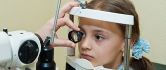
Diagnosis by symptoms
Only a qualified specialist - an ophthalmologist or ophthalmologist - can make a correct diagnosis of a patient. The main diagnosis is determined by testing visual acuity using test optical glasses and tables. The research methods of these doctors are as follows:
- Visometry - determination of visual acuity without and with correction;
- Perimetry – determination of visual fields;
- Tonometry - determination of intraocular pressure;
- Ophthalmoscopy – examination of the fundus of the eye;
- Skiascopy is a study of the refraction of the eye using a special ruler with lenses;
- Keratometry - determination of the optical power of the cornea using a device;
- Refractometry - determination of the clinical refraction of the eye using a special device (keratometry and refractometry are often combined);
- Biometrics – ultrasound or optical examination and measurement of the size of the anteroposterior segment;
You can only trust your health to a specialist - an ophthalmologist.
Diagnostics
Diagnosis of myopia begins with an examination in the office of an ophthalmologist. As part of the inspection, the following studies are carried out:
- Visometry (procedure for determining visual acuity using the Sivtsev table);
- Ophthalmoscopy (a method of examining the eyeball to identify retinal pathologies and various types of dystrophies);
- Biomicroscopy (detailed study of eye structures using a slit lamp);
- Control of intraocular pressure.
According to indications, an ultrasound examination of the eyes is performed to determine the anterior-posterior axis (length of the eye), B-scan to visualize the internal structures of the eye.
The diagnostic results allow the doctor to determine the form and extent of the disease, prescribe the necessary treatment, including assessing the need for laser coagulation of the retina, which poses a risk of developing retinal detachment. At the Federal Scientific and Clinical Center FMBA you can undergo a full range of diagnostic procedures in just one day!
Treatment of myopia
Confirmation of the diagnosis of myopia requires immediate initiation of treatment.
- Drug therapy. Effective with any degree of disease when carried out in courses. Pharmaceutical preparations are used: B vitamins; calcium; drugs that stimulate cerebral circulation, tissue therapy.
- Correction with contact lenses and glasses. The choice between contact lenses and glasses is up to the patient and depends on his personal preferences. The strength of the lenses is selected strictly individually. As myopia progresses, a good result is achieved by using specially designed contact lenses at night.
- Hardware therapy. It is carried out using a laser, accomodo trainer, color pulse treatment.
- Surgical techniques. When myopia develops at a rate of 1 diopter per year, scleroplasty surgery is used, the main task of which is to stop the progress of the pathological process.
- Laser correction. A modern procedure that effectively restores vision by changing the shape of the cornea.
Varieties
It is important not only the degree of the disease, but also the determination of its type. Based on the nature of its occurrence, myopia is divided into:
- to a landline. The identified pathology does not worsen over time;
- progressive. Vision is steadily declining. Every year it decreases by about one diopter;
- malignant. It is not so much a refractive error as a severe type of disease that quickly leads to disability. A person loses the ability to perform usual professional and everyday activities.
Diagnostics
Mild myopia may initially not manifest itself in any way and may not affect the quality of life. To determine the pathology and take timely corrective measures, you need to visit an ophthalmologist annually. The level of acute vision is checked using Sivtsev or Golovin tables. For young children, a specially designed Orlova table is sometimes used. Surveys are carried out using:
ophthalmoscopy. Allows you to evaluate the retina, optic nerve head, and fundus vessels. With the advent of retinophot, you can not only examine, but also get a high-quality image;- skiascopy. “Shadow test” occurs by illuminating the pupil with a beam of light reflected from the mirror. The position of the spatial optical phenomenon depends on the refraction of the eye being examined;
- refractometry. A non-invasive objective diagnostic method that allows you to determine abnormal changes in the refractive power of the eye, leading to various diseases;
- ophthalmometry. Determines the radius of curvature of the cornea, which is involved in ensuring visual acuity along with other elements of the optical system of the eye. It is carried out by three methods: manual, mechanical and using a computer;
- keratotopography. Allows you to study in detail the condition of the cornea of the eye. The result of the scan is a color map of its surface;
- Ultrasound of the eye, or ophthalmoechography. Allows a detailed study of the structures of the ocular apparatus. The technique is considered highly informative because it provides a detailed image of the orbit on a computer monitor.
Physical education for myopia
A diagnosis of myopia is not a reason to limit sports activities. Physical activity plays an important role in preventing the development of pathology. In addition, physical activity contributes to a general beneficial effect on the body, activates the functioning of organs, increases the performance of the ciliary muscle, and strengthens the scleral membrane.
Aerobic exercises: running, cycling, swimming, skiing have a beneficial effect on the accommodative function of the eye, as well as on the circulatory system as a whole. It is recommended to adhere to moderate intensity training, in which the heart rate does not exceed 140 beats per minute. More intense loads can negatively affect the performance of the ciliary muscle.
The permissible intensity of physical activity also depends on the stage of the disease.
For mild myopia, sports games with muscle stretching, such as basketball, tennis, and volleyball, are indicated. By changing the focus of the gaze from distant objects to near ones, an improvement in accommodation is achieved.
In case of moderate disease, moderate physical gymnastics and jumping from a height of more than 1.5 meters are prohibited.
For high myopia, only exercises from the group of special physical therapy are allowed.
Classification
According to the official classification, low myopia is divided into the same types and forms as high myopia. For reasons of occurrence it can be:
- false - caused by a spasm of the visual muscles responsible for accommodation, changes in the shape of the lens;
- true - caused by congenital or acquired changes in the size of the eyeball (axial myopia), thickening of the cornea or lens (refractive myopia).
It is believed that mild false myopia can be cured fairly quickly. To do this, doctors will have to remove the muscle spasm and prevent its reappearance. The causes of true myopia cannot be eliminated using conservative methods, so treatment requires more time and money.
Based on the nature of development, low myopia is divided into 4 types:
- Stationary. The mildest and most stable form, which practically does not progress, even if observed for years. Does not require serious therapy.
- Progressive. A more serious form of the disease, which is accompanied by a gradual decrease in vision up to 1 diopter per year.
- Transitional. A form that progresses when taking certain medications. In some cases, concomitant diseases can provoke a sharp deterioration in vision.
- Malignant. The most aggressive and rapidly progressing disease. In ophthalmology, cases have been recorded when vision deteriorated to 1 diopter per day, and within a month after its discovery the person received disability.
Twilight myopia has been classified as a separate subspecies. At any degree, myopia of this form manifests itself only in conditions of lack of light. During the day and in well-lit rooms, signs of visual impairment are not observed.
Gymnastics for the eyes
The initial stage of myopia can be easily eliminated by performing special therapeutic exercises for the eyes. The exercises should be performed only in a sitting position and preferably in the specified order.
Close your eyes tightly for five seconds, then open them for five seconds. Repeat the exercise 8-10 times.
Blink both eyes for two minutes: try to blink quickly, but without causing discomfort.
Close your eyelids. Use your fingertips to gently massage them, making circular movements.
Close three fingers tightly and press lightly on the upper eyelids with three, release after two seconds and remove your fingers from the eyelids. Repeat 4-5 times.
Use your index fingers to press the skin of your eyebrows, then slowly close your eyes. Hold the skin of the brow ridges firmly: this trains the muscles well. Repeat 10 times.
Complications
Let's sum it up
Myopia often begins to progress in childhood, so early prevention makes it possible to keep the development of the pathology under control. Preventive measures include regular eye exercises, proper organization of the educational process and space, a varied and nutrient-rich diet, physical activity, as well as regular visits to the ophthalmologist.
Non-surgical methods for correcting and stabilizing myopia play an important role in childhood and adolescence. The most important rule is that the correction corresponds to the true amount of myopia. Orthokeratology, or the use of special contact lenses at night, allows not only to correct vision, but also to control the progression of the process.
Laser vision correction, as the most common way to get rid of myopia, is performed when the form is stabilized and after the age of 18 years.
Prevention
Preventive measures for myopia consist of preventing further progression of the disease. This can be achieved through a set of measures:
- Dosing of visual load.
Ophthalmologists recommend giving your eyes rest every 45 minutes when watching TV, working at the computer or with documents. The duration of breaks should be at least 15 minutes for children and 5 minutes for adults. All this time, it is advisable to look at distant objects or close your eyes altogether. - Strengthening the eyes with gymnastics
. It is recommended to do a special set of exercises daily. The essence of gymnastics is to stimulate blood supply and engage the eye muscles, which remain static for a long time. This measure helps to curb the development of hereditary myopia. - Strengthening vision with nutrition and food supplements
. To maintain eye health, it is enough to include in your diet sources of antioxidants (vitamins A, C and E), anthocyanins (blue-colored vegetables and fruits) and lycopenes (red-colored vegetables and fruits). If it is difficult to meet the body’s needs with food, you can take vitamin and mineral complexes, but before using them it is advisable to consult a doctor.
Another measure that can preserve your vision is to regularly visit an ophthalmologist. Annual monitoring of visual acuity will help to identify myopia in time and take steps to eliminate it.