Hypermetropia - a visual impairment in which a person sees well in the distance, but poorly near. It is also called “weak refraction”, since the image is focused in the imaginary space behind the eyeball.
To correct vision, glasses or contact lenses with a plus sign are used. They are often also called “senile”, although hypermetropia also occurs in children, adolescents, and adults. At a young age, a small degree of this disorder is compensated by the reserves of the body, the eye itself. And often people with hypermetropia do not use glasses. However, over the years, often after 30, complaints arise about poor vision when reading and visual work at close range. Then distance vision begins to suffer.
Farsightedness in children
In all cases, hypermetropia is a congenital condition. A child at birth normally has hypermetropia, since his eyeball is still small. With age, the eye enlarges and focusing becomes normal. In some children, the eyeball “loses” its growth rate, and then farsightedness occurs as a persistent deviation from normal development. At school age, this deviation can be compensated by excessive stress of accommodation, and the patient can ignore glasses. However, the presence of strabismus or amblyopia may again require the use of glasses.
Causes of hypermetropia
Most often, the development of hypermetropia is caused by the irregular shape of the eyeball, which is reduced in its longitudinal size. All children are born farsighted. As babies grow and, accordingly, their eyeballs, vision function gradually returns to normal. By the age of four, children usually have good vision both far and near.
Hypermetropia is often observed in older people. This is due to the fact that with age, the lens of the eye loses its ability to change curvature, i.e. to accommodation.
This process begins at the age of 25, but leads to a deterioration in visual function only after a person reaches the age of 45–50 years. By the age of 60, the ability of the lens to accommodate is completely lost. In this case, they talk about senile farsightedness or presbyopia.
Symptoms of farsightedness
Contrary to the name “farsightedness,” good distance vision occurs only with a weak degree of hyperopia and in relatively young subjects: in other cases, vision is poor both near and far. There is also the concept of “hidden hypermetropia”. It is discovered by chance, during a routine examination when special drops are instilled that dilate the pupil.
In clinical manifestations, hypermetropia is characterized by often occurring strabismus, which is detected at an early age in a child and does not go unnoticed. But the most important problem is amblyopia or “lazy eye”, that is, low visual acuity that does not reach normal levels even with glasses!
The appearance of any symptom requires immediate contact with a qualified specialist who will conduct a competent diagnosis and prescribe treatment.
Prevention
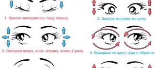
The principles of preventing further progression of farsightedness are as simple as they are effective. They do not require any special investment of time or financial expenses, but, at the same time, they really “work” to preserve vision. We are talking, first of all, about maintaining visual hygiene (lighting, distance, work-rest cycle), performing basic eye exercises and regular visits to an ophthalmologist, who will inform you in the most detailed manner about what measures are a priority in your particular case.
Treatment of farsightedness
Confirmation of the diagnosis of hypermetropia requires timely correction of this visual impairment.
- There is no drug therapy.
- Correction with contact lenses and glasses. The choice between contact lenses and glasses is up to the patient and depends on his personal preferences. The strength of the lenses is selected strictly individually. In some cases, it is possible to use nighttime orthokeratology lenses for a reversible correction effect during the day.
- Laser correction. A modern procedure that effectively restores vision by changing the shape of the cornea. Compared to a similar method for myopia, high efficiency applies only to hypermetropia up to +4.0 diopters. In all cases, before recommending surgery, it is necessary to conduct a specialized examination.
- In cases where laser correction is not indicated or is ineffective in the prognosis, surgical intervention inside the eye is recommended - implantation of an intraocular lens while preserving its own lens (phakic lens implantation) or implantation of a lens to replace the removed lens of the eye (phacoemulsification with implantation). The latter operation is performed when phakic implantation is contraindicated and/or in “aged” patients.
Symptoms and diagnosis of hypermetropia
With mild hypermetropia, people experience difficulty only when looking at nearby objects. Distance vision is usually preserved. Therefore, they only need glasses to work.
As the disease progresses, distance vision also begins to suffer. In this case, doctors prescribe glasses to their patients with farsightedness not only for work, but also for constant wear.
If hypermetropia is not corrected, frequent and persistent headaches occur, often associated with high visual loads. Many patients complain of increased eye fatigue when reading.
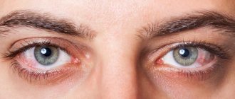
When working and reading, people with hypermetropia try to strain their vision. This leads to a feeling of discomfort and burning in the eyes. A person involuntarily begins to rub his eyes with his hands, thereby introducing various infections into them. As a result, he often develops various inflammatory diseases: barley, chalazion, blepharitis, conjunctivitis.
With hyperopia, patients tend to move their book further away while reading, thereby improving the visible image of the letters. However, in some cases, farsightedness does not manifest itself in any way and is diagnosed only by an ophthalmologist during an examination and determination of visual acuity.
Let's sum it up
Plus glasses today remain the most common method of vision correction for hypermetropia at any age.
Contact lenses allow you to increase the degree of freedom of a patient with clinical manifestations of hypermetropia.
The best way to relieve a patient of poor vision is only through refractive surgery and intraocular (intraocular) implantation. Operations are performed in adults, sometimes in adolescence.
If there are contraindications to laser correction, an alternative to surgery is night (orthokeratology) lenses, which reversibly eliminate hypermetropia.
It is recommended to monitor vision from an early age in order to identify hypermetropia and astigmatism; it is necessary to take measures in the form of glasses or contact lenses as early as possible.
Pathogenesis
In a young healthy adult, optimal visual acuity and the ability to see objects both at close and long distances is ensured by independent physiological correction - self-correction. Both eyes, due to natural accommodation (the ability to refract light rays for equally good viewing of objects at different distances), with tension in the ciliary muscle, increase the refractive optical power and allow images to be perceived correctly, that is, without blurriness and cloudiness.
Hypermetropia refers to a type of ametropia - a change in the refractive physiological ability of both human eyes, their refraction. Another visual impairment is astigmatism. But with astigmatism, the cornea and lens lose their natural shape and become curved. Astigmatism can affect 1 eye: left or right. With this pathology, images become distorted, partially blurred and unclear. With astigmatism, a person may also see objects close up incorrectly, but the picture will also be distorted. With hypermetropia, which some people confuse with astigmatism, objects are uniformly poorly viewed only at close distances.
According to statistics, weak, moderate or high complex degrees of hypermetropia occur in approximately 30-45% of the adult population of the planet over eighteen years of age. In children under 7-12 years of age, farsightedness is physiological in nature, therefore it is diagnosed in 85-90% of children under three years of age, as well as in approximately a third of adolescents 13-14 years old.
For your information!
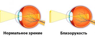
Another type of refractive error is myopia, which in ophthalmological practice is called myopia. With mild, moderate or high myopia, a child or adult is not able to fully see objects located at long distances. The focus is located in front of the retina due to a spasm of accommodation or elongation of the eyeball.
The development of weak, moderate or high degrees of hypermetropia is based on two mechanisms:
- Changes in the size of the eyeball - its longitudinal, that is, anterior-posterior axis. When the eye shortens, its cornea becomes more convex, which is why the focal point is localized not on the surface of the retina, but behind it. But at the same time, accommodation and functioning of the refractive apparatus of the eye remain absolutely normal.
- Insufficiency of the functions of the ocular refractive apparatus. With this mechanism for the occurrence of hypermetropia, a refractive type of farsightedness develops. The dimensions of both eyes are physiologically correct, the anterior-posterior axes remain normal. But the refractive apparatus with its main ciliary muscle, which ensures correct accommodation, weakens and loses the ability to fully and sufficiently refract and focus light rays emanating from objects. As a result, the focus is also located not on the retina itself, but in its posterior plane. Objects also appear blurry and indistinct.
The first mechanism, as a rule, is characteristic of children and is physiological, that is, normal and natural. The vast majority of infants (about 90%) have congenital hypermetropia, but as the eyeball grows, its anterior-posterior axis increases and natural vision correction occurs.
The second mechanism of weak, moderate or high hyperopia is associated either with weakening of the muscle fibers of accommodation (ciliary muscle), or with reduced elasticity of the tissues of the natural lens of the lens. In this case, hypermetropia is caused by natural and inevitable age-related aging processes, and therefore is usually found in adults and older people.
Classification
Hypermetropia has several classifications.
So, according to the mechanism of development, farsightedness is distinguished:
- Axial (axial), caused by the shortened anterior-posterior axis of the human eyeball.
- Refractive, associated with a weakening of the functions of the optical apparatus, namely with a decrease in its refractive physiological ability.
- Hypermetropia is also classified according to the functioning of the optical eye apparatus into:
- Hidden. With this form, the refractive error can be hidden and compensated due to the tension of the ciliary muscle. With this type, unlike the obvious one, accommodation is enhanced, which allows you to see objects normally even with a shortened eyeball. But, unfortunately, the human eye is not always able to hide a refractive error.
- Explicit. The obvious form is characterized by the inability to hide the defect, that is, to carry out self-correction due to increased work and increased tension of the muscles of accommodation. But with age, the hidden form often turns into an obvious one, as the muscle fibers lose their elasticity and they no longer manage to remain in a tense state in order to hide and compensate for the refractive error.
Classification of hypermetropia according to the age of patients assumes two forms:
- Congenital. The infancy period in most cases is accompanied by natural physiological hypermetropia from +2 to +4 diopters, which determines the maturity of the fetus and is determined by the small longitudinal dimensions of both eyes. The axis length is about 16-17 millimeters, and farsightedness has an average degree of 4 diopters. But then, as a healthy child grows older, the eyeball grows, its anterior-posterior axis lengthens and reaches 24-25 millimeters by the age of 12. The refraction becomes proportional, called emmetropia. As visual overgrowth progresses and continues, both eyes lengthen, resulting in myopia. And with developmental delay, hypermetropia persists. But many adults manage to hide their weakened refraction through constant tension of the accommodating muscle until about 35 or 40 years of age. The lens is held in a convex state by muscle fibers and normally refracts light rays, but then the optical apparatus of the eye weakens due to age-related changes, and hyperopia progresses.
- Acquired or age-related, which in ophthalmology has a separate name - presbyopia. This type of disease occurs in many people, since even a healthy and well-seeing adult, due to age-related changes that weaken the ciliary muscle and affect the structure of the lens, is deprived of the opportunity to compensate for weak refraction through self-correction of vision.
Diagnostic measures
Get a complete vision examination at the Lege Artis Eye Clinic
It's time to correct your vision!
Make an appointment by phone:
8(804) 333-02-14 Free call
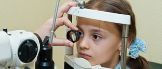
Diagnosis of hypermetropia in children is carried out by an ophthalmologist. Visual acuity testing is carried out using different methods, depending on the age of the child. Also, before the study begins, drops are instilled into the eye to dilate the pupil. This leads to relaxation of the ciliary muscle and allows you to correctly measure the light refractive ability of the eye. For timely detection of farsightedness, it is recommended to visit a pediatric ophthalmologist at least once a year.
Diagnosis consists of collecting an anamnesis, parents provide complete information about the child’s behavior and present complaints: a child with problems with visual perception quickly gets tired, becomes withdrawn, has a predominant bad mood, school-age children are characterized by slow reading, but bright pictures at a distance (billboards, posters) they look at with great interest.
To identify farsightedness, different diagnostic methods are used:
- visometry - tables with pictures are used for children, tables with letters are used for middle school children;
- determination of violations of the refractive power of the eye is carried out using autorefractometry, necessarily in conditions of cycloplegia: drug dilation of the pupil and switching off accommodation;
- skiascopy and retinoscopy.
Forecasts
If the disease is detected in a timely manner, and after diagnosis, treatment is prescribed, but the progression of the disorders can be stopped, especially when the hypermetropia indicator is weak - no more than three to four diopters.
If vision is not corrected, it will continue to be impaired, and hypermetropia will progress. In this case, a complication may develop, for example, glaucoma, chronic recurrent blepharitis, conjunctivitis or keratitis, strabismus (left, right or both eyes at once) or amblyopia.
Diagnostic methods
Diagnostics is very important, because the success of hypermetropia correction and the risks of complications depend on its timeliness and reliability. The diagnosis is made by an ophthalmologist, who should be contacted at the first signs of disturbances. But you should regularly visit an ophthalmologist in any case, even with seemingly normal visual acuity. Sometimes deviations are discovered by chance during routine examinations.
Diagnostics includes the following procedures:
- Visometry. This diagnostic method involves checking visual acuity using special tables. Both visual organs are assessed, but one at a time: one eye is closed when checking the second left or right.
- Refractometry. This is a computer diagnostic method that helps examine refraction and accurately measure sharpness in diopters.
- Ultrasound of the eyeball. The left, right, or both eyes are subjected to ultrasound diagnostics to determine the exact dimensions of the axis.
- Skiascopy. This diagnosis is carried out to assess weak, medium or high refraction when observing the movements of shadows.
- Ophthalmoscopy involves examining the fundus of the eye. A special fundus lens or ophthalmoscope device is used.
- Gonioscopy is prescribed to people over 35 to analyze the condition and structure of the anterior eye chamber.
- Biomicroscopy is the examination of tissues under a microscope equipped with a slit lamp. The retina and the natural lens, the crystalline lens, change pathologically.
- Tonometry is performed to detect intraocular pressure.
- Perimetry is prescribed to determine the visual fields of both eyes.
An examination is necessary when a person notices the first obvious symptom and his vision is impaired. The weak type requires a thorough examination of the ocular structures, and with moderate or high age-related or acquired hypermetropia, making a diagnosis is usually less difficult.