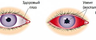
In every person, the larynx is located at the level of the fourth and sixth cervical vertebrae. The paths of the respiratory and digestive systems converge at this point. The larynx borders the pharynx on top, the esophagus on the side, and the trachea on the bottom, forming a kind of “crossroads.”
The larynx consists of several elements:
- Hyoid bone and muscles;
- The lateral part adjacent to the thyroid gland;
- Various paired and unpaired cartilages connected by ligaments, muscles and joints;
- The mucous membrane inside the organ, it is often the target of damage for viruses and other various pathogens (bacteria, fungi);
- Lymphoid tissues that form the laryngeal tonsils.
Each breath, the amount of air necessary for the body passes through the larynx, which then enters the trachea, bronchi and lungs. In addition to participating in the respiratory process, the human voice apparatus is contained in the larynx1.
What is laryngeal stenosis
Forms of stenosis
Taking into account the reasons that provoked laryngeal stenosis, the following forms are distinguished:
- Paralytic (results from a disturbance in the conduction of nerve impulses (innervation), for example, if the nerve supplying the larynx is compressed by a tumor of a neighboring organ).
- Scar. There are three types: - post-infectious (occurs after an infection, for example, inflammation of the middle ear, pneumonia); — post-traumatic (consequence of injury to the larynx, surgical operations); - post-intubation (diagnosed due to prolonged incubation during resuscitation measures (during artificial ventilation of the lungs, a special tube is inserted into the lumen of the trachea).
- Tumor (a malignant or benign tumor forms in the larynx).
Based on the criterion of localization and prevalence of the pathological process leading to insufficient ventilation of the lungs, doctors distinguish stenosis:
- subvocal space;
- glottis;
- extended (narrowing also affects the trachea);
- anterior (the anterior wall of the larynx narrows);
- posterior (the posterior wall of the larynx narrows);
- total (all departments are involved in the pathological process);
- circular (stenosis is the result of circular compression of a section of the tube).
Etiology, prevalence, pathogenesis
Post-intubation injuries of the trachea are understood as a violation of the anatomical integrity or functional state of the trachea that occurs after intubation. Military conflicts, urbanization, the increase in road traffic injuries, natural and industrial disasters increase the number of people in need of resuscitation measures. As a result, the number of patients with concomitant trauma who require long-term artificial pulmonary ventilation (ALV) and tracheostomy increases annually by 3-5% [37, 52, 68]. The need for long-term mechanical ventilation also arises after surgical operations in patients with diseases of the heart, brain, and abdominal organs [23, 26, 36]. The pathological condition of the trachea, which develops during prolonged intubation through the larynx or tracheostomy, is proposed to be called post-intubation disease [2, 36]. According to the literature, the frequency of injuries to the larynx and trachea accompanying long-term mechanical ventilation ranges from 14 to 80% [42, 44, 57]. The main ones are erosions and ulcers (50-80% of observations), granulations (up to 30%) and cicatricial stenosis [8, 17, 19, 39, 41]. The frequency of the latter varies very widely - from 0.2 to 25% [5, 9, 29, 33]. Such a serious complication as tracheoesophageal fistula develops in 0.5-5.4% of patients [7, 21, 35]. The main cause of damage to the larynx and trachea during intubation is mechanical impact on its wall. The literature considers two leading mechanisms of such damage. The first includes direct mechanical trauma to the larynx and trachea, caused by rough and repeated attempts at intubation, the use of rigid conductors, mismatch of the size of the endotracheal tube, its displacement inside the lumen of the trachea during coughing, barotrauma during jet ventilation, as well as significant overinflation of the sealing cuff of the endotracheal tube. In this case, the mucous membrane of the larynx and trachea often suffers, which leads to the development of fibrinous granulation laryngotracheitis. In rare cases, tracheal perforation or its complete rupture occurs with the development of mediastinitis [3, 30, 59, 61, 64, 66]. Perforation usually occurs in the membranous part of both the cervical and intrathoracic trachea. Longitudinal tears ranging from 2 to 13 cm in length are more often observed [26]. According to a number of authors [58, 63], the incidence of tracheal rupture during intubation ranges from 0.05 to 0.19%.
Another mechanism of damage to the larynx and trachea is impaired blood circulation in the mucosa associated with prolonged compression in the area of fixation of the inflatable cuff of the endotracheal and tracheostomy tubes, as well as along the edges of the tracheotomy wound. If the pressure on the tissue exceeds 25-30 mmHg, ischemia occurs in the mucous membrane and cartilage of the larynx and trachea, followed by inflammation and necrosis [2, 5, 19, 23, 26, 36, 44]. In the future, this can lead to their cicatricial stenosis.
An important factor causing damage to the tracheal mucosa with the formation of its scar deformation is the reflux of gastric and duodenal contents into the pharynx, followed by aspiration. More often, this complication occurs in patients with severe traumatic brain injury, accompanied by damage to the brain stem, central and peripheral respiratory disorders. In 75% of such patients, a significant decrease in cough and swallowing reflexes is determined, up to their complete loss, which leads to overflow of the airways with products of aspiration and bronchial secretion [15, 19, 45, 60, 62]. Aggressive contents can quickly cause inflammation of the intact mucous membrane of the larynx and trachea, aggravate damage to their walls in the area where the endotracheal tube is located with the addition of a productive component in the form of growth of granulations into the lumen of the organ and the development of chondroperichondritis [21, 62].
Great importance in the development of post-intubation complications is attached to bacterial inflammation, the rate of occurrence of which depends on the type of microorganism and the contamination of the larynx and trachea [44, 56]. On days 2-6 of mechanical ventilation, 36-40% of patients develop nosocomial pneumonia, and 6% develop pulmonary atelectasis. Bilateral endobronchitis of II-III degree is diagnosed when the duration of mechanical ventilation is more than 7 days in all patients [19]. The experiment found that acute bacterial inflammation of the mucous membrane, cartilage of the larynx and trachea provokes autoimmune aggression against these tissues, which leads to chronic inflammation and scar-fibrous deformation [1, 23, 34, 55]. M.A. Shuster and E.K. Onufriev [53] established that the autoimmune process is a delayed-type reaction to collagen localized in cartilage.
Considering that the long stay of the endotracheal tube leads to the obligatory development of chondroperichondritis of the larynx with the subsequent formation of cicatricial stenosis, patients in intensive care units and in need of respiratory support, especially if its duration is unpredictable, a tracheostomy is applied on the 3-5th day from the moment of intubation [11, 38-41].
Endoscopic diagnostics
Trophic damage to the wall of the larynx and trachea is determined by the duration of exposure to the endotracheal and tracheostomy tubes. The range of their visual manifestations ranges from minimal changes in the form of edema and hyperemia of the mucous membrane to maximum changes - the formation of a tracheoesophageal fistula [7, 8, 17, 35].
Most often, changes in the larynx and trachea during naso- or orotracheal intubation develop at the level of the vocal and subvocal parts of the larynx, in the cervical trachea at the level of fixation of the inflatable cuff of the endotracheal tube, as well as in the area of the distal end of the latter [3, 16, 18, 29, 54].
In the first 3 days of orotracheal intubation, catarrhal and erosive lesions of the mucous membrane of the anterior and lateral walls of the larynx and trachea predominate. Less commonly, the damage is circular in nature [8, 17, 23]. In 62% of patients, catarrhal and erosive laryngitis is detected [19]. Unilateral and bilateral paresis with degree II-III laryngeal stenosis were diagnosed in 23% of patients [41]. It is believed that laryngeal paresis is caused by swelling of the mucous membrane, prolonged limitation of mobility of the laryngeal joints due to the installed endotracheal tube, as well as its exclusion from respiratory and vocal function [8].
V.E. Kokorina [21] found that most often during orotracheal intubation the posterior wall of the larynx at the level of the tube cuff is affected - in 80.2% of patients on the 3rd day of mechanical ventilation. The author explains this localization by compression of this zone between the inflated cuff and the gastric tube and the development of ischemia. With a decrease in pressure in the cuff of the endotracheal tube, 28.8% of them showed epithelization of acute erosions within 24 hours.
On the 3-5th day of orotracheal intubation, 11-17% of patients develop ulcerations of the posterior part of the vocal folds, interarytenoid region and areas of the trachea in the area where the endotracheal tube cuff is located [19]. During these periods of mechanical ventilation, unilateral and bilateral paresis with laryngeal stenosis of II-III degree were detected in 31% of patients, proliferation of granulations in the subglottic cavity of the larynx - in 10% and in the cervical trachea - in 8% of patients [41].
When the duration of mechanical ventilation was from 6 to 10 days, post-intubation laryngitis was found in all cases, unilateral and bilateral paresis with laryngeal stenosis of II-III degree - in 46% of patients, proliferation of granulations in the subglottic region of the larynx - in 7% and in the cervical trachea - in 10% of patients [23, 41].
Orotracheal intubation lasting more than 10 days is accompanied by post-intubation laryngitis in all patients, unilateral and bilateral paresis with laryngeal stenosis of II-III degree - in 63%, proliferation of granulations in the subglottic region of the larynx - in 8%, in the cervical trachea - in 14% and in the thoracic trachea - in 4% of patients [23, 41].
After tracheostomy, damage to the trachea mainly develops along the upper edge of the tracheostomy and at two levels of the thoracic trachea - at the site of fixation of the inflatable cuff of the tracheostomy tube and along the lower edge of its position. Subsequent tracheal stenoses are localized there [10, 12, 16, 18]. Damage to the mucosa distal to the tube, as a rule, is a consequence of traumatization by the catheter during sanitation of the trachea and bronchi [8].
In the first 3 days after tracheostomy, catarrhal and erosive lesions of the tracheal mucosa predominate [8, 21].
On days 3-7 after tracheostomy, tracheal ulcers are found in 16.1% of patients. Superficial, round-shaped ulcerative defects with a diameter of 0.5 to 1.5 cm are detected along the side walls of the trachea at the level of the distal end of the tracheostomy tube. Their bottom is represented by a loose, dirty gray coating of fibrin. Tracheal ulcers, in contrast to laryngeal ulcers, are characterized by greater depth and size, progressive course and rapid development of granulations [2, 36].
Epithelization of these ulcers in most patients was observed within 28 to 42 days after tracheostomy. In a number of observations, hypergranulations were detected [36]. The growth of granulations was observed, as a rule, in the area of fixation of the inflatable cuff of the tracheostomy tube [41].
In the long term after prolonged tracheal intubation, 0.2-25% of patients develop cicatricial stenosis of the larynx and trachea [5, 9, 29, 33]. The location of the stenosis depends on the type of intubation, and the degree depends on its duration. During orotracheal intubation, the level of stenosis extends from the vestibular larynx to the cervical trachea. During intratracheal intubation (after tracheostomy), the cervical and thoracic parts of the trachea are affected. In 43-80% of patients, combined stenosis is detected [41, 43].
In some patients, intraepithelial granulomas of the larynx and trachea are detected within 14 days to 2 years from the date of intubation. During endoscopic examination, granulomas look like smooth, brightly hyperemic tumor-like formations with a poorly defined stalk, ranging in size from 0.5 to 1.5 cm [2, 19, 36].
Morphology
A morphological study of fragments of the tracheal wall obtained during the application of a tracheostomy revealed dystrophic changes already by the 1st-2nd day of mechanical ventilation. They were characterized by poor staining of the ground substance, impregnation of it with plasma proteins, and pyknosis of chondrocyte nuclei [23, 41].
By the 3rd day of mechanical ventilation, perichondrial detachment, usuration of the surface of the cartilage and fibrinous deposits on it were detected. Cartilage cells in the cartilage disappeared [19, 43]. Thus, already 3-day intubation caused pathological changes not only in the mucous membrane of the larynx and trachea, but also destructive and dystrophic changes in the tracheal cartilage.
By days 4-7 of mechanical ventilation, dystrophic and destructive changes in the tracheal cartilage progressed. There was no perichondrium in them, the surface of the cartilage was urinated, and foci of necrosis were identified. In some patients, replacement of cartilaginous tissue with granulation tissue was noted. Focal accumulation of leukocytes and hemorrhages were detected in the peritracheal connective tissue [19, 23, 41, 43].
With mechanical ventilation for more than 7 days, along with the death of cartilage and its replacement with granulation tissue, regeneration processes were detected, which was manifested by focal proliferation of chondrocytes, thickening and fibrosis of the perichondrium, as well as sequestration of dead cartilage [23, 43].
The results of histological examination of biopsy material from the tracheal mucosa obtained during bronchoscopy generally correlated with the data from studies of fragments of the tracheal wall taken during tracheostomy. In the bottom of tracheal ulcers that developed on days 3-7 after orotracheal intubation, necrotic masses and destructively altered leukocytes were determined, and in their edges - the presence of granulation tissue with leukocyte infiltration and detritus on the surface. When the defect existed for up to 21-35 days, 21.4% of patients developed squamous metaplasia of the epithelium, destruction or focal ossification of cartilaginous structures [36].
Morphological studies of biopsy material from the area of granulation stenosis on days 21–49 after orotracheal intubation or tracheostomy revealed granulation tissue with a vascular-poor stroma, consisting of macrophages and neutrophilic granulocytes, sometimes with areas of necrosis [2].
With the development of cicatricial stenosis in the period from 3 months to 6.5 years after long-term mechanical ventilation, excessive growth of connective tissue with lympholeukocyte infiltration around the glands in the submucosal layer was detected, and massive growths of coarse fibrous connective tissue were detected near destructively altered cartilaginous half-rings [2, 36].
Histological confirmation of the diagnosis of intraepithelial granuloma was the presence in the tissue of the formation of inflammatory sclerotic changes with neutrophilic leukocytes, macrophages and fibroblastic elements [36].
Thus, endoscopic and morphological studies have shown that prolonged tracheal intubation in combination with artificial ventilation initiates the formation of ulcerative tracheitis already on days 3-7. The existence of an ulcer for 2 months leads to excessive growth of granulations, destruction of cartilaginous structures and the formation of connective tissue in the submucosal layer and in the area of pathologically altered cartilage. The course of chronic inflammation and continuous stimulation of fibrogenesis for 3 months to 2 years lead to the formation of a stenosing tracheal scar.
Based on endoscopic and morphological studies D.V. Trishkin (2007) identified 4 clinical and pathogenetic variants of post-intubation tracheal disease: ulcerative tracheitis (4.6±3.2 days), granulation tracheal stenosis (36±9 days), cicatricial process in the trachea, presented in the form of inflammatory pseudotumors (9, 2±6.6 months) and cicatricial stenosis (4.9±3.6 months) [2, 36].
Endoscopic prevention and treatment
The indication for surgical treatment of stenoses that have developed as a result of complications of long-term mechanical ventilation is a violation of tracheal patency. The incidence of postoperative complications ranges from 6.2 to 28%; postoperative mortality, according to some data, can reach 10% [6, 43]. In this regard, more and more attention is currently being paid to the prevention of post-intubation complications. It primarily includes the treatment of early manifestations of post-intubation disease - erosions and ulcers.
One of the accessible and effective methods for preventing post-intubation injuries of the larynx and trachea is adequate sanitation of the trachea and bronchi. In this case, antiseptic substances and antibiotics are used, which are injected directly into the lumen of the trachea and bronchi [13, 24]. As an antiseptic, a 0.5-1% solution of dioxidine in a 2% sodium bicarbonate solution or a 0.01% solution of miramistin is often used [8, 17, 25]. If the patient has a large amount of mucopurulent secretion, 1-2 ml of a mucolytic (ambroxol, acetylcysteine, mistabron) is added to the sanitizing solution. If the patient is prone to bronchospasm, 5 ml of a 2.4% aminophylline solution is added to the injected solutions [26, 51]. However, this method has a number of disadvantages. Firstly, the method of introducing sanitizing solutions remains the same - through the lumen of the endotracheal and tracheostomy tubes, which eliminates the sanitization of the “dead” space formed by the glottis cranially, the tracheal cuff caudally, the walls of the trachea and the walls of the tube enclosed in it laterally [26]. Secondly, sanitation of the trachea and bronchi with an antiseptic does not affect the repair of ulcers and erosions, but only allows the removal of purulent secretions.
Among other therapeutic endoscopic methods used for erosive and ulcerative tracheobronchitis, laser photostimulation is the best known.
The use of a helium-neon laser during therapeutic bronchoscopy is described in the publications of E.V. Klimanskaya et al. (1988-1990), S.I. Ovcharenko et al. (1990) and V.X. Sosyury (1991) [51]. The authors noted good results in patients with chronic bronchitis and pulmonary suppuration due to improved sputum production, increased concentration of antibiotics in the bronchi and improved local immune defense of the respiratory tract. A good result of the endobronchial use of a low-intensity helium-neon laser in patients with erosive-ulcerative tracheobronchitis was obtained by N.E. Chernekhovskaya [46, 49, 50].
The effect of laser radiation is manifested by a multi-level effect on the body. There are changes in the rate of synthesis of protein, RNA, DNA, collagen, changes in oxygen balance and the activity of redox processes, changes in the membrane potential of the cell and the pH of the intercellular fluid, and improvement in microcirculation. The result of this is the formation at the body level of complex adaptive neuroreflex and neurohumoral reactions with activation of the immune system [20, 51, 65]. The therapeutic effect of helium-neon laser radiation is manifested by stimulation of epithelization of the mucosa in erosive and ulcerative injuries, normalization of lipid peroxidation processes, ensuring a physiological regime of tissue regeneration, limiting scarring processes and to a certain extent prevents the repeated excessive formation of fibrous tissue at the site of scar dissection [14, 22 , 27, 31, 46].
A fairly new method of treating erosive and ulcerative tracheitis is the endobronchial use of nitric oxide supplied from the Plazon apparatus through the endoscope channel [40, 61]. It has been established experimentally and clinically that the use of NO therapy leads to a significant acceleration of wound healing [4, 32, 67]. Nitric oxide activates the function of macrophages and fibroblasts and stimulates tissue regeneration [28, 47].
NOT. Chernekhovskoy et al. The possibility of re-epithelialization of tracheal ulcers under ozone therapy conditions was investigated [48]. In the experiment, the action of ozone caused acceleration of re-epithelialization and proliferation of the population of basal and immunocompetent cells, which in turn contributed to more rapid closure of the mucosal defect. The result of clinical trials was a decrease in edema, mucosal hyperemia and epithelization of ulcers in all patients [46, 48, 49]. An ozonated isotonic sodium chloride solution with an ozone concentration of 5 mg/l is injected under the mucosa into the edges of bronchial ulcers and additionally into the lumen of the trachea and damaged bronchus in an amount of 60-80 ml. It is also recommended to apply Ozonide oil to ulcers of the trachea and bronchi [46, 49].
In case of severe inflammation in the bronchi, intrabronchial immunotherapy with the immunomodulator T-activin is used in combination with the endolymphatic administration of antibiotics into the interbronchial spurs [46, 49-51].
Thus, at present, despite the improvement of endotracheal tubes, the frequency of post-intubation trophic complications in patients requiring long-term mechanical ventilation is increasing. Since these injuries are localized mainly at the level of the larynx, tracheostomy and tube cuff, it is impossible to detect them in a timely manner during bronchoscopy performed through the lumen of the tube. At the same time, there is successful clinical experience of local endoscopic treatment of purulent and erosive-ulcerative tracheobronchitis, which can be used in the early stages of trophic injuries of the larynx and trachea in patients undergoing long-term mechanical ventilation. In this regard, the task of improving endoscopic techniques for examining and treating patients with prolonged tracheal intubation in order to prevent the development of laryngotracheal strictures and tracheoesophageal fistulas becomes urgent.
Causes of laryngeal stenosis in children and adults
The main cause of laryngeal stenosis is resuscitation measures (artificial ventilation). The disease can also be caused by:
- traumatic brain injury (the innervation of the muscles of the larynx is disrupted, their paralysis occurs, causing the occurrence of stenosis);
- foreign bodies entering the larynx;
- performing surgery on the larynx or its injury;
- tumor of the thyroid gland or mediastinum;
- diseases of the upper respiratory tract occurring with complications (purulent inflammation of the throat, sore throat);
- allergic reactions;
- thermal/chemical burn of the larynx (ingestion of acids into the respiratory tract, inhalation of caustic smoke);
- presence of neoplasms;
- radiation therapy;
- scars of the larynx.
Also, laryngeal stenosis in adults can cause complications of infectious diseases such as syphilis and diphtheria.
Causes of laryngeal stenosis in children
In children, the development of stenosis is facilitated by the anatomical and physiological characteristics of the larynx, so symptoms of the disease can appear with allergies or a common cold. So, they have:
- the larynx has a funnel-shaped shape (in adults it is cylindrical);
- the narrowest point of the tube is the subglottic space, limited by the cricoid cartilage;
- the subglottic space contains a large number of glands responsible for the production of mucus (they become less and less over the years);
- in the area of the cricoid cartilage, submucosal tissue is well defined;
- the mucous membrane in the subglottic space is covered with cylindrical epithelium, when the cells of which are exfoliated, a significant area of the reflexogenic zone is exposed.
Causes and course of the disease
The causes of acute laryngeal stenosis can be both general and local in nature, and they are very diverse and numerous.
These include: Local inflammatory diseases - false croup, acute catarrhal laryngitis, acute phlegmonous laryngitis, laryngeal tonsillitis, laryngeal abscess, laryngeal diphtheria;
Non-inflammatory processes in the larynx - foreign bodies of the hypopharynx, congenital defects in the development of the larynx, gunshot wounds of the larynx, as well as mechanical injuries to the larynx, thermal and chemical injuries to the larynx received both at home and at work;
- Infectious diseases of an acute nature - scarlet fever, diphtheria, typhoid, measles, etc.;
- General diseases - pneumonia, heart and kidney disease, syphilis, tuberculosis, allergies to medications, etc.;
- Laryngeal nerve palsy;
- Retropharyngeal abscess and pathological processes in nearby areas;
- Neoplasms in the esophagus and mediastinum;
- Hypertrophic changes in the thyroid gland, which is sometimes called a goiter.
The early stages of development of acute laryngeal stenosis are characterized by the occurrence of hypoxia (acute oxygen starvation) and hypercapnia (carbon dioxide poisoning). Excessive levels of carbon dioxide in the human body leads to excitation in the respiratory and vasomotor centers. Then this causes excessive activity of the inhibitory fibers of the vagus nerve, a drop in pressure and a decrease in heart rate.
Friends! Timely and correct treatment will ensure you a speedy recovery!
Symptoms of laryngeal stenosis
There are four degrees (stages) of laryngeal stenosis:
- Compensated.
- Subcompensated.
- Decompensated.
- Asphyxia.
Each of them has its own symptoms.
At the first (I) stage:
- breathing becomes rare and deep;
- short pauses are heard between inhalation and exhalation;
- heartbeat slows down;
- shortness of breath occurs when walking (or even at rest).
At the second (II) stage, the following symptoms of stenosis appear in adults and children:
- difficulty breathing;
- even at rest, breathing remains wheezing;
- the skin turns pale;
- blood pressure rises;
- auxiliary muscles begin to take part in breathing (the supraclavicular fossa and intercostal spaces are retracted during inspiration);
- shortness of breath does not go away with rest.
Laryngeal stenosis of the third (III) degree in children and adults manifests itself:
- noisy whistling and frequent shallow breathing;
- rapid heartbeat;
- increased sweating;
- decreased blood pressure;
- persistent shortness of breath.
The patient is in a forced position - sitting with his head thrown back. His skin takes on a pale bluish tint (only his cheeks may remain red).
The fourth (IV) stage of stenosis (asphyxia) is the most dangerous. The patient has:
- intermittent breathing or its complete stop;
- increased heart rate (may not be palpable);
- involuntary urination/defecation;
- convulsions;
- loss of consciousness;
- heart failure.
Sometimes in the fourth stage of laryngeal stenosis it may seem that the patient’s condition is improving. So, shortness of breath may decrease, body temperature normalizes. However, this is a misleading impression. Hypoxia in this situation reaches its extreme values, and combined acidosis develops.
If you notice similar symptoms, consult a doctor immediately. It is easier to prevent a disease than to deal with the consequences.
Treatment
Patients with acute laryngeal stenosis need emergency treatment and are urgently sent to an ENT hospital. If laryngeal stenosis is in the stage of compensation, then conservative treatment is possible. However, constant monitoring of the patient’s ENT condition is necessary, since surgical intervention—tracheotomy—may be required at any time. Initially, patients are prescribed distraction and degritation (removal of excess fluid) measures. Foot baths, intravenous administration of desensitizing (decongestant) drugs and glucose solution, warm drinks in small portions, and magnesium are indicated. Bromides, antihistamines and corticosteroids that have a sedative effect are prescribed.
Diagnosis of laryngeal stenosis
Diagnosis of laryngeal stenosis includes:
- Analysis of patient complaints, study of anamnesis. The doctor determines the breathing pattern, looks to see if there is shortness of breath, and clarifies whether the patient has had illnesses in the past that could have caused swelling of the larynx.
- General inspection. Symptoms of respiratory failure are examined. It is checked whether there are tumors in the neck area.
- Study of external respiration functions.
- Laryngoscopy. An instrumental diagnostic method that makes it possible to determine how narrowed the larynx is, as well as to identify possible causes of the pathological condition.
- Endoscopy. A flexible endoscope is inserted into the larynx, with which you can determine the location, degree and extent of the narrowing.
If it is not possible to make a diagnosis immediately, the following may be additionally carried out:
- Ultrasound of the thyroid gland;
- CT scan;
- Magnetic resonance imaging;
- bacteriological examination of a throat smear.
During diagnosis, laryngeal stenosis is differentiated from laryngospasm, tracheal stenosis, bronchial asthma, and tongue retraction (a consequence of traumatic brain injury).
Pathogenesis
In the pathogenesis of stenosis, allergic inflammation is important, which occurs with excessive production of inflammatory cytokines, accumulation of immune complexes and release of serotonin and histamine . These phenomena are triggered by the influence of viruses or bacteria. Allergic inflammation occurs with swelling of the larynx, which is accompanied by a reflex spasm - laryngeal stenosis (croup syndrome). At risk for the development of stenosis are children with atopic anomalies ( food allergies , atopic dermatitis , hay fever ), those who are often ill, who are sensitized by previous infections, as well as those who have undergone vaccination.
mucosal ischemia occurs in the pressure zone of the intubation tube. What matters is the trauma to the mucosa during intubation, the shape of the endotracheal tube, the pressure exerted by the inflatable cuff, bacterial inflammation, the duration of intubation, damage to the cricoid cartilage if a tracheostomy is performed, as well as an atypical lower tracheostomy. When the passage of air becomes difficult, oxygen deficiency occurs, which disrupts the functioning of the entire organism. The reaction of breathing to the development of stenosis is to slow down and deepen breathing. Respiratory failure leads to the development of hypoxia and acidosis . Hypoxia does not equally affect the functions of various organs; the central nervous system is primarily affected.
How to treat laryngeal stenosis in children and adults
First aid for laryngeal stenosis in children and adults includes:
- calling an ambulance;
- giving the patient a semi-sitting position;
- opening the window;
- freeing the chest from constricting clothing;
- alkaline inhalations (for laryngeal stenosis, before the ambulance arrives, it is advisable for the patient to breathe saline solution).
Also, to improve the patient’s well-being, you can immerse his legs in warm water or rub them (this will reduce swelling for a while).
Treatment tactics for laryngeal stenosis depend on the cause of the disease:
- If the problem is caused by an allergic reaction, antihistamines and glucocorticoids are indicated (relieve swelling and inflammation).
- If the matter is a blockage of the larynx with a foreign body, you need to remove it immediately.
- If the pathology occurs as a result of infection, medications are needed to improve respiratory function and relieve swelling. Afterwards, antibacterial/antiviral therapy is carried out.
- In case of paralysis of the larynx, removal of the vocal cord along with the adjacent cartilage is indicated.
In case of asphyxia, doctors perform a tracheotomy - they make an incision on the front surface of the neck and insert a tube into the respiratory tract through which the patient can breathe. Intubation is also possible - a tube is inserted into the larynx, expanding its lumen. The duration of this procedure is no more than three days. But within a day you need to try to remove the mechanism that expands the larynx and see if the patient can breathe on his own.
Chronic, long-term, as well as congenital stenosis is treated with surgical methods:
- excision of tumor, scars;
- implantation of stents (tubes that prevent the larynx from narrowing).
There are specialized otolaryngology centers in Moscow
, which successfully cope with this problem.
When to see a doctor
If symptoms of laryngeal stenosis are observed in a child, then before taking first aid measures yourself, you must call emergency medical help. Such services are offered by JSC “Medicine” (clinic of academician Roitblerg), located in the center of Moscow. Competent doctors work here. All studies are carried out in the shortest possible time using modern equipment.
First aid from parents in this case may include only non-specific actions. If possible, it is necessary to calm the child (if he is conscious), pick him up or help him take a position in which he is most comfortable breathing. It is important not to panic, so that the child feels supported and confident from adults that everything will end soon and everything will be fine. Under no circumstances should you delay contacting a doctor, otherwise the consequences may be irreversible.
Also, an undoubted advantage of contacting “Medicine” is the presence of highly qualified doctors and experienced medical staff who have many years of experience and always apply an individual approach.
How to prevent laryngeal stenosis in children and adults - prevention
Prevention of laryngeal stenosis consists of:
- competent and timely treatment of diseases that can cause narrowing of the airways;
- exclude neck injuries;
- timely diagnosis and treatment of upper respiratory tract infections;
- refusal of long intubation (no more than 3 days);
- strict adherence to the timing of tracheostomy;
- observation by an otolaryngologist after laryngeal surgery;
- avoiding inhalation of caustic smoke, ingestion of acids and alkalis into the respiratory tract;
- carrying out allergen-specific and immune therapy;
- avoiding contact with allergens.
This article is posted for educational purposes only and does not constitute scientific material or professional medical advice.