What kind of disease is this
An esophageal diverticulum is an unpleasant defect caused by a limited protrusion of the mucous membrane. The defect looks like a pouch on the outer wall of the esophagus. Pathology can form in any area of the digestive organ, but mainly appears in the chest.
In most cases, a single location of the diverticulum is visible, but it happens that there are several defects on the wall at once. Diverticula are classified into types:
- Traction - characterized by an inflammatory process of surrounding tissues, pulling the walls of the esophagus towards the lesion.
- Pulsion - caused by the formation of a defect as a result of intra-food pressure that occurs during contraction of the esophagus.
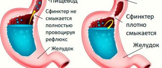
Initially, the mechanism of diverticulum development is characterized by traction factors, then by pulsion factors. Sometimes the two types combine, and a traction-pulsion diverticulum appears. Esophageal diverticulum can be congenital or acquired. The birth defect is associated with weakness in the muscular wall of the esophagus. Formed inside the mother's womb. It may not bother the patient for many years. If a person is relatively healthy, then the diverticulum does not manifest itself in any way. If the patient experiences inflammatory processes in the digestive system, an exacerbation of the diverticulum may occur.
The acquired defect is associated with various pathologies of the gastrointestinal tract and other organs:
- chronic inflammation in the mediastinum;
- histoplasmosis (fungal infection of the lungs);
- pathologies of the lymph nodes;
- pulmonary tuberculosis;
- pathology of the duodenum;
- esophageal injuries;
- condition after surgery;
- chemical burn of the esophagus;
- motor dysfunction;
- other inflammations of the digestive system.
According to the type of structure, diverticula are classified as true and false. True ones consist of layers of the esophageal wall, and pseudodiverticula (false) do not have a muscular membrane. They appear at the moment of contraction of the esophageal wall, and at the moment of relaxation they disappear.
Features of the esophagus affecting the course of diverticulitis
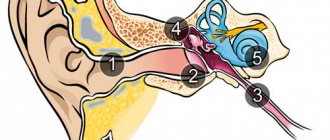
The upper boundary of the thoracic section of the esophageal tube is the conventional line of the second thoracic vertebra from the posterior mediastinum (the space surrounding the heart). The lower one coincides with the esophageal opening of the diaphragm. The entire segment is 16–18 cm long in an adult. It is separated from the spine by a thin layer of fatty tissue.
It is in close contact with the inner layer of the pleura (mediastinal region). Passing from top to bottom, the esophagus is first located to the left of the trachea, then passes the area of the aorta and azygos vein, at the level of the fourth thoracic vertebra, located next to the left main bronchus and the bifurcation of the trachea.
Here, the left atrium of the heart and the pericardial wall, the aortic arch, and the subclavian artery are adjacent to the esophagus in front. Along its entire course, the esophagus is accompanied by the recurrent nerve and multiple groups of lymph nodes. The close proximity to important organs of the chest leads to their damage due to diverticula of the esophagus.
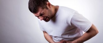
Symptoms
Symptoms of esophageal diverticulum manifest differently, depending on the location of the defect. If a diverticulum grows in the upper part of the esophagus, then it is characterized by the following symptoms:
- swallowing disorder (dysphagia);
- regurgitation of undigested food;
- dry cough;
- soreness or lump in the throat;
- nausea;
- voice change;
- increased salivation.
Signs of a diverticulum located in the middle part:
- chest pain;
- night cough;
- dysphagia;
- nausea and belching.
- Moreover, small diverticula are asymptomatic, while large defects are manifested by the above symptoms.
- Signs of a diverticulum located in the lower part:
- cardiopalmus;
- reflex shortness of breath;
- bronchospasm;
- heart pain.
These symptoms are supplemented by signs of a middle location of the defect.
The mechanism of development of complications of diverticulum
Diverticular disease is a progressive disease. Diverticula do not undergo reverse development. The risk of developing diverticulitis after a diverticulum persists for 5 years is approximately 10%. If the disease lasts more than 10 years, the risk increases to 25%.
Diverticulitis:
With diverticular disease, inflammatory changes develop in the wall of the diverticulum. The absence of a muscle layer leads to the fact that intestinal contents stagnate in them without the possibility of evacuation. This leads to the formation of fecalitis (fecal stone) in the lumen of the diverticulum, followed by inflammation in the wall of the diverticulum.
Diverticulum perforation:
Inflammatory changes may be limited to the diverticulum wall with its swelling and infiltration. With aggressive microbial flora, inflammation takes a malignant course and can lead to perforation of the diverticulum wall, which in turn can be delimited by the adjacent fatty tissue of the intestinal suspension or mesentery with the formation of an abscess.
Violation of the integrity of the diverticulum wall can lead to serious complications in the form of peritonitis when the diverticulum is localized on the free edge of the intestine.
Relapses of diverticulitis:
When acute inflammation is relieved, restoration of the diverticulum wall does not occur. The damaged mucosa is replaced by granulation tissue, coming into close contact with the tissues surrounding the diverticulum, creating favorable conditions for a chronic inflammatory process and subsequent relapses of acute diverticulitis.
Bleeding from a diverticulum:
Damage to the inflamed mucous membrane of the diverticulum by fecalitis when it comes out of the mouth or the development of a bedsore can lead to bleeding.
Fistula formation:
When inflammatory changes spread to nearby abdominal organs and/or the anterior abdominal wall, fistulas may form. Through such anastomoses, intestinal contents can spread into the lumen of the bladder, the uterine cavity, and even onto the anterior abdominal wall.
Which doctor should I contact?
If the described symptoms occur, you need to urgently make an appointment with a gastroenterologist. Our gastroenterologists are true professionals. They will listen to your complaints, conduct an examination, and prescribe therapy. At the Kuntsevskaya clinic you will receive truly qualified assistance.
IMPORTANT! Esophageal diverticulum is a dangerous disease that requires careful medical supervision and control. Often the diverticulum does not manifest itself for a long time.
However, if a person experiences symptoms of esophageal diverticulum, it is imperative to consult a gastroenterologist. Make an appointment with a gastroenterologist at the Kuntsevo Medical and Rehabilitation Center, who will conduct a full diagnosis and prescribe a complete detailed treatment plan for the person. This is followed by the rehabilitation phase, during which you will also be accompanied by our gastroenterologist.
Sign up
Diverticulum diagnosis – accuracy within 3 days
A comprehensive diagnosis of esophageal diverticulum in most Israeli clinics takes from 3 to 4 days. During this time, the patient undergoes all tests and receives an individual treatment program.
- Day 1
- Day 2
- Day 3
First day – arrival in the country
During the first day, the patient, accompanied by a curator, goes from the airport to the hotel, is accommodated in it and arrives at the hospital for the first consultation with a leading specialist. The doctor examines the medical history and prescribes a number of necessary tests.
Day two – diagnostics
During the second day, a diagnostic program is carried out. It usually includes the following studies:
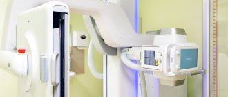
- radiographic examination using barium contrast. This diagnostic method is the most common and accurate when examining patients with suspected diverticulum;
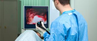
- endoscopic examination can also be effectively used in the diagnosis of medium and large diverticula. A flexible endoscope with a miniature camera at one end is inserted into the esophagus. Using this camera, the surgeon examines the condition of the mucous membrane and walls of the esophagus on a computer screen.
- Esophageal manometry is a study of the contractile activity of the esophagus, allowing one to measure the pressure in its lumen.
Third day – treatment program
On the third day, a medical council analyzes the diagnostic results and determines the final diagnosis. On the same day, an individual treatment program is developed - when drawing it up, doctors take into account the patient’s condition, his age, the presence of concomitant diseases, etc.
Diagnostics
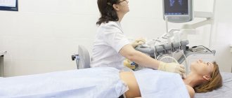
During the examination, the doctor will ask the patient about the signs of the disease - when the symptoms began and how they manifest themselves. The patient then undergoes blood and urine tests. Of the instrumental methods, the most effective is gastroscopy. But in this case, it is important to undergo a study under the guidance of an experienced doctor, since you need to do the procedure carefully so as not to hurt the diverticulum.
The Kuntsevo Medical Center uses Japanese endoscopes of the latest generation, with an ultra-thin tube. In addition, the gastroenterologist who will perform the procedure is an experienced doctor who has performed the procedure more than once. The doctor irrigates the patient's mouth with an anesthetic solution and inserts a cable into the esophagus. The entire process is displayed on the monitor screen. The location of the diverticulum, its size and other subtleties are visible.
If the patient turns out to be hypersensitive, then anesthesia is used and gastroscopy is performed “in sleep”. So the patient will not feel anything.
In addition, magnetic resonance imaging of the esophagus is used. The diverticulum is examined in all projections.
Anatomy of the colon
The colon consists of the following sections: cecum, ascending colon, transverse colon, descending colon, sigmoid colon. The cecum has the widest lumen, and the sigmoid colon has the smallest. The junction of the sigmoid colon and the rectum is designated as the rectosigmoid junction. It is important to note that this section of the intestine has the narrowest diameter.
The wall of the colon consists of three layers: serous, muscular and mucous. The function of the mucous membrane of the colon is the absorption of water, the formation of feces, preparing them for evacuation by secreting a large amount of mucus and the synthesis of vitamins B and K. Under the epithelium of the colon mucosa there is a submucosa, represented by loose fibrous connective tissue in which blood vessels and lymphatic vessels and submucosal nerve plexus.
The muscular layer has a frame function and is responsible for the progressive movement of feces to the rectum. The muscular layer consists of a continuous inner circular layer, divided into three ribbons of the outer longitudinal layer.
The serous membrane consists of a connective tissue base covered with mesothelium, into which outgrowths of adipose tissue, the so-called fat pads, penetrate from the muscular layer.
Treatment
Esophageal diverticulum should be treated based on the causes of the disease and the characteristics of the patient. First, we eliminate the factors that led to the development of the disease. Then we look at how much the defect interferes with everyday life. If the diverticulum is small and does not manifest itself in any way, then we do not touch it. You just need to stick to a diet so as not to provoke a “bag”, and constantly be monitored by a specialist.
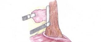
If the diverticulum is large and causes discomfort, it is removed and the walls of the esophagus are sutured. There is a possibility that the defect will form again if the root cause is not eliminated. Therefore, it is necessary to visit your doctor.
The diet should be based on the exclusion of fried, salty, heavy foods. After a meal, be sure to drink a glass of warm water and spray your mouth with a weak antiseptic solution. Maintain temperature conditions - food and drink should be warm. Drug therapy is used in case of inflammation of the digestive system. Painkillers, anti-inflammatory, and antibacterial drugs are prescribed. It all depends on the concomitant disease.
Pharyngeal-esophageal (Zenker's) diverticulum.
The main role in the pathogenesis of the formation of this type of diverticula is achalasia (inability to relax) of the cricopharyngeal muscles, which results in a violation of the opening of the upper esophageal sphincter in response to swallowing. Excessive intraluminal pressure pushes the submucosal layer into the resulting muscle defect. The diverticulum then descends between the posterior wall of the esophagus and the spine. The inner surface of the diverticulum is covered with a mucous membrane; it may have superficial erosions, foci of inflammation and scars.
Treatment results and lifestyle after recovery
The effectiveness of treatment will depend on how well the patient follows the points of the therapeutic program. An esophageal diverticulum is like a time bomb; anything can happen. Therefore, after treatment, strictly follow the recommendations of the gastroenterologist. Visit a specialist from time to time.
Learn a few rules for yourself:
- walk more in the fresh air;
- follow a diet;
- establish a work and rest schedule;
- get rid of bad habits;
- play sports.
We are talking about light exercises. Kuntsevo has its own exercise therapy room. Under the guidance of experienced instructors, do gentle exercises.
What complications does diverticulitis cause?
Inflammation of the diverticulum over a long period of time can lead to:
Hiatus hernia
- to attacks of suffocation;
- phlegmon of the neck;
- chronic bronchitis;
- aspiration pneumonia, lung abscess;
- bleeding;
- abscess formation of a diverticulum;
- mucosal erosion;
- perforation into surrounding tissues;
- mediastinitis with esophageal-mediastinal fistula;
- esophageal polyps;
- cancerous degeneration.
Bifurcation diverticulitis has the rarest tendency to complications.
Why is it better to seek treatment from us?
Early detection of esophageal diverticulum and a complete assessment of the risk of the disease for a particular patient allows doctors at the Kuntsevo Treatment and Rehabilitation Center to fully assess the person’s condition and choose the most effective treatment tactics in each individual case. In addition, modern diagnostic methods available at the Kuntsevo Treatment and Rehabilitation Center (gastroscopy, MRI of the esophagus, MRI of the esophagus “in sleep”) allow the doctor to monitor the patient’s dynamics, evaluate the effectiveness of therapy and make changes to speed up the person’s recovery process.
IMPORTANT! You should not self-medicate if you discover this symptom. Cold hands can be a manifestation of both the physiological norm of your body and a pathological process that develops in the body.
The Kuntsevo Multidisciplinary Center has doctors of the highest category and doctors of medical sciences. Using modern equipment and high-tech devices, diagnosing esophageal diverticulum has become easier and more effective. You will be pleasantly surprised by the best prices and friendly staff. Don't delay treatment - make an appointment!
The contents of this article have been checked and confirmed for compliance with medical standards by gastroenterologist-nutritionist Tatyana Vladimirovna Lucheninova
Causes of diverticulosis
The reasons for the development of diverticular disease remain a topic of much debate to this day. There are several theories about the occurrence of diverticula.
The most likely and most often discussed reasons include: increased intraluminal pressure in the intestine, increasing weakness of the intestinal wall, impaired colon motility and congenital predisposition.
Increased intraluminal pressure in the intestine:
Currently, the most recognized factor in the occurrence of diverticulosis is food. A decrease in the proper amount of plant fiber in the diet reduces the volume of feces, which in turn leads to a disruption in their evacuation with an increase in intraluminal pressure in the intestine. The evidence base is based on observations of vegetarians and residents of agricultural countries who consume significant quantities of fiber, in whom the incidence of diverticula is 42% lower than in a group of people who do not consume plant fiber in sufficient quantities.
Increasing weakness of the intestinal wall:
The fact that diverticula appear in old age confirms the theory of weakness of the intestinal wall, which is a consequence of the aging process of the body, when degenerative changes in muscle tissue and collagen fibers develop in the intestinal wall.
Colon motility disorders:
Impaired intestinal motility leads to constipation and increased pressure in the intestinal lumen when it is necessary to evacuate feces. Loss of elasticity of the intestinal wall leads, when it is stretched, to micro-tears of the circular muscles, through which diverticula begin to form.
Vascular disorders in the intestinal wall:
It is impossible not to indicate the vascular component in the formation of a diverticulum. Impaired blood supply leads to structural changes in the intestinal wall, while at the site where the vessel passes through the muscular layer to the intestine, an expansion is formed, which over time transforms into the mouth of a diverticulum.
Congenital predisposition:
Various congenital systemic connective tissue diseases and collagenoses are a provoking factor in the development of diverticular disease.