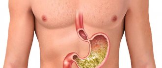
The cardia, or cardiac part of the stomach, is a cylindrical segment covering the esophagogastric junction and the space 2 cm above and below it. Cancer of the gastric cardia (cardioesophageal cancer, CER) differs in anatomical and clinical features, and requires a special approach to treatment. Many experts even identify it as a separate nosology, and there are several reasons for this:
- Tumors of this location tend to spread to the esophagus much more often than cancer of the underlying parts of the stomach.
- Such tumors metastasize to the abdominal and mediastinal lymph nodes.
- In addition, they have a poorer prognosis than gastric or esophageal cancer.
Cardioesophageal cancer is characterized by aggressive growth and a tendency to metastasize to the mediastinal lymph nodes.
- Causes
- Symptoms of gastric cardia cancer
- Classification
- Diagnostics
- Treatment of gastric cardia cancer
- Complications and relapses
- Prognosis and prevention
Causes
The causes of cardiac cancer are not reliably known. Currently, it is customary to talk about risk factors - circumstances under which the likelihood of developing a particular pathology increases. For stomach cancer they are as follows:
- Helicobacter pylori infection.
- The presence of atrophic gastritis of the proximal stomach.
- Hereditary predisposition.
- Smoking and alcohol abuse.
As for gastroesophageal reflux and Barrett's esophagus, these conditions are the main predisposing factors for the development of esophageal cancer, but they do not affect the incidence of cardia cancer.
Achalasia cardia
Clinical manifestations of the pathology are dysphagia, regurgitation and chest pain. Dysphagia is characterized by difficulty swallowing food. In some cases, a violation of the act of swallowing develops simultaneously and is stable; Dysphagia is usually preceded by influenza or another viral disease or stress. In some patients, dysphagia is initially episodic (for example, when eating hastily), then it becomes more regular, making it difficult to pass both solid and liquid food.
Dysphagia with achalasia cardia can be selective and occur when only a certain type of food is consumed. Adapting to impaired swallowing, patients can independently find ways to regulate the passage of food masses - hold their breath, swallow air, wash down food with water, etc. Sometimes, with achalasia cardia, paradoxical dysphagia develops, in which the passage of liquid food is more difficult than solid food.
Regurgitation in achalasia cardia develops as a result of the reverse reflux of food masses into the oral cavity during contraction of the esophageal muscles. The severity of regurgitation can be in the form of slight regurgitation or esophageal vomiting, when profuse regurgitation “mouth full” develops. Regurgitation can be periodic (for example, during eating, simultaneously with dysphagia), occur immediately after eating or 2-3 hours after eating. Less commonly, with achalasia cardia, reflux of food can occur during sleep (the so-called nocturnal regurgitation): in this case, food often enters the respiratory tract, which is accompanied by a “night cough.” Slight regurgitation is typical for stages I – II of achalasia cardia, esophageal vomiting – for stages III – IV, when overflow and overstretching of the esophagus occurs.
Pain with achalasia cardia can be bothersome on an empty stomach or during eating and swallowing. Pain sensations are localized behind the sternum, often radiating to the jaw, neck, and between the shoulder blades. If in stages I–II of achalasia cardia the pain is caused by muscle spasm, then in stages III–IV it is caused by developing esophagitis. For achalasia cardia, periodic paroxysmal pain is typical - esophagodynic crises, which can develop against the background of excitement, physical activity, at night and last from several minutes to one hour. A painful attack sometimes goes away on its own after vomiting or passing food masses into the stomach; in other cases it is relieved with antispasmodics.
Symptoms of gastric cardia cancer
Cardia cancer is most characterized by weight loss, food aversion, pain and dysphagia. The pain is usually localized in the epigastric region, is constant, pressing in nature and does not depend on food intake. Sometimes it radiates to the lumbar region or behind the sternum. In the latter case, the disease can simulate heart pathology, such as angina.
The severity of dysphagia depends on the size of the tumor and obstruction of the digestive tract. It is mainly manifested by malnutrition, which leads to hypovolemia and changes in electrolyte balance.
Anatomy of the stomach, structure of the stomach, treatment of the stomach
The stomach is a hollow organ that is adapted for filling with food, initial digestion of food, partial absorption of nutrients with further evacuation of the contents into the duodenum. The stomach is located in the upper part of the abdominal cavity, under the diaphragm, mostly to the left of the midline.
The shape and volume of the stomach depend on the tone of its muscles, on its filling with food, on the condition of neighboring organs, and on the position of the body. In the upper part of the stomach the esophagus flows into it, in the lower part the duodenum departs from the stomach.
The stomach has four parts:
- The cardial part of the stomach is located above and adjacent to the opening from the esophagus to the stomach, which is called the “cardia”
- The fundus or fornix is the part of the stomach that is located at the top and forms a kind of dome.
- The body of the stomach is the main middle part of the stomach
- The pyloric or pyloric part is located at the entrance to the duodenum, where the sphincter is located, which regulates the flow of food into the duodenum - pylorus
The stomach wall consists of four layers:
- mucous membrane
- submucosal layer
- muscle layer
- outer serous membrane
Stomach mucosa
The gastric mucosa is a layer on top of which there are cylindrical epithelial cells, under which there is loose connective tissue and then a thin layer of smooth muscle. The loose connective tissue of the mucous membrane contains the glands of the stomach.
There are three types of cells that form these glands. Some of them are called the main ones. These glands produce pepsinogens and chymosin. The next type of cells is called parietal or parietal cells. They produce the synthesis of hydrochloric acid and gastromucoprotein. The third type of cells are accessory cells or mucocytes. They produce mucoid secretion. In the area of the pylorus (pylorus) there are hormonally active cells. These cells synthesize gastrin.
The gastric mucosa also contains a huge amount of other producing biologically active substances. The role of some of them still remains not fully understood. A very important function of the glandular cells of the stomach is the formation of a protective mucous barrier. Continuous synthesis of gastric mucus is required, which is produced by mucus-forming cells.
This function is stimulated by the activating effect of the autonomic nervous system, insulin, serotonin, and prostaglandins. The secretion of mucus increases under the mechanical influence of parts of the food bolus that irritate the gastric mucosa. Some medications reduce the mucus-forming function: aspirin (acetylsalicylic acid), non-steroidal anti-inflammatory drugs, etc.
There are contraindications. Read the instructions or consult a specialist.
Cost of ultrasound of the stomach at the EMC clinic.
Classification
The classification of cardia cancer is based on the location of the center of the tumor. The anatomical reference point is the Z-line - the line of the esophagogastric junction.
- Type I cancer - the center of the tumor is located at a distance of 1-5 cm above the Z-line towards the esophagus.
- Type II cancer - the center of the tumor is located within 1 cm orally and 2 cm below the Z-line.
- Type III cancer - the center of the tumor is located at a distance of 2-5 cm below the Z-line.
Diagnostics
- Endoscopic examination of the stomach and esophagus. Allows you to examine the tumor and take a biopsy for morphological examination.
- Endoscopic ultrasound. It is carried out to determine the degree of tumor invasion, its growth into neighboring organs and structures, as well as to search for regional metastases.
- Abdominal ultrasound. It is carried out to search for metastases.
- X-ray contrast study. Allows you to determine the extent of the tumor and assess the degree of stenosis of the cardia and esophagus. The method is especially relevant for diffuse-infiltrative forms of cancer, when false-negative biopsy results can be obtained due to submucosal localization.
- CT scan is used to look for distant metastases.
- Diagnostic laparoscopy. Allows you to detect the spread of the tumor to the serous membranes, as well as diagnose intraperitoneal dissemination.
- Determination of tumor markers - CEA, CA 19-9, CA 72-4.
Stomach - anatomical and physiological information
The stomach (Ventriculus, gaster) is a hollow organ shaped like an inverted retort or an old post horn. It is located asymmetrically in the upper floor of the abdominal cavity, with about 5/6 of it located to the left of the midline (Fig. 94) in the left hypochondrium. The wide left part of the stomach is located under the diaphragm, and the narrowed right part is under the liver.
Stomach - anatomical and physiological information 1
The shape and position of the stomach largely depend on its filling, the tone of the muscular lining and the condition of other organs. The stomach capacity ranges from 1.5 to 3-7 liters. In a collapsed state, it takes on a cylindrical shape, and during fasting it sharply decreases and becomes like an intestine. The length of the stomach is 21-25 cm, and the distance between the inlet and outlet holes varies from 7 to 16-17 cm. In diameter in the widest section it is 10-12-13 cm. The distance between the front and back walls depends on the degree of filling stomach. On an empty stomach, the walls of the stomach are in contact, with the exception of the sections containing air.
In the stomach, there are anterior and posterior walls, connected on the right along a concave edge called the lesser curvature (curvatura minor), and on the left along a convex edge (curvatura major). The stomach is connected to the esophagus by an opening (ostium cardiacum) or inlet (cardia). The junction of the stomach and duodenum is called the pylorus (pylorus or ostium pyloricum). Here, under the serous membrane on the anterior surface, the vena pylorica runs almost transversely.
From the lesser curvature of the stomach departs the lesser omentum, consisting of two layers of peritoneum and including three ligaments: gastrodiaphragmatic (lig. gastrophrenicum), hepatogastric (lig. hepatogastricum) and hepatoduodenal (lig. hepatoduodenale). The greater omentum originates in the area of greater curvature. It is a continuation of the gastrocolic ligament (lig.gastrocolicum), running from the greater curvature of the body and the pyloric part of the stomach to the transverse colon.
The gastrocolic ligament is connected to the transverse colon by thin plates, almost along its entire length, which makes it possible to bloodlessly separate the omentum from the intestine and thereby quickly mobilize the greater curvature of the stomach during operations for gastric cancer. From the greater curvature of the fundus of the stomach to the spleen there is a gastrosplenic ligament (lig. gastrolienale), which is often very short and makes it difficult to mobilize the fundus of the stomach when performing gastrectomy. The greater and lesser omentum contains blood and lymphatic vessels, regional lymph nodes and nerves.
Of great practical importance is the gastropancreatic ligament (lig. gastropancreaticum), which passes between the cardiac part of the stomach and the upper edge of the pancreas. This ligament contains the left gastric artery with the vein of the same name and the lymph nodes (gastropancreatic and pancreasplenic), into which cardiac gastric cancers metastasize. The pyloropancreatic ligament is also distinguished, but it often merges with the gastropancreatic ligament or is not expressed.
In gastric pathology, especially acute, the omental bursa plays an important role. It is located behind the stomach and lesser omentum, bounded above by the liver and diaphragm, below by the mesentery of the transverse colon, and on the left by the spleen. The posterior wall of the omental bursa is the peritoneum, covering the pancreas and other organs and tissues. The right part of the omental bursa narrows and, through the omental foramen (foramen epiploicum s. Winslowi), located under the hepatoduodenal ligament, connects with the free abdominal cavity. There are several sections in the stomach (Fig. 95)
Stomach - anatomical and physiological information 2
- 1) pars cardiaca (inlet section) - the section of the stomach closest to the cardia,
- 2)fundus. fornix ventriculi (fundus of the stomach) - located to the left of the cardia under the dome of the diaphragm,
- 3)corpus ventriculi (body of the stomach),
- 4) pars rulorica (pyloric section) - the narrowest section of the stomach adjacent to the pylorus.
In the pyloric section, two parts are distinguished: antrum pyloricum and canalis pyloricus .
At the border of the body of the stomach and the antrum of the pylorus there is a sac-like expansion of the stomach - sinus ventriculi.
Along the lesser curvature, on the border of the mentioned sections of the stomach, there is an angular notch (incisura angularisj, which surgeons call the angle of the stomach. Along the greater curvature, on the border of the cardia and the fundus, there is an entrance notch - incisura cardiaca.
The stomach is also divided into two parts (Fig. 96), according to the most characteristic function of each section:
- 1) digestive section (saccus digestorius) - includes the cardiac section, fundus and body of the stomach,
- 2) evacuating (canalis egestorius)—corresponds to the pyloric section.
Stomach - anatomical and physiological information 3
The entrance to the stomach is located to the left of the spine at the level of the X-XI thoracic vertebrae. There are cases when the cardia of the stomach is located in the midline. Access to the cardiac part of the stomach is not always the same, which depends on the anatomical structure of the chest wall, the epigastric angle, the structure of the internal organs and the length of the ligaments.
In thin people with a large epigastric angle, a small distance between the sternum and the spine and a long abdominal section of the esophagus, the cardiac part of the stomach is easily accessible for inspection; after dissection of the peritoneum and mobilization of the esophagus located above it, it is brought out into the wound.
More often there are more difficult conditions when approaching the cardiac part of the esophagus, especially in obese people, with short gastric ligaments and an almost complete absence of the abdominal part of the esophagus. In such cases, the cardia lies high under the diaphragm and is determined only by palpation. Operations in such patients present great difficulties. To mobilize and remove the cardiac part of the stomach in such anatomical relationships, it is necessary to widely dissect the diaphragm, cross the legs of the diaphragm, and sometimes resort to sagittal sternotomy.
The fundus of the stomach is located under the left dome of the diaphragm, tightly adjacent to it. Here, above the diaphragm, is the heart, whose activity is influenced by the condition and filling of the stomach. This circumstance must be taken into account both before surgery in cases of stagnation in the stomach and paresis, intestines, and in the postoperative period.
The body of the stomach is located downward and anterior to the bottom, and the lesser curvature initially runs parallel to the spine and to the left of it, and then the pyloric section of the stomach crosses the spine at the level of the XII thoracic or I lumbar vertebrae. The greater curvature, depending on the shape and filling of the stomach, changes its position, sometimes descending to the level of the iliac crests even under normal conditions.
The projection of the stomach onto the chest and abdominal walls is clear from Fig. 97. However, it must be borne in mind that the position of the stomach is not stable and depends on its tone, filling and, to a certain extent, on its shape (retort-shaped, horn-shaped, hook-shaped, etc.). The most constant position of the stomach is in the area of entry and exit. The stomach is in close connection with many organs: the liver, spleen, anterior abdominal wall, left kidney and adrenal gland, pancreas, transverse colon and sometimes gall bladder.
Stomach - anatomical and physiological information 4
Close to the stomach are the aorta, the celiac artery with its branches, the portal and inferior vena cava. This justifies the need for great attention and thoroughness in performing operations on the stomach. Knowledge of the anatomy and topography of the stomach is of great importance during X-ray examination of the stomach; and duodenum.
The stomach is covered on all sides with a serous membrane and only small parts of it in the area where the bottom is connected by the diaphragm and along the attachment of the ligaments are devoid of peritoneal cover in thin strips. The muscular layer is represented by three layers: outer (longitudinal), middle (ring-shaped) and deep (oblique).
The mucous membrane, 1.5-2 mm thick, is assembled into multiple folds due to the muscular lamina propria and thick, loose submucous membrane. Along the lesser curvature, the folds run in the longitudinal direction, forming a gastric track.
The gastric mucosa is rich in glands that produce gastric juice. The mucosa contains main cells (produce enzymes), accessory cells (produce mucoid secretions) and parietal cells (produce hydrochloric acid). In the area of the fundus and body of the stomach there are all three types of cells, but in the pyloric part there are no parietal cells. Consequently, the juice secreted by the pyloric region does not contain hydrochloric acid and has an alkaline reaction. The cardiac glands are similar in morphology to the pyloric glands, but they contain separate parietal cells. The boundaries between the glandular zones are unclear. At the border of the body and the pyloric part, an intermediate zone is distinguished, in which there are both types of glands (Fig. 99).
Stomach - anatomical and physiological information 5
The blood supply to the stomach is provided by the celiac artery (a. coeliaca) and its branches (Fig. 100). Only the left gastric artery originates independently from the celiac artery. The right gastric artery is a branch of the hepatic artery, the left gastroepiploic artery is a branch of the splenic artery, and the right gastroepiploic artery is a branch of the gastroduodenal artery.
Stomach - anatomical and physiological information 6
The efferent vessels from these lymph nodes accompany the blood vessels and go in three main directions (Fig. 101):
- 1) from the lesser curvature, the upper half of the pylorus and cardia with the corresponding sections of the anterior and posterior walls of the stomach to the celiac lymph nodes,
- 2) from the fundus of the stomach to the lymph nodes of the hilum of the spleen and
- 3) from the lymph nodes of the greater curvature and from the subpyloric nodes with the corresponding parts of the walls of the stomach to the celiac lymph nodes.
Stomach - anatomical and physiological information 7
Both gastric arteries follow the lesser curvature, and the gastroepiploic arteries follow the greater curvature. There are also short gastric arteries that originate from the distal splenic artery and pass in the gastrosplenic ligament to the fundus of the stomach.
All of these arteries give off numerous branches, widely anastomosing with each other. The veins follow the arteries and empty into the portal vein. Lymph from the stomach flows into the upper gastric lymph nodes (lesser curvature), lower lymph nodes (greater curvature), pericardial, supra- and subpyloric and gastro-pancreatic (along the course of the left gastric artery from its beginning to the stomach).
Innervation of the stomach is provided by sympathetic nerves, originating from the celiac plexus (plexus coeliacus) and running along with blood vessels, and parasympathetic, represented by two vagus nerves, entering the abdominal cavity along with the esophagus and giving branches to the stomach, liver, pancreas, small intestine , to the celiac and inferior mesenteric plexuses.
The duodenum (duodenum) is a continuation of the stomach and has a horseshoe-shaped, highly variable shape (Fig. 102). The duodenum is divided into an upper horizontal part, a descending part and a lower part, which includes the lower horizontal part and the ascending part. The latter passes into the jejunum. The duodenum has three bends—superior, inferior, and duodenojejunal.
Stomach - anatomical and physiological information 8
. The duodenum begins with expansion (bulbus duodeni). It is 25-30 cm in length and 4-5 cm in diameter. Only the initial section of the intestine is surrounded on all sides by the peritoneum for 2-5 cm. The rest of the intestine is located in the retroperitoneal space. On the medial wall of the descending part of the duodenum there is a longitudinal fold ending in a tubercle (papilla duodeni major s. Vateri) with the mouth of the common bile duct and the mouth of the pancreatic duct. An important anatomical formation is the hepatoduodenal ligament, which runs from the porta hepatis to the upper part of the pancreas and contains the hepatic artery, portal vein and common bile duct.
In the horseshoe of the duodenum is the head of the subgastric gland, to the right and below is the right kidney with the adrenal gland, behind is the aorta and inferior vena cava, above is the liver and gall bladder, in front is the transverse colon with the mesentery. The superior mesenteric vessels pass in front of the ascending part of the duodenum.
The duodenum receives blood supply from the branches of the celiac and superior mesenteric arteries. The outflow of lymph goes to the lymph nodes of the pancreas, and through them to the main collector (celiac lymph nodes).
The stomach and duodenum perform secretory, motor, absorption and, to a certain extent, endocrine functions. The stomach, in addition, acts as a reservoir that holds the food eaten and retains it for several hours. In the stomach and duodenum, incoming food undergoes chemical changes under the influence of hydrolytic enzymes and hydrochloric acid. Gastric juice secretion in humans occurs almost continuously and only during sleep it stops for a short time or sharply decreases.
After eating, gastric secretion increases, and the gastric juice produced varies in composition and quantity depending on the food taken. Thus, after eating meat, gastric juice contains more hydrochloric acid than after eating bread or milk, and bread causes a greater release of enzymes. Fats inhibit gastric secretion for several hours and then stimulate it. The amount of gastric juice produced depends to a certain extent on the amount of food eaten.
Gastric secretion is usually divided into three phases:
- 1) complex reflex phase - begins under the influence of conditioned and unconditioned reflexes that arise at the sight of food, smell, sounds accompanying food preparation, when chewing and swallowing it;
- 2) the gastric phase of secretion begins at the moment of mechanical and chemical irritation of the gastric mucosa, food entering it, as well as the humoral effect on gastric secretion of substances (gastrin, histamine) absorbed from the stomach;
- 3) intestinal phase of gastric secretion—excitation of gastric secretion, caused by substances formed from food entering the small intestine and absorbed from it (enterogastrin, etc.). It is generally accepted that 45% of gastric juice is released in the first and second phases of gastric secretion, and 10% in the third.
Gastric secretion is inhibited by enterogastron, produced in the duodenum under the influence of sharply acidic contents (pH below 2.5), and gastron, formed in the pyloric part of the stomach.
In the first phase, gastric secretion, according to Gszacki (1968), is stimulated not only by the vagus nerve system, but also by irritation of the posterior part of the hypothalamus, which stimulates the function of the anterior pituitary gland. An increase in adrenocorticotropic hormone, in turn, stimulates the function of the adrenal cortex. As a result, the level of 17-hydroxycorticosteroids, which stimulate the function of the gastric mucosa, increases (Fig. 103).
Stomach - anatomical and physiological information 9
. Gastric juice is produced by the glands of the gastric mucosa. There are main, accessory and parietal cells of the gastric glands. The main cells secrete enzymes of gastric juice, the accessory cells secrete mucoid secretion, and the parietal cells secrete hydrochloric acid. In the area of the fundus of the stomach, the glands contain all three types of cells, the glands of the lesser curvature of the stomach contain the main and parietal cells, and the pyloric glands contain the main and accessory cells. Therefore, it is clear that the glands of the lesser curvature of the stomach secrete juice containing a lot of pepsin and hydrochloric acid, and the glands of the pyloric region produce alkaline juice containing a lot of mucus.
In the pyloric part of the stomach, progastrin is formed, which, under the influence of digestive products, is converted into a gastrin-physiologically active hormone, which is absorbed into the blood and stimulates the function of the cells of the gastric glands. Gastrin is very active. Subcutaneous administration of gastrin at a dose of 0.002 mg per 1 kg of human weight causes maximum gastric secretion. Hydrochloric acid entering the pyloric region inhibits the formation of gastrin. The mediator in the formation of gastrin is acetylcholine, which is released when the vagus nerve is stimulated.
The second powerful chemical stimulant of gastric secretion (stimulates the secretion of hydrochloric acid) is histamine, which is released from tissues under the influence of acetylcholine (vagal influence). Subcutaneous administration of 0.04 mg (per 1 kg of weight) of histamine causes maximum gastric secretion. Histamine inhibits the secretion of enzymes.
Gastric juice is acidic. The concentration of hydrochloric acid in it. is 0.4 - 0.5%, and pH - 0.9 - 1.5. It contains proteases (pepsin, gelatinase, chymosin) and lipase, under the influence of which the breakdown of proteins and fats occurs. In the stomach, the breakdown of polysaccharides continues, which began in the oral cavity under the influence of ptyalin and salivary maltase.
The second function of the stomach is motor; it ensures the mixing of food and its movement into the duodenum. Contractions of the stomach occur due to mechanical, chemical and humoral influences (gastrin, histamine and choline stimulate contraction of the stomach muscles, and zenterogastron, adrenaline and norepinephrine inhibit them).
Of great importance in the motor activity of the stomach are irritations traveling along the vagus (stimulates the strength and frequency of contractions) and sympathetic (inhibits muscle contractions) nerves. This explains the role of mental stressors in the etiology and pathogenesis of gastric ulcer.
Under normal conditions, the absorption function of the stomach is small. The absorption function increases with gastritis, impaired pyloric patency and other pathological processes that cause disruption of the motor function of the stomach and retention of food masses in it.
The entry of food into the duodenum stimulates the function of the pancreas, liver and gall bladder. Food here is exposed to bile, pancreatic juice and juice produced by the duodenal mucosa, and quickly moves to the small intestine. Research by A. M. Ugolev (1973) established that the duodenum not only plays a subordinate (stomach) role in continuing the digestive process with the involvement of the work of the two most important digestive glands, but also has, in connection with the hormones it produces, an independent influence on digestion and metabolism . A. M. Ugolev established that removal of the duodenum leads to disruption of nitrogen, mineral and lipid metabolism.
Treatment of gastric cardia cancer
Treatment of cardia cancer requires a combined approach and includes chemotherapy and surgery. For unresectable tumors, the main treatment method is chemotherapy, and operations are performed for life-threatening indications.
Surgical treatment
The choice of the extent of the operation and surgical approach is determined based on the type of cancer and its local prevalence. For type I CER, proximal gastrectomy is performed
| KER type I Proximal gastrectomy, subtotal esophageal resection. | KER type II Gastrectomy (the entire stomach is removed), resection of the lower thoracic esophagus | KER type III Gastrectomy, resection of the lower thoracic esophagus. |
The minimum volume of lymph node dissection involves the removal of 1st and 2nd order lymph nodes (extended lymph node dissection is performed in the volume of D2). In CER I, bilateral mediastinal lymph node dissection is performed.
Chemotherapy
Chemotherapy is carried out using one of the following options:
- Perioperative chemotherapy. Performed in CF or ECF mode for 8-9 weeks before and after surgery.
- Adjuvant chemotherapy. It begins 4-6 weeks after surgery, if there are no severe complications, and is carried out for 6 months. XELOX or CAPOX schemes are used.
- Adjuvant chemoradiotherapy. Currently, it is rarely used and only in case of non-radical surgery.
Treatment of unresectable and disseminated tumors is carried out using chemotherapy according to the CF, CX, XELOX regimens in 6-8 three-week courses, or 9-12 two-week courses. After this, treatment is stopped and observation is carried out until the disease progresses. For HER2-positive gastric cancer, treatment with trastuzumab is possible.
After disease progression, chemotherapy is resumed, and the treatment regimen is selected taking into account the existing medical history.
Metastasis
With CER types I and II, metastasis mainly occurs in the lymphatic collectors of the paracardial zone, celiac trunk and lesser curvature of the stomach. In type I CER, the mediastinal nodes may also be involved in the process. Such features of metastasis are taken into account when planning surgery and lymph node dissection.
Treatment
Treatment of gastric stenosis is surgical. The goal of surgical treatment of gastric outlet stenosis is to eliminate the stenosis and restore passage through the stomach and duodenum.
Patients with sub- and decompensated stenosis require comprehensive preoperative preparation, including correction of water-electrolyte metabolism, protein composition, volemic disorders, and activity of the cardiovascular system; as well as gastric lavage and decompression.
Patients without pronounced disorders of gastric motility (stages of developing and compensated stenosis) can be operated on after a relatively short (5-7 days) period of preoperative preparation (anti-ulcer therapy, gastric decompression).
The choice of surgical intervention is determined by the disease that led to the formation of obstruction of the gastric outlet tract.
- In case of malignant etiology of the disease, examination and treatment are carried out in accordance with oncological recommendations for gastric cancer. For tumor stenosis, subtotal proximal resection or gastrectomy in combination with lymphadenectomy is performed.
- For cicatricial stenoses of benign origin, it is necessary to perform either organ-preserving drainage operations (pyloroplasty in various modifications, gastrojejunostomy bypass) or distal gastrectomy. In case of ulcerative stenosis, a standard resection of 2/3 of the stomach is performed with an anastomosis according to Billroth 1 or Billroth 2 in various modifications, depending on the conditions for the anastomosis and the formation of the duodenal stump. All surgical treatment options are possible using minimally invasive technologies in the absence of contraindications.
Pyloroplasty according to Heineke-Mikulicz
Complications and relapses
The main complications of cardioesophageal cancer:
- Tumor disintegration and bleeding. In this case, urgent endoscopic examination and bleeding control are required. If the problem cannot be solved endoscopically, surgical intervention is performed.
- Tumor stenosis. This complication leads to dysphagia and, as a consequence, to the development of nutritional disorders. To eliminate it, recanalization, balloon dilatation, and stenting are used. In severe cases, bypass anastomosis or gastrostomy removal is indicated.
- Pain. To reduce pain, radiation therapy, drug treatment and, in some cases, locoregional anesthesia are used.
- Ascites is an accumulation of fluid in the abdominal cavity. To eliminate it, diuretics and intracavitary chemotherapy are prescribed. When large volumes of fluid accumulate, laparocentesis is indicated - evacuation of the contents through puncture.
Prognosis and prevention
The 5-year survival rate for any type of CER averages 23.6%. Type III tumors have the most unfavorable course, since they are predominantly represented by poorly or undifferentiated neoplasms. The 5-year survival rate for this location is about 14%. The prognosis is significantly worsened by the presence of metastases in the mediastinal lymph nodes.
Euroonko employs doctors with extensive experience in treating stomach cancer. We are guided by current European standards. For this, the clinic has everything necessary - from the latest medications to modern equipment, thanks to which we are ready to provide assistance in the most severe cases.
Book a consultation 24 hours a day
+7+7+78


