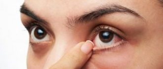
The eyes are called the mirror of the soul, so it is not surprising that many people first of all pay attention to them. Circles forming under the eyes cause women many problems and worries. They try to disguise the flaw with the help of correctors and foundations. But this way cannot solve the problem. You can only hide it, and only temporarily. In order to radically eliminate the defect, it is necessary to first determine the cause, conduct a diagnosis and, only based on the results obtained, select treatment methods.
Diseases
Not always, swelling and bruising under the eyes are a sign of an unhealthy lifestyle. Often, the reason is the presence of certain diseases:
- Allergies. Accompanied by lacrimation, sneezing, redness of the eyes, difficulty breathing and coughing;
- Inflammation of the sinuses (sinusitis and sinusitis). They occur with painful sensations localized in the forehead and under the eyes, as well as copious discharge from the nose;
- Kidney diseases. When they occur, fluid retention occurs in the body, which causes not only swelling of the eyes, but also lower back pain and headaches;
- Diseases of the thyroid gland. Due to dysfunction of the thyroid gland, hormonal imbalance occurs - general weakness increases, body weight increases, the menstrual cycle changes, edema occurs;
- Heart failure. It is accompanied by shortness of breath and fatigue. Swelling and bruising under the eyes, almost invisible in the morning, appear in the late afternoon;
- Skin diseases - eczema and dermatitis.
Treatment methods
If the diagnosis does not reveal internal diseases, then circles under the eyes are a cosmetic defect that appears as a result of an incorrect lifestyle or hereditary predisposition.
In such cases, the most popular and effective treatment methods are:
- Mesotherapy. Typically, meso-cocktails are used, which contain arbutin, ascorbic, kojic and phytic acids. The therapeutic course includes 6-10 procedures, between which there is a one-week break.
- Bioreparation with ascorbic acid (vitamin C). Thanks to the combination of ascorbic and hyaluronic acid, the skin around the eyes is well moisturized and noticeably brightened. The course of treatment consists of 2-4 sessions, with a 2-week break between them.
- Plasmolifting. The procedure restores metabolism, normalizes tissue respiration, activates blood microcirculation, thereby increasing local immunity. A similar effect will appear after 4-6 sessions at weekly intervals.
- Biorevitalization. The skin around the eyes shows a pronounced reaction to dehydration and, due to dryness, often takes on an unaesthetic appearance, creating the effect of “tired eyes”. A course of birevitalization, consisting of 2-4 procedures with a 2-week interval, will help solve the problem.
- Contour plastic. If the blood vessels are located close to the skin and “see through” through it, then the filler forms an additional layer, making the vessels almost invisible. In addition, the drugs smooth out the nasolacrimal groove.
- Laser peeling. Using a laser, the top layer of skin is removed, which stimulates the regeneration process and promotes rejuvenation. The treatment course involves 2-5 procedures with a one-month break.
Preventive actions
To prevent the formation of dark circles under the eyes, it is recommended:
- adhere to a regime of wakefulness and rest;
- eat a balanced diet;
- maintain water regime;
- take vitamin and mineral supplements;
- to refuse from bad habits;
- do eye exercises;
- avoid direct sunlight to reduce insolation;
- use high-quality perfumes and cosmetics;
- walk in the fresh air more often;
- avoid stressful situations.
Bleeding in the eye
Atherosclerosis
Diabetes
Fungus
27084 March 22
IMPORTANT!
The information in this section cannot be used for self-diagnosis and self-treatment.
In case of pain or other exacerbation of the disease, diagnostic tests should be prescribed only by the attending physician. To make a diagnosis and properly prescribe treatment, you should contact your doctor. Hemorrhage in the eye: causes of occurrence, what diseases it occurs with, diagnosis and treatment methods.
Definition
Hemorrhage in the eye is the accumulation of blood under the conjunctiva, in and between the membranes of the eye, in the chambers of the eye or in the vitreous body. Hemorrhage is caused by a violation of the integrity of the blood vessels of the eye.
The eyes continuously perceive, transform and send a huge amount of information to the visual analyzer.
The eyes are sensitive to changes in the body and, although quite protected, are easily damaged by external influences.
The eyeball is located in the cavity of the orbit - a bone frame; the orbit protects the eyeball from above, below, behind and on the sides. The eye in the orbit is located in fatty tissue - a kind of pillow. In front, the eyeball is protected by eyelids and eyelashes.
Eyeball
consists of three shells:
- external, represented by a convex transparent cornea, turning into the sclera - the frame of the eyeball;
- middle - in front is the colored iris, then the ciliary body, and in the back is the choroid itself, represented by a network of small arteries and veins. At the center of the iris is the pupil, which allows light to enter the eye. The ciliary body produces intraocular fluid, which is needed to maintain the shape of the eye and help with metabolism. The lens is attached to the ciliary body with the help of muscles and ligaments. By contracting and relaxing the muscles, the curvature of the lens changes, thus the eye “switches” the distance and focuses the image;
- the inner retina, which contains rods and cones that perceive light and convert it into an electrical impulse, which is then sent to the brain.
Vitreous body
fills the space between the lens and the retina and is a gel-like substance.
The eye has two spaces filled with intraocular fluid: the anterior chamber of the eye (between the cornea and the iris) and the posterior chamber of the eye (between the iris and the vitreous body, which contains the lens).
The cameras communicate with each other through the pupil. Conjunctiva
- This is the mucous membrane, it lines the inside of the eyelids and extends to the visible part of the eye. The conjunctiva is nourished by a network of very small vessels located directly underneath it. Externally, it is constantly washed with tears produced by the lacrimal glands.
Types of hemorrhages in the eye
Hemorrhages in the eye are divided depending on the place where the blood accumulates:
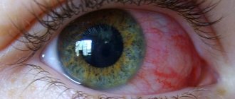
- With hyposphagma, blood accumulates between the conjunctiva and sclera due to damage to the conjunctival vessels. It looks like a bright red spot on the white of the eye. This is the least dangerous type of hemorrhage and does not lead to visual impairment, but you should be wary if it occurs regularly;
- With hyphema, blood flows into the anterior chamber of the eye. Hyphema has 4 degrees: 1st degree is characterized by filling the chamber by no more than 1/3, 2nd – by no more than 1/2, 3rd – by more than 1/2, but not completely, 4th – complete filling of the anterior chamber of the eye with blood. If blood reaches the pupil, the vision becomes blurred, vision decreases, and fear of bright light appears. With this type of hemorrhage, pain occurs;
- with hemophthalmia, blood fills the vitreous body. There are complete and incomplete hemophthalmos. Vision drops sharply, the degree of impairment of which depends on the volume of blood shed. A very dangerous type of hemorrhage that can lead to filling of the vitreous with connective tissue, permanent vision loss and possible retinal detachment;
- In retinal hemorrhages, blood accumulates just in front of the retina (preretinal hemorrhages), inside the retina (intraretinal hemorrhages), or just after the rod and cone layer (subretinal hemorrhages).
This is the most serious type of hemorrhage, most often leading to decreased or even loss of vision.
Possible causes of hemorrhage in the eye
The most likely causes of hemorrhage in the eye include:
- mechanical impacts, eye injuries, head injuries - the degree of damage depends on the force of the impact;
- intraocular operations;
- rupture of newly formed vessels;
- diseases and conditions that affect the composition and properties of blood, disrupting the blood clotting process (taking blood-thinning drugs, hematological diseases);
- diseases affecting the vascular wall (hypertension, atherosclerosis, diabetes mellitus);
- eye diseases: glaucoma (with increased intraocular pressure), high degree of myopia, retinal detachment, thrombosis (blockage) of the central retinal vein;
- various infections (viral, bacterial, fungal, etc.) leading to inflammation of the eye structures. Conjunctivitis (inflammation of the conjunctiva), iritis (irises), iridocyclitis (iris and ciliary body), chorioretinitis (choroid and retina), retinitis (retina only);
- increased intracranial pressure due to excess volume in the cranial cavity due to intracranial hematomas, brain tumors, cerebral edema;
- an increase in intra-abdominal and, as a consequence, venous pressure during severe physical exertion, debilitating vomiting, and during childbirth.
For non-penetrating blunt eye injuries
The cause of vascular rupture is a sharp increase in intraocular pressure, the same mechanism is triggered in glaucoma.
Newly formed vessels have a rather fragile wall; they can appear without good reason or to replace damaged ones, for example, in diabetes mellitus
. In addition, an increase in blood sugar levels leads to damage to the smallest vessels - capillaries.
For infectious diseases
the inflammatory process leads to increased vascular permeability, which can cause damage.
Blood diseases that most often lead to hemorrhages include hemorrhagic diathesis
.
This is a group of diseases in which the processes of stopping bleeding are disrupted.
For example, in hemophilia, there is a hereditary decrease in blood clotting factors, which prevents a blood clot from forming. In thrombocytopenia, there are not enough platelets to initially block the lesion. Oncological diseases of the blood. The most striking example is acute leukemia
. Tumor cells displace bone marrow cells, then enter the bloodstream and replace all healthy cells, including those responsible for stopping bleeding.
Atherosclerosis
manifested by the deposition of cholesterol plaques in blood vessels.
A constant or periodic increase in blood pressure
damages the inner layer of blood vessels - the endothelium, while the middle, muscular layer thickens, and the vessel loses its elasticity. These changes sooner or later lead to a violation of the integrity of any vessel.
Retinal disinsertion
from the choroid with hemorrhage occurs with eye injuries, inflammatory diseases of the retina, and disturbances in its nutrition. The risk of detachment increases in people with a high degree of myopia.
Symptoms of retinal detachment include a sharp decrease in vision, narrowing of the visual field or loss of a portion of the image, the presence of spots in front of the eyes, flashes of light.
Which doctors should you contact if there is a hemorrhage in the eye?
The treatment of hemorrhages in the eye is carried out by an ophthalmologist; to correct concomitant conditions, a consultation with a general practitioner may be required.
Diagnosis and examinations for hemorrhage in the eye
The doctor conducts a survey and examination of the patient and his eyes, determines visual acuity, measures blood pressure, intraocular pressure, studies the structures of the anterior chamber of the eye, and examines the fundus, if possible.
A number of additional examination methods may be required:
- ultrasound examination of the eye;
- electroretinography to assess the functional activity of the retina;
- general blood analysis;
Price for cheekbone correction with fillers
| Name | Price |
| Preparations: 1st group - Biorevitalizants | |
| Administration of the drug - MesoEye C71 /USA/ (1.0 ml) | 14 500₽ |
| Administration of the drug - NEAUVIA ORGANIC Hydro Delux XL /Italy/ (0.5 ml) | 5 000₽ |
| Administration of the drug - Hyalual 1.1% (1.0 ml) | 10 500₽ |
| Administration of the drug - Hyalual 1.1% (2.0 ml) | 13 900₽ |
| Preparations: 2nd group - Hyaluronic fillers | |
| Administration of the drug - Belotero Balance /Switzerland/ (1.0 ml) | 18 500₽ |
| Administration of the drug - Juvederm Volite /France/ (1.0 ml) | 19 000₽ |


