Skin characteristics and immunity
The human skin is an independent organ that performs various functions.
The dense surface layer of the skin primarily prevents pathogens from penetrating into the underlying tissues. Also on the surface of the skin there are more receptors that inform the brain about environmental temperature, tactile contacts, pain and other types of sensitivity. Damage to the skin is always a risk factor for infection or inflammation. Additional functions of the skin:
- protection of underlying tissues from external influences;
- maintaining a fixed body temperature;
- removal of metabolic products through sweat;
- maintaining a constant internal environment of the body;
- external breathing;
- formation of vitamin D, necessary for metabolism and organ development;
- blood deposition.
The outermost portion of the skin is the stratified epidermis. The keratinized cells of the upper layer of the epidermis perform a barrier and protective function. The lower layers of the epidermis regularly renew the cellular composition of the tissue. The middle layer of the skin is represented by the dermis, containing hair follicles, blood vessels, smooth muscles, sweat and sebaceous glands. The lowest part of the skin is the subcutaneous tissue. Adipose tissue prevents hypothermia of the body and protects internal organs from mechanical stress.
Cells of the immune system are present in the skin. The task of immunity in this anatomical region is to quickly prevent the invasion of pathogenic microorganisms into internal organs. In addition, the protective system protects the body from harmful chemicals. At the same time, due to congenital abnormalities, the immune system can react to certain neutral substances and cause an inflammatory response.
Autotoxic type of toxicoderma
Autotoxic toxicoderma occurs due to the influence of allergens and various toxins that are formed in the body due to metabolic disorders. It can manifest itself in gastritis, stomach ulcers, pancreatitis, hepatitis and kidney diseases - pyelonephritis, hydronephrosis, chronic renal failure, in the case of malignant processes such as kidney adenocarcinoma, lung cancer, and intestinal cancer. Autotoxic toxicoderma tends to develop into a chronic form. Toxicoderma can also be caused by various metals used in dentures and various types of metal structures, which are used in traumatology and orthopedics. The reaction occurs due to the content of chromium, cobalt, nickel, molybdenum in them; they enter the patient’s blood and sensitize the body.
Selected species
The term “toxicoderma” refers to any type of skin damage that occurs when an allergen spreads through the bloodstream. At the same time, doctors are aware of individual syndromes characterized by such a pathological reaction.
The most dangerous forms of the disease:
- Stevens-Johnson syndrome is a severe lesion of the skin and mucous membranes of the genitourinary organs, mouth and eyes. Multiple exudative erythemas form on the surface of the patient's skin. The disease is manifested by an increase in body temperature, tachycardia, weakness, headache and other symptoms. In children, this condition often develops against the background of infectious diseases.
- Lyell's syndrome is a life-threatening condition manifested by damage to the skin, mucous membranes and internal organs. External symptoms of the disease include peeling of the skin and the appearance of blisters filled with exudate. General intoxication of the body and the addition of infection can lead to the death of the patient. The nervous system is affected.
It is important for a doctor to recognize a dangerous form of the disease in time and begin treatment. Often the patient requires intensive care to maintain the functions of vital organs.
Toxicoderma
Toxicoderma, or toxic-allergic dermatitis, is an acute inflammation of the skin, and sometimes mucous membranes, developing under the influence of an allergenic, toxic or toxic-allergic factor that enters the body through the respiratory tract, digestive tract, with intravenous, subcutaneous, intramuscular administration . Thus, the etiological factor does not act directly on the skin, as in dermatitis, but penetrates it hematogenously
.
Recently, the problem of toxicoderma has worsened, which is associated with the expanding introduction of household chemicals, deteriorating environmental conditions, and the emergence of new drugs.
Etiology
In the etiology of toxicoderma, the main role is played by exogenous causes, less often - endogenous ones. To exogenous
causes include
medications, food products, industrial and household chemicals
that enter the body through the digestive and respiratory tract. In addition, drugs can cause toxicoderma by any method of administration: intravenous, intramuscular, subcutaneous, as well as as a result of absorption through the skin when used externally.
Endogenous causes
– this is autointoxication with unusual metabolic products that appear in the body due to dysfunction of the gastrointestinal tract, liver, kidneys, thyroid gland; neoplasms, metabolic diseases, helminthic infestations.
The basis of the pathogenesis of toxicoderma is an allergic reaction.
In most cases, we are talking about drug toxicoderma. Drug toxicoderma is a manifestation of the sensitizing effect of the drug. Its pathogenesis often combines toxic and allergic components, which causes the development of various lesions of the skin, mucous membranes, nervous and vascular systems, and internal organs characteristic of the drug-induced disease.
Drug toxicoderma
may occur as a result of long-term administration of a drug and is a variant of a drug-induced disease. The cause of the development of toxicoderma can be a variety of drugs: antibiotics, sulfonamides, barbiturates, amidopyrine, halogens, tranquilizers, vaccines and serums, vitamins, organic arsenic preparations, iodine, aminazine, ACTH, quinine compounds and many others. There are even known cases of the development of toxicoderma to antihistamines and corticosteroids.
Drug toxicoderma is observed in 2–3% of hospitalized patients and accounts for 19% of all complications of drug treatment. For many commonly used drugs, the incidence of toxicoderma exceeds 1%. It is usually impossible to predict the occurrence of toxic-allergic reactions. Risk of toxicoderma
considered
high
(3 – 5%) when treated with penicillins, carbamazepine, allopurinol, gold preparations;
average
in treatment with sulfonamides, oral hypoglycemic agents, diuretics, non-steroidal anti-inflammatory drugs (NSAIDs), isoniazid, chloramphenicol, erythromycin, streptomycin;
low
when treated with barbiturates, benzodiazepines, phenothiazines, tetracyclines.
A toxic reaction can be caused by eating poor-quality food, as well as the ingestion of arsenic and mercury. The cause of toxicoderma may be idiosyncrasy to any substance. Toxicoderma can be caused by certain allergenic foods
.
These include: eggs, honey, nuts, strawberries, citrus fruits, chocolate, coffee, garlic, many spices, smoked meats, marinades, seafood, etc. Among the factors of household chemicals,
it is necessary to highlight washing powders, detergents, paints, varnishes, solvents, gasoline, kerosene, insecticides, dyes for fur and clothing (especially those containing ursol), synthetic polymer compounds, epoxy resins, etc. Pathogenesis
Allergen
, penetrating into cells skin and other tissues, enters into contact with the functional structures of the cytoplasm (nucleoproteins, mitochondria).
Skin damage can occur as a result of drug suppression of enzyme systems, toxic damage to tissues, blood vessels, and changes in the body's reactivity. With allergic toxicoderma, antibodies appear in the blood serum. Sensitization, i.e. the acquisition of hypersensitivity to any substance is determined by the amount of allergen entering the body, the frequency of its exposure and antigenic activity; immune reactivity of the body. Previous and existing allergic diseases and hereditary predisposition to allergic processes contribute to sensitization. Immune mechanisms of drug toxicoderma
1. Allergic reactions of immediate type.
IgE mediated. Antigen is a medicine. The interaction of the drug with IgE leads to the release of histamine, prostaglandins and other substances from mast cells. Manifestations: urticaria, Quincke's edema, anaphylactic shock.
2. Cytotoxic allergic reactions.
The drug (antigen) is fixed on the cell membrane. In case of complement-mediated cytotoxicity, the binding of a drug (penicillins; cephalosporins; sulfonamides; rifampicin) with a cytotoxic antibody leads to complement activation and lysis of platelets and leukocytes. In case of antibody-dependent cellular cytotoxicity, the combination of an antibody with a drug (quinine, quinidine, salicylamide, isoniazid, chlorpromazine, drugs containing a sulfonamide group) activates cell lysis or phagocytosis.
3. Immune complex allergic reactions.
The drug (antigen) in the bloodstream forms immune complexes with antibodies, which are deposited in the walls of small vessels, activating complement and neutrophil migration. This is the pathogenesis of serum sickness. The latent period of the reaction is 5 – 7 days. Manifestations: vasculitis, urticaria, arthritis, nephritis, alveolitis.
4. Delayed allergic reactions
mediated by T lymphocytes.
Antigen is a medicine. Sensitized lymphocytes, after binding to antigen, release cytokines that trigger an inflammatory response. Drug rash may develop through this mechanism. Non-immune mechanisms of drug toxicoderma
1.
Hereditary enzyme deficiency
(idiosyncrasy in the narrow sense of the word).
2. Cumulation (for example, melanosis during treatment with gold preparations or amiodarone). 3. Local irritant effect of the drug. 4. Individual intolerance to the drug (idiosyncrasy in the broad sense of the word).
5. Combined effect of the drug and ultraviolet radiation ( phototoxic reactions
).
It is also possible to develop immune photoallergic reactions. Clinical picture
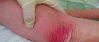
Toxicoderma in most cases occurs acutely
and is characterized by widespread, disseminated, symmetrical and monomorphic rashes consisting of macular, papular, nodular, vesicular, bullous, pustular and papulopustular pruritic elements (see figure on page 657). The process may involve mucous membranes.
Among spotted toxicoderma
can be distinguished: hyperemic, hemorrhagic (purpura) and pigmentary (toxic melasma from arsenic, metacycline, petroleum hydrocarbons or coal).
Spots of hyperemia can be located isolated from each other (roseola toxicoderma) or merge, forming large areas of hyperemia, sometimes reaching a universal skin lesion ( erythroderma
). The rash may be ring-shaped. When resolved, peeling is often observed; in the case of damage to the palms and soles, complete rejection of the stratum corneum is possible. When peeling occurs only in the center of roseola spots, the clinical picture resembles Zhiber's pityriasis rosea. Severe itching, connection with taking a drug or a poor-quality product, profuse rashes on the extremities, the presence of rashes on the face, relapses of the disease - all these symptoms are characteristic of toxicoderma.
For papular toxicoderma
characterized by disseminated lesions. A rash of flat polygonal papules appears, reminiscent of lichen planus; its appearance can be caused by long-term use of hingamine, quinine, phenothiazines, PAS, streptomycin, tetracycline, iodine preparations, mercury, etc.
Nodular toxicoderma
characterized by the formation of painful acute inflammatory nodes, slightly elevated above the skin level, with vague outlines (acute erythema nodosum).
For vesicular toxicoderma
the rash consists of large vesicles bordered by a narrow rim of hyperemia. The process is disseminated. Limited manifestations of vesicular toxicoderma in the area of the palms and soles (in the form of dyshidrotic vesicles) are possible.
Pustular toxicoderma
usually associated with exposure to drugs such as
bromine
,
iodine
, chlorine, and fluorine (halogens). Staphylococci located in the pilosebaceous apparatus, which are activated under the influence of the listed drugs, also play a certain role in their development. These substances are excreted from the body with sebum, so the manifestations of toxicoderma are more pronounced in areas richer in sebaceous glands (chest, face, upper back). The rash consists of pustules or acne (bromide acne, iodide acne). The cause of the development of acne toxicoderma may be vitamins B6, B12, isoniazid, barbiturates, steroids, lithium.
Among bullous toxicodermas
localized and disseminated forms are distinguished.
The localized form appears in a limited area and is called fixed. Fixed toxicoderma
is characterized by the appearance of one or several round-shaped spots, their diameter reaches 2 - 3 cm, after a few days they acquire a bluish and then brown tint.
A bubble will form in the center of some spots. The favorite localization of fixed toxicoderma is the genitals and oral mucosa
; rashes can also be localized in other areas of the skin.
When rashes appear on the oral mucosa, the blisters quickly open, forming erosions. Fixed toxicoderma develops as a result of taking sulfonamides, barbiturates, salicylates, antibiotics, chloral hydrate, arsenic
and other drugs. With each repeated dose of the corresponding drug, the process recurs in the same places, increasingly intensifying pigmentation, and gradually spreads to other areas of the skin. If the drug is stopped, the process resolves within 7–10 days; in case of relapse, the process may take longer.
Clinical picture of common bullous toxicoderma
may be similar to the manifestations of exudative erythema multiforme. The latter is supported by: predominant damage to the dorsum of the hands and feet, mild itching, seasonality of relapses (spring and autumn), general catarrhal phenomena, and lack of connection with medication use.
The most severe form of exudative erythema multiforme is Stevens-Johnson syndrome
– usually begins suddenly and acutely, with high fever. False films, yellow or white-yellow, form on the conjunctiva of the eyelids, which can be completely removed. They can occupy the conjunctiva of the eyeball and the cornea. Disappears within 3–6 weeks. In complicated cases, conjunctival scars and corneal cataracts remain. Almost simultaneously with changes in the conjunctiva, erythematous spots, blisters or tubercles, swelling and bloody-serous exudate appear on the skin on the lips, oral mucosa to the palatine arch with multiple films, with foul-smelling purulent discharge from the mouth, external genitalia.
In severe cases of toxicoderma
Among the general symptoms, functional disorders of the nervous system may be observed (irritability followed by a depressive state, insomnia, emotional lability, etc.), fever, accompanied by general malaise, fatigue, transient arthralgia, symptoms of damage to the cardiovascular system, including small vessels (which causes the development of a hemorrhagic component), as well as the liver and kidneys (drug-induced disease). Subjective symptoms are mainly reduced to a feeling of itching, burning, tension and soreness of the skin of the affected areas.
In addition to those listed, there are other, rarer forms of toxicoderma
.
Drugs such as sulfonamides, antibiotics, anticoagulants, barbiturates, salicylates can cause allergic vasculitis
.
Arsenic, with long-term use, can contribute to the appearance of toxicoderma in the form of hyperkeratosis of the palms and soles
.
Bromine and iodine cause bromoderma and iododerma
, which are characterized by the appearance of soft bluish-red plaques covered with purulent crusts; after their removal, the papillomatous surface of the infiltrate is exposed.
Some medications cause the clinical picture of toxicoderma that is characteristic of them. At the same time, the same drug substance can cause morphologically different forms of toxicoderma in different people. Almost all patients with infectious mononucleosis and cytomegalovirus infection develop a drug rash when ampicillin is prescribed. In 50–60% of HIV-infected people, when treated with drugs containing sulfonamides, a rash occurs. Ampicillin often has side effects during treatment with allopurinol.
If a drug has previously caused toxicoderma in a patient, then when this drug is re-prescribed, it will most likely occur again.
In approximately 10% of patients allergic to penicillins, cephalosporins cause a rash due to cross-sensitization. In 20% of patients sensitized to any drug containing sulfonamides, cross-sensitization to other such drugs occurs. Course
The course of drug toxicoderma, provided that the drug is quickly discontinued, is usually short-lived. However, in case of damage to the cardiovascular system and internal organs, the prognosis may be unfavorable. In terms of the severity of the prognosis and the difficulty of diagnosis, allergic myocarditis ranks first among lesions of internal organs. Sometimes a drug allergic reaction manifests itself in the form of drug-induced lupoid syndrome, similar to systemic lupus erythematosus, but which resolves quickly after the drug is stopped.
The course of toxicoderma caused by exogenous causes is usually acute. Toxicoderma of endogenous origin often occurs chronically.
Diagnostics Diagnostics
Establishing the cause of toxicoderma is not easy, and often impossible. The presence of symptoms of damage to internal organs, the nervous system, and blood vessels during drug toxicoderma has differential diagnostic significance. Differentiate bullous toxicoderma
on the genitals (in the erosive stage) follows from chancroid, genital herpes, erosive balanoposthitis;
roseolous, papular, pustular
toxicoderma - from the corresponding syphilides of the secondary period of syphilis.
Roseola
manifestations must also be differentiated from rashes associated with rubella, measles, and scarlet fever; the bullous form is distinguished from chickenpox, the pustular form from pyoderma.
When diagnosing drug toxicoderma, all other causes of the rash, in particular infectious ones, are excluded.
. This is not always easy to do: a rash due to a viral infection can be indistinguishable from a rash caused by any drug prescribed before treating this infection. The time that passed from the start of drug treatment to the onset of symptoms and the effect of drug withdrawal (whether there is an improvement) are assessed. Find out whether similar reactions to this drug have been observed previously.
To confirm the causal role of the suspected factor, various diagnostic tests are used, including skin tests. Provocative tests provide more convincing results
, in which the etiological factor is administered in the same way as the drug that caused toxicoderma was taken. So, if toxicoderma occurred when taking the drug orally, then the test should be carried out orally, prescribing the minimum therapeutic dose of the drug. The test is considered positive if a relapse of toxicoderma develops. Skin tests with the same substance may be negative. With fixed toxicoderma, only tests performed at the site of resolved lesions can be positive. Provocative tests require special caution and should not be performed in patients who have suffered a severe form of toxicoderma.
There are diagnostic tests performed outside the body
human, also allowing to confirm the etiological factor: leukocyte agglomeration reaction according to Fleck, basophil degranulation test according to Shelley, blast transformation reaction of lymphocytes, platelet reactions, hemolytic tests, etc. These tests are based on the reaction of the patient’s blood cells with the chemical substance that caused sensitization. Both false-positive and false-negative reactions are possible.
At biopsy
rashes most often show perivascular lymphocytic and eosinophilic infiltration.
Treatment
The most important condition for the treatment of toxicoderma is interruption of the effect of the etiological factor
(prescribed medications are canceled, released from work associated with exposure to occupational hazards, contact with household chemicals is excluded, a hypoallergenic diet is prescribed). To accelerate the removal of toxic substances from the body, diuretics and laxatives are prescribed; They restore the function of the gastrointestinal tract, liver, and kidneys in case of toxicoderma of endogenous origin. Intravenous administration of polyvidone is possible, as well as plasmapheresis and hemosorption.
It is better to hospitalize patients with toxicoderma, accompanied by an increase in body temperature and symptoms of general intoxication.
As desensitizing drugs
solutions of calcium gluconate, sodium thiosulfate intravenously or calcium gluconate intramuscularly are used.
It is more effective to alternate the two drugs every other day. Prescription of antihistamines
. In more severe cases, drugs are administered parenterally.
It is advisable to carry out enterosorption
(activated carbon and other sorbents). To improve the functioning of the gastrointestinal tract, enzymes and eubiotics (bifidum-, coli-, lactobacterin) are prescribed.
External treatment
usually carried out with antipruritic talkers, corticosteroid ointments (betamethasone, flumethasone, budesonide, etc.), creams and aerosols.
For severe toxicoderma, corticosteroid drugs are prescribed orally and intravenously (prednisolone, dexamethasone, hydrocortisone). Toxic-allergic bullous epidermal necrolysis
Toxic-allergic bullous epidermal necrolysis or Lyell's syndrome (LS) is the most severe form of drug-induced toxicoderma, described by Lyell in 1956. LS develops as a reaction to the combined effects of toxic, medicinal and infectious agents, occurring against the background of a high degree of hypersensitivity body.
Etiology and pathogenesis
The most common causes of LS are drugs
: sulfonamide drugs, antibiotics (erythromycin, penicillins, tetracyclines, streptomycin, etc.), barbiturates, analgesics (phenylbutazone, salicylates, paracetamol, piroxicam, diclofenac, cephalosporins, fluoroquinolones, etc. The disease can develop as a result of the combined use of several drugs. Other reasons SI can be caused by intoxication with chemicals or spoiled food.
The basis of the pathogenesis of LS is an allergic reaction.
As a rule, patients have a history of hypersensitivity to any drugs or substances. Often the development of the disease is preceded by an acute infectious process, for which a drug is taken, which subsequently causes acute epidermal necrolysis. Immune mechanisms include the cytotoxic effect of lymphocytes on epidermal cells. Infiltration of the epidermis with activated lymphocytes and macrophages is observed, releasing cytokines that cause cell death, fever and other manifestations. Clinical picture
The disease develops several hours or days after the first dose of the drug (from 1 day to 3 weeks). Sometimes there is a prodromal period
in the form of fever, weakness, headache, muscle pain, skin soreness, burning or itching of the conjunctiva.
The temperature suddenly rises to 39–40°C, a macular and/or petechial rash appears, urticaria or blisters may be observed
, often the rash resembles exudative erythema multiforme.
Often the first rashes appear on the mucous membranes
of the mouth, nose, genitals and eyes.
Over the course of several days painful erythroderma
, against which
the epidermis begins to peel off
.
A symptom of “wet underwear” is observed when, when touched, the skin begins to wrinkle, is easily pulled back and then rejected with the formation of painful erosions. Sometimes blisters with flabby tires appear. With light pressure on them, the area of the latter increases. A positive Nikolsky sign
is noted (when rubbing an apparently healthy skin, erosions appear in areas adjacent to the rash). Patients complain of severe skin pain, burning, increased sensitivity, and paresthesia.
Subsequently, there is a sharp deterioration in the general condition
.
The temperature persists, severe headaches, drowsiness or anxiety are noted, prostration is possible; symptoms of dehydration are observed - severe thirst, dry oral mucosa; as well as blood circulation due to blood thickening. Swallowing is difficult due to pain, patients refuse to eat. Kidney function is impaired (acute tubular necrosis), urination is painful. Ulceration of the mucous membrane of the trachea, bronchi, and gastrointestinal tract is possible. Diagnosis
At the onset of the disease, differential diagnosis is made with exudative erythema multiforme, scarlet fever, phototoxic reactions, maculopapular toxicoderma, and graft-versus-host disease. Late manifestations of Lyell's syndrome must be differentiated from thermal burns, exfoliative erythroderma, Stevens-Johnson syndrome, and acute febrile pemphigus. It is difficult to make a correct diagnosis before the development of necrolysis of the epidermis. An important diagnostic sign is the appearance of severe pain in both the rash and healthy skin.
In general blood test
Anemia, lymphopenia, and sometimes eosinophilia are observed. The occurrence of neutropenia is a poor prognostic sign. An increase in the number of band neutrophils (up to 16–55%) and toxic granularity may be observed.
During pathomorphological examination
Vacuolization and necrosis of keratinocytes of the basal layer, necrosis of individual cells in the thickness of the epidermis are detected.
Acantholysis leads to detachment of the upper layers of the epidermis. Edema and polymorphic perivascular infiltrates are noted in the dermis. To exclude other diseases accompanied by the formation of blisters, immunofluorescence testing is used. Course and prognosis
The disease progresses during the first 3 days. The further course is in many ways similar to the course of burn disease. The prognosis depends on the extent of necrosis. The larger the affected area, the higher the loss of fluid through the skin and the greater the electrolyte disturbances. Frequent complications include kidney failure, bacterial infections and sepsis. Cachexia (due to increased catabolism) and diffuse interstitial pneumonia may develop.
Mortality in Lyell's syndrome reaches 30%
, older patients die more often.
The causes of death are sepsis, gastrointestinal bleeding, and fluid and electrolyte disorders. In survivors of Lyell's syndrome, repeated administration of the same drug causes a relapse; the reaction develops faster and is significantly more severe than the first. Treatment
Treatment is carried out in the intensive care unit or burn department. It is necessary to maintain water-electrolyte and protein balance
, careful care and treatment of erosive surfaces to avoid infection.
Glucocorticoids and antibiotics are prescribed, as well as symptomatic treatment. It is extremely important to quickly identify and discontinue the drug that caused toxicoderma
.
When administering drugs that cause the disease per os, you must first rinse the stomach. The patient must be given intravenous drips of up to 2 or more liters of fluid per day (dextrans, polyvidone, plasma, saline), correct electrolyte disturbances, and provide parenteral nutrition. Anabolic steroids are prescribed. Glucocorticosteroids
are administered parenterally.
The initial dose corresponds to 120–150 mg of prednisolone. Choose an antibiotic with a broad spectrum of action
that does not have a nephrotoxic effect and a prolonged effect. Penicillins and tetracyclines should not be prescribed. It is necessary to check sensitivity to the selected antibiotic using medical history and the results of in vitro allergy tests.
Aqueous solutions of aniline dyes are used externally
(fucorcin, methylene blue),
corticosteroid aerosols
antibacterial lotions
on wet areas , then solcoseryl ointment or cream is prescribed. If the oral mucosa is damaged, astringents, disinfectants and analgesics (chamomile infusion, solutions of boric acid, potassium permanganate, nitrofural) are prescribed for rinsing. It is possible to lubricate the affected oral mucosa with novocaine, sodium tetraborate, and egg white. For the eyes, rinsing with boric acid solution, zinc drops, hydrocortisone ointment or drops is used.
Patient care is very important
.
The patient should be in a warm room equipped with bactericidal lamps. It is necessary to change underwear and bed linen (sterile) 2–3 times a day; instead of bandages, it is better to use gauze “shirts”; analgesics must be prescribed before dressings. Necrotic areas of skin are removed. Budesonide – Apulein (trade name)
(Gedeon Richter)
Flumethasone – Lorinden (trade name)
(Jelfa)
Betamethasone – Beloderm (trade name)
(Belupo)
| Applications to the article |
| Classification of toxicoderma: • medicinal; • amental; •professional; •autotoxic; • the substances caused by you are of flax and animal origin. |
| Drug toxicoderma accounts for 19% of all complications of drug treatment |
| Toxicoderma may be a consequence of another disease, incl. neoplasms |
| Signs of life-threatening drug toxicoderma Clinical picture: confluent erythema; swelling of the face or its central part; pain; palpable purpura; skin necrosis; blisters, epidermal detachment; Nikolsky's symptom (detachment of the epidermis with light pressure on the skin); erosion of mucous membranes; hives; swelling of the tongue. General condition: high fever (temperature above 40°C); enlarged lymph nodes; arthralgia, arthritis; shortness of breath, wheezing; arterial hypotension. Blood test: eosinophilia, lymphocytosis, atypical lymphocytes, changes in biochemical parameters of liver function. |
| With toxicoderma, the rashes are widespread, symmetrical, monomorphic |
| The course of drug toxicoderma, provided that the drug is quickly discontinued, is usually short-lived |
| An important diagnostic sign of Lyell's syndrome is severe pain in rashes and healthy skin. |
| Prevention The patient should be aware of his hypersensitivity to a particular drug and cross-reactions to other drugs. This drug cannot be re-prescribed. An appropriate entry must be made in the patient’s chart; it is recommended to wear an identification bracelet. Prescription of drug cocktails and polypharmacy should be avoided. |
| Rice. 1. Toxicoderma |
| Rice. 2. Atopic dermatitis |
| Rice. 3. Ichthyosis |
| Rice. 4. Lymphoma of the skin |
| Rice. 5. Mycosis of the feet caused by T.rubrum |
| Rice. 6. Onychomycosis |
Causes
The disease occurs when allergens enter the body along with food, injection solutions and inhaled air. The allergen can also enter the bloodstream through skin contact. The substance that causes an allergic reaction quickly spreads through the body and enters the skin. In rare cases, substances that cause a response from the immune system are formed in internal organs. Doctors include medical alcohol, bleaches, detergents, shampoos, dust, pollen and pesticides as common pathology triggers.
Possible reasons:
- Intoxication of the body with unusual metabolic products formed in the digestive system, kidneys or liver.
- Penetration of a drug that causes an allergy into the body. This can be any route of administration of the medication, including oral and injection.
- Alimentary penetration of the allergen (with food). Most often, foods contain additives that cause a reaction in the immune system.
- Air pollution from certain chemicals. In this case, the provocateurs of the disease quickly penetrate the blood vessels through the respiratory tract.
Congenital allergic reactions are caused by the transmission of certain genes from parents. In this regard, the study of family history plays an important role in diagnosing the disease.
Causes of toxicoderma
- Taking medications. Drug toxicoderma develops as a side effect during therapy. The disease can occur when using any medications. Often toxicoderma is provoked by:
- antibiotics;
- sulfonamides;
- B vitamins, ascorbic and nicotinic acids;
- blood serum products;
- antiepileptic drugs. In some cases, a reaction may occur to antihistamines and corticosteroids, gamma globulins, and anti-inflammatory drugs.
- Food allergens. Individual hypersensitivity to food is more often observed in pediatric patients. Common food allergens include egg whites, bananas, strawberries, chocolate, nuts, citrus fruits, sea fish and seafood, sausages, and canned food.
- The influence of industrial irritants. Occupational acute toxicoderma is a manifestation of toxic chemicals entering the body. It is observed when using kerosene, insecticides, dyes for clothing and fur, and medications.
- Accumulation of endogenous toxins. The autotoxic form of the disease is caused by metabolic disorders and is observed in the following conditions:
- chronic pathologies of the digestive tract (gastritis, peptic ulcer of the stomach and duodenum, liver disease);
- kidney damage (chronic pyelonephritis, glomerulonephritis, renal failure);
- malignant neoplasms of various localizations;
- helminthiases.
Risk factors
Dermatologists take into account not only the ways in which allergens enter the patient’s body, but also certain forms of predisposition to such a disease. Risk factors may be related to a person's lifestyle, personal history, and genetics.
Possible risk factors:
- Chronic diseases of internal organs, such as gastritis, pancreatitis or glomerulonephritis.
- The presence of diseases associated with pathological reactions of the immune system. These are Crohn's disease, urticaria, bronchial asthma, allergic rhinitis and other ailments.
- Use of untested vaccines and serums.
- Diseases of metabolism and endocrine system.
- Malignant neoplasms.
- Taking antibiotics, aminazine, quinine, corticosteroids and other medications that often cause allergies.
- Features of professional activity that lead to frequent contact with chemicals, such as dyes, varnishes and household chemicals.
- Frequent contact with plants and animals.
- Unfavorable family history due to diseases of the immune system in close relatives.
Taking into account risk factors is important for disease prevention.
Why does toxicoderma occur?
The main mechanism of disease development is toxic-allergic. This means that the substance circulating in the blood has poisonous (toxic) and allergic properties. However, it's not just about the external factor. The patient’s body initially has an allergic “mood,” or episodes of damage to other organs have already occurred before. The main prerequisites for the disease are hereditary enzymatic deficiency, individual intolerance, cumulation (accumulation) of drugs, interaction with the ultraviolet spectrum of solar radiation and others.
The immediate causes of toxicoderma are most often exogenous, that is, located in the external environment. The most common powerful allergens are:
- medications, they account for up to 20% of all cases;
- household chemicals or industrial hazards;
- food products;
- autotoxic compounds.
Among drugs, the most allergenic are antibiotics, vaccines and serums, non-steroidal anti-inflammatory drugs, sulfonamide compounds, barbiturates, vitamins and others. In some cases of the disease, there is a reaction even to corticosteroids and antihistamines, i.e. to substances that are usually used specifically to combat allergies.
Household chemicals are an equally common cause of skin lesions. This is especially true for compounds that have a benzene ring in their chemical formula. Most often, the skin reacts to washing powder, solvents and paints, and epoxy resin. Among the substances used in production, arsenic and mercury cause the greatest harm.
Among food products, those containing preservatives and dyes are dangerous. In this sense, even bread is dangerous if it is baked with the addition of a large amount of baking powder. Snacks, sausages, canned goods, and some semi-finished products are usually suspect. Chocolate, chicken eggs, seafood, honey, citrus fruits and coffee have natural allergenic properties.
Autotoxic toxicity is a skin lesion that occurs under the influence of its own pathological metabolic products. This happens with diseases of the kidneys and digestive tract, malignant tumors. Often autotoxic dermatitis develops during pregnancy, especially during its pathological course.
Increasingly in recent years, toxic dermatitis occurs in children; it is caused by a respiratory viral infection coupled with poor nutrition. Children who are prescribed many medications and pharmaceutical vitamins at the same time suffer.
It is important that with toxicoderma the irritating substance does not directly contact the skin, but enters the bloodstream.
Pathogenesis of toxicoderma
Allergens penetrate the body, overcoming natural barriers. Thus, substances that come into contact with the mucous membranes of the respiratory and digestive systems are easily absorbed into the capillaries and distributed throughout all tissues. Also, small organic molecules (haptens), characterized by chemical reactivity, bind to proteins and cause an allergic reaction. In skin cells, haptens activate special receptors and provoke an innate immune response.
In tissues, the immune system recognizes allergens and provokes the production of inflammatory mediators. Cells of the body's defense system also fight allergens and produce anti-inflammatory cytokines that help destroy haptens. The process of destroying allergens is accompanied by characteristic skin reactions, such as the formation of blisters and spotty rashes.
Mechanisms of disease formation that are not related to the body’s defense system:
- A local reaction of a certain substance, characterized by tissue irritation.
- Gradual accumulation of chemicals in the body.
- A deficiency of certain enzymes caused by a genetic mutation.
- Intolerance to a certain drug.
Only through careful diagnosis can the true mechanism of skin lesions be identified in a particular patient.
Fixed toxicoderma
Symptoms of fixed toxicoderma include the following:
- round or oval spots reaching a diameter of 3 centimeters;
- spots that have a brown tint on the 5th day from the moment of appearance;
- the bubble in the middle is an optional feature.
In order to stop the appearance of these rashes, you must stop taking the causative drug. In this case, after a week the skin rash will disappear without a trace. But if a person starts taking this medicine again, the rash will appear again. Moreover, in most cases they appear in the same places where they were before.
Sometimes a rash appears on the oral mucosa, but without inflammatory signs. In this situation, subjective symptoms are less pronounced. Often with toxicerma, disorders associated with the central nervous system are observed. Therefore, the following symptoms may appear:
- irritability;
- depression;
- insomnia;
- weakness;
- central itching of the skin.
Symptoms
Toxicoderma can manifest itself with a variety of symptoms. Different patients may have different areas of skin lesions and types of pathological formations. Most often, the first signs of inflammation appear in the mucous membrane of the mouth and eyes. Blisters filled with cloudy liquid, papules, vesicles and warts may appear on the surface of the skin. Skin reactions depend on the mechanism of the disease and the specific allergen.
Other symptoms and signs:
- redness and swelling of the skin;
- increase in body temperature;
- breathing problems;
- cardiopalmus;
- dizziness and weakness;
- burning of the skin and mucous membranes;
- pain in joints and muscles;
- anxiety and sleep disturbance;
- headache.
As complications arise, the patient may develop more dangerous symptoms.
Types of toxicodermy
Toxidermia is classified according to the type of course:
- Erythroderma is an inflammation of the skin, accompanied by redness, swelling, rash of papules and pustules, their erosion, formation of crusts, peeling, a tendency to coalescence and peripheral growth.
- Exudative erythema multiforme – characteristic red spots and blisters on the limbs, body, and mucous membranes. Sometimes accompanied by itching.
- Erythema nodosum is a painful, palpable subcutaneous nodule of red or purple color, often in the lower leg area.
- Lichen planus - itchy small purple papules that can coalesce into scaly plaques and can affect the mucous membranes of the mouth and genitals.
- Allergic vasculitis - subcutaneous capillary hemorrhages.
- Keratoderma is excessive keratinization mainly in the area of the palms and soles.
- Stomatitis is inflammation of the oral mucosa.
- Toxic epidermal necrolysis - intoxication, elevated temperature, formation of extensive weeping blisters with serous-hemorrhagic contents. Accompanied by secondary lesions of internal organs. Life-threatening condition. Source: I.Yu. Melnikova, V.M. Shaytor Toxic-allergic dermatitis in children // Issues of modern pediatrics, 2008, vol. 7, no. 4, pp. 68-74
Diagnostics
If symptoms of toxicoderma appear, you must make an appointment with a dermatologist or immunologist-allergist. During the consultation, the doctor will ask the patient about the complaints and carefully study the anamnestic data to suggest the cause of the skin changes. Then an initial examination of the skin is carried out, making it possible to assess the nature of the rash. To confirm the diagnosis, the doctor prescribes instrumental and laboratory tests.
Additional diagnostic methods:
- Blood analysis. In the treatment room, a nurse collects venous blood and sends the material to the laboratory. The immunologist will be interested in the ratio and quantity of blood elements. The biochemical composition of plasma also allows us to clarify the form of the disease. If necessary, serological diagnostics for infection are carried out: specialists look for specific antibodies in the blood that are produced by the immune system to fight pathogens.
- Skin scraping or exudate collection. The resulting material is studied in the laboratory. The use of special nutrient media allows a specialist to identify the causative agent of the disease or, conversely, to exclude the infectious nature of the disease. Often, a dermatologist will need to rule out sexually transmitted infections such as syphilis.
- Skin tests. The dermatologist places a small amount of the allergen on the surface of the patient's skin and observes the tissue reaction. The appearance of characteristic signs of allergy indicates the body's sensitivity to a certain substance. With toxicoderma, skin tests often do not reveal pathology.
- Provocative tests are the introduction of a suspected provocateur of the disease into the body. The doctor uses the minimum dosage. When symptoms of toxicoderma appear, the patient is immediately given medicine. This diagnostic method is not performed if there is a risk of a severe allergic reaction.
- Allergy tests outside the patient's body. For this purpose, blood is drawn. A test for basophil degranulation when exposed to a specific substance can confirm the diagnosis.
- Biopsy of skin lesions. The dermatologist removes a small area of damaged skin. Histological examination of the material reveals a large number of lymphocytes and eosinophils in the tissue.
- Examination of internal organs. If indicated, the doctor prescribes electrocardiography, ultrasound examination and computed tomography. In most cases, it is necessary to evaluate the condition of the cardiovascular system, kidneys and liver.
Diagnosing toxicoderma can be quite a complex undertaking, requiring the exclusion of many other diseases with similar symptoms. If necessary, the doctor prescribes the patient a consultation with a cardiologist, nephrologist or infectious disease specialist. It is important to suggest a differential diagnosis in time.
Our doctors
Orlova Tatyana Vladimirovna
Doctor - allergist-immunologist, pulmonologist, doctor of the highest category
Experience 37 years
Make an appointment
Shundeva Oksana Veniaminovna
Allergist, doctor of the highest category
Experience 38 years
Make an appointment
Treatment
The primary goal in treatment is to stop contact of the allergen with the patient's body. In severe cases, it is also necessary to eliminate the symptoms of the disease by suppressing the allergic reaction. Often, additional therapeutic procedures are required to cleanse the body of toxins. Maintaining the functions of vital organs allows you to eliminate the consequences of the most dangerous forms of the disease.
Main methods of treatment:
- Cleansing the body of toxins. Depending on the indications, the doctor prescribes diuretics and desensitizing drugs to the patient. These drugs, administered by injection, help relieve symptoms of the disease. If necessary, the patient is prescribed a cleansing enema. It is important to consider the cause of the disease, since in case of drug-induced toxicoderma, desensitizing solutions may be contraindicated.
- Suppression of an allergic reaction. The dermatologist prescribes antihistamines to the patient to prevent the effect of the inflammatory mediator on the tissue.
- Efferent therapy for severe intoxication of the body. The patient's circulatory system is connected to a special device that removes unwanted chemicals. At the same time, solutions of corticosteroids, dextran and blood plasma proteins are administered intravenously to the patient. Antibiotics are prescribed if necessary.
- Topical medications for treating damaged skin. Depending on the indications, the dermatologist may prescribe topical medications based on corticosteroids and other anti-inflammatory medications. Gels and ointments improve the condition of the skin, but do not affect the root cause of the disease.
In severe cases, treatment must be carried out in a hospital. Dangerous forms of pathology, such as Lyell's syndrome, may require resuscitation measures. After the condition has stabilized, the doctor prescribes the patient a hypoallergenic diet, antihistamines and other medications for constant use.
Common toxicoderma
This form of the disease is more serious. Its symptoms can be life-threatening. Common toxicoderma is characterized by the following symptoms:
- the appearance of a rash;
- temperature increase;
- chills;
- digestive disorders.
If you stop taking the allergen immediately after the allergy appears, you can easily break the vicious circle. The symptoms of this type of disease are similar to other processes - urticaria, lichen Giber, erythema, lupus. Therefore, it is very important to contact qualified specialists.
Complications
The hematogenous mechanism of spread of allergens is dangerous due to severe intoxication of the body and disruption of the functions of internal organs. If not treated promptly, complications may occur. In most cases, negative consequences are associated with the cardiovascular and excretory systems.
Possible complications:
- Inflammation of the muscular lining of the heart, manifested by shortness of breath, rapid heartbeat, weakness and general malaise.
- Renal dysfunction.
- Damage to liver tissue.
- Peeling of skin, as well as loss of hair and nails.
- Impaired blood supply to internal organs.
- The occurrence of an infectious disease.
- Toxic-septic shock.
Stopping an allergic reaction in the first hours after the onset of the disease usually prevents the development of dangerous complications.
Popular questions about toxicoderma
How does toxicoderma manifest?
The disease is characterized by the appearance of a rash and itching due to the entry of allergens into the body. In severe cases, general health worsens and symptoms of damage to internal organs occur.
How dangerous is toxicoderma?
With an increase in the activity and prevalence of the disease, internal organs may be affected with the development of severe complications requiring emergency specialized care.
What happens if toxicoderma is not treated?
In the absence of diagnosis and treatment, generalized toxicoderma and its serious complications, including Lyell and Stevens-Johnson syndromes, and shock conditions, may develop.
Prevention
Any diseases of the skin and internal organs that arise due to sensitization of the immune system can be successfully prevented. An immunologist’s medical recommendations usually relate to the patient’s lifestyle and diet.
Basic preventive recommendations:
- consumption of hypoallergenic foods;
- taking medications only under medical supervision;
- timely treatment of chronic diseases, including infection;
- ensuring quick access to antihistamines for diagnosed allergies;
- examination by an allergist and immunologist in the presence of pathologies of the immune system.
Compliance with all of the above recommendations significantly reduces the risk of initial occurrence of the disease and relapse. It is important to know the symptoms of an allergic reaction and always have emergency medications with you. If allergies are seasonal, the patient should be especially careful.
Treatment of toxicoderma in children
Therapeutic measures are aimed at prompt removal from the body of the substance that provoked this condition. Antihistamines, desensitizing, laxatives and diuretics are prescribed for internal use. For topical use, ointments, mash, and cooling creams are prescribed.
Extensive skin damage requires external treatment, similar to the treatment of burns.
A patient with toxidermia is prescribed a hypoallergenic diet and plenty of fluids to remove toxins and allergens from the body.
Toxidermia treatment should be carried out by an experienced dermatologist together with an allergist .
The information in this article is provided for reference purposes and does not replace advice from a qualified professional. Don't self-medicate! At the first signs of illness, you should consult a doctor.
Sources:
- I.Yu. Melnikova, V.M. Shaytor. Toxic-allergic dermatitis in children // Issues of modern pediatrics, 2008, vol. 7, no. 4, pp. 68-74.
- Federal clinical recommendations for the provision of medical care to children with exudative erythema multiforme and toxicoderma // Ministry of Health of the Russian Federation, Union of Pediatricians of Russia, 2015.
The information in this article is provided for reference purposes and does not replace advice from a qualified professional. Don't self-medicate! At the first signs of illness, you should consult a doctor.
Causes of toxic dermatitis
Toxic dermatitis can be caused by almost any drug, but the most common causes are antibiotics, nonsteroidal anti-inflammatory drugs, and drugs used to treat epilepsy. A drug rash appears a short time after starting to take the medication, and in most cases goes away immediately after discontinuation, but sometimes it takes two to three weeks. Some types of rashes are severe and may even be life-threatening and require hospital treatment, even if the patient is no longer taking the drug that caused the dermatitis.
Most often the reaction is caused by:
- Amoxicillin
- Biseptol
- Ampicillin
- Penicillin
- Blood products
- Cephalosporins
- Quinidine
- Gentamicin
- Diuretics
- Heparin
Cross-reactions occur between penicillins and cephalosporins, as well as between some anticonvulsants. For example, if a patient is allergic to ampicillin, cefazolin will also cause pathological changes in his skin.
Toxic skin damage is based on processes such as:
- Allergic reaction,
- Accumulation of toxic drugs in the dermis,
- Increased skin sensitivity to sunlight due to the influence of the drug,
- An interaction between two or more substances.
Drug rashes are more common in older people and affect women more than men. Other risk factors include:
- Taking antibiotics for a viral infection
- Weakening of the immune system due to some current disease, due to taking a drug,
- Malignant disease.
Symptoms of toxicoderma
The main manifestation of toxicerma is various polymorphic types of rashes on the skin, and sometimes on the mucous membranes:
- spotted, macular, and the spots can be isolated or merge, forming large areas of hyperemia , hemorrhage or pigmentation ;
- papular, nodular - the presence of dense flat inflammatory formations in the thickness of the skin;
- urticaria, with blisters - caused by swelling of the middle layer of skin - the dermis, disappear without a trace;
- pustular, with pustules - superficial or deep;
- vesicular - with the formation of blisters 1-5 mm in size - cavity elements containing cloudy or bloody fluid, their covers can open and cause erosion or turn into an abscess;
- bullous - large cavitary blisters up to 5 cm in size (as in the photo of toxicoderma caused by antibiotics), which can have serous, bloody or purulent contents, can subside and form a crust or open.
Types of skin rashes
In addition, toxicoderma may be accompanied by systemic manifestations:
- fever;
- weakness and malaise;
- joint and headaches;
- abdominal pain.
Symptoms of toxicoderma when interacting with the same substances can vary significantly from person to person.