Natural focal infections
According to the International Bureau of Epizootics, in recent years there has been a deterioration in the epizootic situation regarding foot-and-mouth disease in the world. Large outbreaks of foot and mouth disease have been reported in Mongolia, North and South Korea, Japan, Taiwan, Afghanistan, India, China, Kyrgyzstan, Tajikistan, Kazakhstan, and Turkey. There is a tendency to complicate the epizootic situation on the territory of the Russian Federation. Young artiodactyl farm animals (cattle, pigs, goats, sheep, deer) are most susceptible to infection. Foot and mouth disease causes great economic damage to livestock. In a matter of hours, one sick animal can infect hundreds. Sick animals must be destroyed.
Foot and mouth disease (snout-hoof disease , aphthous fever, epizootic stomatitis) (Aphtae epizooticae) is an acute viral highly contagious disease from the group of zoonoses (infectious animal diseases). The causative agent of foot-and-mouth disease is a filterable ultramicroscopic virus that belongs to the Picornoviridae family and is one of the smallest RNA-containing viruses. This infectious agent is characterized by high variability and stability in the external environment.
History of the study. The first report of foot-and-mouth disease in animals was made in Italy in 1546 by the Venetian physician Fracastoro. The first scientific information about foot-and-mouth disease in humans was published in Norway in 1764 by Sagar, who observed more than 1,500 cases of foot-and-mouth disease (aphthous foot-and-mouth disease). In 1897, F. Leffler and P. Frosch proved the viral nature of the pathogen; they established that the fluid of foot-and-mouth disease vesicles passes through bacterial filters, maintaining its virulence.
The name of the disease is apparently of Russian origin, borrowed from the local dialect. One of the leading domestic experts on foot and mouth disease, Professor A.L. Skomorokhov in his monograph “Foot and Mouth Disease” (1952) reports that in Russian literature, starting from the first half of the 19th century, this disease is described under the name “lizard-hoofed” or “foot-and-mouth disease” of livestock. In the Explanatory Dictionary of V.I. Dahl (1882) states: “Lizard - rough inflammation of the tongue in livestock, cracks along the tongue.” And further: “A lizard is rough skin, skin covered with a rash.” Translated from English, the name of the disease sounds like “damage to the limbs and oral cavity”, from German - as “damage to the mouth (muzzle) and hooves.” In this regard, in pre-revolutionary literature, foot and mouth disease was often called “snout-hoof disease.”
The main source and reservoir of the virus are domestic and wild ungulates. The pathogen is excreted by sick animals in urine, saliva, feces, and milk. Failure to comply with personal hygiene rules when in contact with sick animals can cause infection in people.
Humans are of great importance in the spread of the foot-and-mouth disease virus, since it most often comes into contact with animals and can travel long distances. The foot-and-mouth disease virus is mechanically transmitted through vehicles, poultry and other species of non-susceptible animals (including wild ones), as well as insects and ticks.
The main route of transmission of foot and mouth disease is through household contact; human infection occurs when the virus comes into contact with damaged skin or mucous membrane. Infection can occur through contact with feed and various animal care items, as well as through consumption of raw milk and undercooked meat from sick animals.
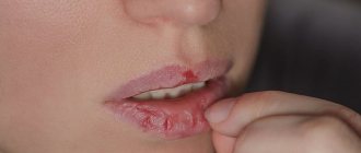
In the clinical picture , the disease in humans occurs with symptoms of aphthous stomatitis (aphthous stomatitis - superficial ulcerations, painful upon contact with any surface, very difficult to chew and swallow, and a concomitant symptom is hypersalivation (increased salivation)); herpetic sore throat against the background of influenza-like conditions (headache and muscle pain, including in the lumbar region); feelings of burning and dry mouth; as well as the appearance of vesicular-erosive (vesicle-ulcerative) lesions of the nail bed and the skin of the interdigital folds. The virus enters the body through the mucous membranes of the oral cavity (less commonly, the digestive and respiratory tracts) and damaged skin. At the site of introduction of the pathogen, a primary affect (focus of lesion) occurs - a small vesicle (bubble) where the virus multiplies and accumulates. The next stage is viremia (penetration of the virus into the blood), leading to intoxication. The pronounced dermatotropism of the virus causes its fixation in the epithelium of the mucous membranes (oral cavity, nose, urethra) and skin (hands and feet), where secondary vesicles are noted. With their appearance, the virus is not detected in the blood. Recovery usually occurs on the 10th - 15th day of illness. Patients with foot and mouth disease, regardless of the severity of the disease, must undergo treatment in a hospital , where they must remain for at least 14 days from the onset of the disease until complete clinical recovery, healing of ulcers on the mucous membranes and skin.
The infection is not transmitted from person to person.
Prevention. Due to its high contagiousness and resistance in the external environment, the virus does not lose its relevance to this day. To prevent the spread of foot and mouth disease among people, it is necessary to eliminate it among animals, which is achieved by establishing strict quarantine measures (fencing, disinfection of vehicles traveling outside the outbreak, etc.). Persons in contact with sick animals must observe a number of personal protective measures: work in protective clothing, do not drink water, do not eat food, do not smoke in the area of infection. Pregnant women, teenagers and people with microtrauma to their hands are not allowed to work in farms unfavorable for foot-and-mouth disease.
In Russia and some other countries, inactivated vaccines are successfully used to prevent foot and mouth disease among animals, which create immunity to the disease 2–3 weeks after vaccination.
Memo to the public:
- if external signs of disease are detected in animals or their sudden death, you should immediately contact the state veterinary service;
- do not self-medicate, do not approach if a corpse is discovered;
- do not purchase animals without accompanying veterinary documents confirming the epizootic welfare of the area and the health of the animals;
- at the request of veterinary specialists, provide animals for clinical examination;
- notify the state veterinary service about newly acquired animals;
- movement of cattle with permission from the head of the state veterinary service.
Pathogenesis
Penetration of the pathogen occurs through the mucous membranes, where the virus remains in the epithelial cells and begins to actively multiply. At the site of reproduction, swelling occurs, turning into an inflammatory focus. Aphthae are formed - a distinctive feature of foot and mouth disease. Otherwise, the animal feels stable, without changes.
Within a day, the virus enters the bloodstream from the primary lesion sites and penetrates into organs and tissues. This leads to an increase in temperature, causes an immune reaction, which leads to the destruction of the virus everywhere except the mucous membranes, which are less well supplied with blood. Subsequently, active reproduction of the pathogen occurs in the tissues of the epidermis.
Thus, new aphthae appear in the mouth, interhoof space, corolla, and udder. In severe cases, the heart is also affected.
Pathological changes and diagnosis
When examining a corpse, aphthae and erosions are found on the mucous membranes of the mouth and nose, less often on the mucous membranes of the intestines and in the proventriculus of ruminants. During autopsy of young animals, inflammatory processes are found, which are accompanied by bleeding in the intestinal area, “tiger heart”.
In the malignant form, cardiac changes are found - the muscle is pale, flabby, covered with gray-red spots. Hemorrhages are found under the epicardium, and signs of tissue degeneration are found in parenchymal organs.
Most often, making a diagnosis of foot and mouth disease, due to the characteristic clinical picture, does not present much difficulty.
Diagnosis of foot and mouth disease is complex. They take into account the epizootic situation of the region, clinical signs, laboratory tests, etc. Excised lymph nodes, aphthae and their contents, blood samples taken during the period of fever or blood serum are sent to the laboratory.
Patent material is sent frozen, in a sterile, hermetically sealed container using a preservative solution. The container is sent with a courier and a cover letter.
Control measures and prevention
First of all, to prevent foot and mouth disease, it is necessary to monitor the veterinary and sanitary condition of farms, to prevent contact between farm animals and wild animals, and not to replenish herds with animals from unfavorable areas. In disadvantaged regions, preventive vaccination is carried out.
If quarantine is established, sick animals are killed and the corpses are disposed of. Visually healthy animals are slaughtered at slaughter stations. In case of widespread spread of the disease, all clinically healthy animals are vaccinated.
Quarantine is lifted three weeks after the last clinical case of the disease has been eliminated, subject to vaccination of live stock and final disinfection. By-products and meat from recovered animals are used in the production of canned food and cooked sausages.
Course and symptoms
Foot and mouth disease occurs in an acute form. In adult animals, an abortive form occasionally occurs, which ends in recovery. In cattle, the incubation time is up to 7 days, more often 1–3 days.
There are four forms of foot and mouth disease:
1. Benign.
2. Malignant.
3. Abortion.
4. Asymptomatic.
Adult animals, as a rule, become ill in a benign form. The clinical picture is accompanied by high fever, tachycardia, depression, a significant decrease in appetite, inability to take food and disruption of the rumination process in ruminants. On the 2nd–3rd day of illness, aphthae appear on the skin of the lips, nose, in the oral cavity, between the hooves, and on the udder. At the beginning of the disease, the aphthae are small, but later they unite and increase in size, first to the size of a pea, and then to the size of a medium-sized apricot. On average, after a day the walls of the aphthae are destroyed and erosions appear. During this period, the animal's temperature returns to normal. During the examination, abundant secretion of foamy saliva is observed, the animal smacks its lips, and often licks the nasal mirror.
Erosion heals within a week, but if the animal is weakened, the process may take a longer period. Aphthae in the area between the hoofs leads to lameness. Animals lie down a lot and refuse to get up. With timely veterinary care and maintenance in dry conditions, erosions from aphthae disappear within a week. If the damage to the legs is extensive, then phlegmon of the corolla, pododermatitis and arthritis occur.
In lactating animals, aphthae on the skin of the udder quickly grow to the tip of the nipple and penetrate the mucous membranes of the nipple canal. Due to inflammation, the milk of such cows becomes slimy, bitter, and sour. Cows develop mastitis and milk production decreases. In some animals, digestion is disrupted, diarrhea and constipation occur.
In small calves, foot and mouth disease occurs without aphthae, but with signs of severe gastroenteritis and, often, in the absence of veterinary care, leads to the death of young animals. Often foot and mouth disease in calves is complicated by bronchopneumonia. In a benign form, the disease lasts up to 10–12 days, with complications up to a month.
The malignant form initially occurs with the classic symptoms of foot and mouth disease, but during the recovery stage the patient’s condition worsens. Animals become weaker, they experience tachycardia, refusal to eat, and impaired chewing. Some begin to experience paralysis. Death occurs due to cardiac arrest.
The incubation time in pigs can last up to 8 days. The disease occurs in an acute form and is accompanied by the death of young animals. In addition to fever and lethargy, the limbs suffer greatly. Lameness is noted, and in severe cases the hooves fall off. Aphthae cover the snout, milk bags, and the oral cavity. FMD is difficult for piglets to tolerate and mortality can reach 100% in the first 48 hours of illness. Severe course is accompanied by hematomas on the mucous membranes and parenchymal organs.
In sheep, the period of development of the disease is up to three days. The disease is milder than in cattle. Sheep practically never drool. Aphthae the size of millet grains, quickly open and heal, lameness is noted. In the flock, classic symptoms of foot-and-mouth disease appear: aphthae and erosions in the oral cavity, on the legs, and the udder. Fever, lack of appetite, lethargy and cessation of chewing gum. However, there are often cases when the disease is asymptomatic; sheep remain carriers of the virus for a long time. Lambs suffer from the disease with difficulty; foot and mouth disease causes the death of most of the young animals.
Goats get sick in a milder form than cattle. On the first day, symptoms are similar to those in large ruminants. Later, the goats close their jaws and teeth grinding is noted. There is no drooling, the animal recovers within two weeks. In rare cases, goats contract malignant foot-and-mouth disease.
In deer, the gastrointestinal tract is affected and diarrhea is observed. Diseases of the limbs are sometimes accompanied by the development of necrobacteriosis. In the absence of additional pathogenic microflora, the animal recovers within two weeks.
Epizootic data
Foot and mouth disease is a disease of animals that belong to the order Artiodactyls. The virus can infect any wild or domestic artiodactyl, but cattle are most susceptible. Pigs, goats and sheep are less frequently infected. A person can also suffer from foot-and-mouth disease, but this is where the chain of transmission of the virus ends. In humans, the disease is rarely transmitted to others and cannot provoke an epidemic. It is treatable and does not cause complications with timely diagnosis and medical care.
The source of the causative agent of foot-and-mouth disease in animals is infected individuals. The virus is contained in biological material that they release into the environment. There is especially a lot of it in saliva, it is also found in urine and feces, milk, and the contents of aphthae. It is also possible for the virus to spread aerogenously, through inhaled air. It can be transported over long distances, remaining on feed, staff clothing, and in transport, thereby ensuring a high degree of infectiousness.
After clinical recovery, animals remain carriers for up to 400 days. They continue to release the virus into the environment, posing a danger to healthy livestock.
The causative agent of the disease
The causative agent of foot and mouth disease in animals is an RNA virus that belongs to the Picornaviridae family. It enters the body of animals with water and food, as well as with inhaled air. The danger lies in the high resistance of the pathogen in the environment. It can persist on animal fur for up to 50 days, and in feed and soil for up to 150 days. However, the virus immediately dies when boiled, as well as when exposed to certain disinfectants (formaldehyde, sodium hydroxide).
In total, there are 8 serotypes of foot and mouth disease virus. They differ in structure, but cause a similar clinical picture of the disease. When diagnosing, the type and subtype of the virus must be determined. This is important data, since animals that have recovered from the disease develop short-term immunity, but only to a specific serotype of the virus. On average, it lasts 12–18 months, after which the animal again becomes susceptible to the virus.


