- Wart removal
- Diagnosis of neoplasms
- Removal of condylomas
Plantar warts look like round or oval skin lesions with a diameter of no more than 1 - 2 cm. In most cases, they are not distinguished by color, but may have a pink or brownish color. Over time, the protrusion becomes rough and rough to the touch due to constant friction.
Treatment methods
Depending on the severity of the problem and individual contraindications, plantar tumors are removed in different ways:
- electrocoagulation with high-frequency current;
- cryodestruction with liquid nitrogen;
- surgery;
- conservative drug therapy;
- radio wave exposure;
- laser technique.
Laser removal is recognized as the most effective and safe in modern medicine. Light radiation burns damaged tissue without injuring nearby ones. At the same time, neighboring cells are “sterilized,” which eliminates the risk of re-formation of plantar warts. The non-invasive, non-contact method eliminates the possibility of infection, does not cause discomfort, and has a short rehabilitation period. Another advantage of the laser is a reduced list of contraindications. The technique is also used in patients with insufficient blood clotting, a tendency to allergies, weakened immunity, and impaired metabolism.
Laser removal of warts on the elbow. Wart removal in progress. No traces remain
Laser removal of warts on the finger. Removing warts on hands with laser. Fast and painless.
Laser removal of multiple warts on the hand. Laser removal of warts
What do plantar warts look like?
Plantar warts, or, colloquially, thorns, are benign manifestations of several strains of the human papillomavirus. Unlike their “brothers”, which usually occupy the more delicate skin of the hands, neck or face, these tend to go where the epidermis is rough and subject to constant friction.
They are discovered by accident. The main symptom is a sharp stabbing pain in the foot or between the toes, as if from a pebble or splinter caught inside. Upon examination, raised round keratinized plaques of white or yellowish color with depressions in the center become visible. Sometimes dark spots appear inside - damaged capillary vessels of the dermis. The surface of the skin in the affected areas is usually rough or glossy smooth. There is no characteristic papillary pattern on it. This is the main external difference between papillomas and ordinary corns. Dry calluses always retain their skin pattern.
When pressing on the spine, a sharp pain is felt. The keratinized cells grow deeper, irritating healthy tissue as they move. In advanced cases or when trying to pick off such a “callus,” blood appears. Damage to warts often provokes the development of secondary infections, which leads to suppuration. Such neoplasms do not pose an oncological danger, but cause severe physical discomfort.
In what cases is tumor removal prescribed?
Experts recommend seeking help if there are indications:
- pain when wearing tight shoes or walking for a long time;
- bleeding in the affected area;
- uneven coloring of the neoplasm;
- rapid growth, association of individual lesions into masses.
If you still have doubts about whether it is worth removing the tumors, seek the advice of a specialist.
Where do plantar warts come from?
HPV enters the body from the outside. Pathogens love moist environments. You can become infected by wearing someone else's shoes, neglecting the rules of hygiene in pedicure salons, baths, saunas or swimming pools. A virus suppressed by the immune system may not manifest itself for years. But in a weakened body, viral particles begin to divide very quickly. The propagation of plantar strains is facilitated by:
- increased sweating of the feet;
- uncomfortable tight shoes;
- microcracks in the skin;
- fungal infections of the feet;
- circulatory disorders due to joint deformation: arthritis, arthrosis, flat feet.
Within the epidermis, the virus first infects the basal layer. Then it provokes the proliferation of keratinized particles, forming characteristic hard plaques on the skin.
The main symptom of this form of HPV is severe pain while walking. The roots reaching the lower layer of the dermis constantly injure the nerve endings. Many patients have to give up their usual shoes and use thick, soft insoles to alleviate the pain.
Contraindications to laser treatment
- infections and exacerbation of chronic pathologies;
- diabetes mellitus in the stage of decompensation;
- epilepsy and mental disorders;
- history of oncological pathologies;
- pregnancy and lactation;
- exacerbation of herpes.
| Cost of removing plantar warts | |
| Plantar warts up to 5 mm | 2950 rub. |
| Plantar warts from 5 to 10 mm | 3750 rub. |
| Plantar wart from 10 mm to 3 cm | 4900 rub. |
Sign up for a consultation
Spread of infection
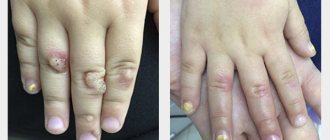
Such warts are usually transmitted through personal contact. Common methods of spread are through common bathing areas. The infection enters the skin if it is damaged - cut, scratched, abraded or ulcerated in any way.
Infection can also occur if there are scratches, damage from nail treatments and shaving. This leads to the spread of the virus and the formation of warts in different parts of the body in the same person.
Treatment of toenails
Preparation and carrying out the procedure
The appointment of a session is preceded by a consultation with a specialist. The doctor examines the tumors, selects the necessary laser parameters, and collects anamnesis to eliminate the risk of contraindications. If necessary, additional laboratory and instrumental studies are prescribed.
Before the procedure, the target area is decontaminated and treated with a local anesthetic. In combination with the laser cooling system, it provides complete patient comfort. Using the device's manipulator, the doctor acts on the affected skin in a targeted manner. High temperature burns damaged cells at a given depth, disinfects the wound surface and coagulates blood vessels. The whole procedure takes only a few minutes.
To completely remove a plantar wart, one session of laser treatment is usually sufficient. After treatment, the affected epidermis becomes covered with brown granulations, which disappear over time and leave clean, young skin.
Symptoms and types of warts on the foot
A plantar wart appears as a callus-like thickening with a stratum corneum layer of skin. It interferes with walking and causes pain. The passive state is characterized by slow reproduction, not reaching the stratum corneum of the epithelium, so this condition does not manifest itself externally.
The active state is characterized by the fact that the virus develops rapidly and, rising to the upper layers of the epidermis, manifests itself in numerous symptoms. The plantar wart is also called the spinous wart, chicken wart. The virus enters during its contact with the skin through cuts and abrasions in the outer layer of the skin:
- First, a small yellowish-gray papule with an uneven surface appears.
- Gradually, the small element becomes dense and acquires a dirty color.
From the inside, a plantar wart looks like fused papillae of different sizes with a pinkish tint. Additional capillary vessels form there, causing bleeding if you catch a wart.
Photos of completed work
Warts - what are they?
Neoplasms of a benign nature of viral etiology that develop on the epithelial and mucous membrane are called warts.
Most often they occur in young people or children, but there are exceptions; they can also appear in older people. All warts clinically have the form of a papule, which in turn is an elevated formation on the skin. Under a microscope it resembles broccoli. It grows deep in the layers of the skin and has enhanced nutrition.
There are the following types of warts:
- vulgar or ordinary type,
- flat type, characteristic of adolescence and childhood,
- plantar warts,
- warts of old age,
- and pointed type (condylomas).
The treatment process affects all types differently, that is, some of them can be quickly treated, and some are eliminated by surgery or laser therapy.
Why you need to get rid of warts
Up to 90% of neoplasms on the soles do not affect health and life. A few growths just appeared. And after a couple of months they disappeared. Sometimes people do not have time to visit a doctor, but the warts already disappear, without medications or therapy. And no problems with unpleasant sensations.
Unfortunately, not everyone is so lucky.
The remaining 10% of growths can make life extremely difficult - sometimes they cause severe pain when walking and limit mobility.
How to get rid of warts on the foot?
The first step is to be examined by a dermatologist. The doctor makes the diagnosis easily.
Treatment is prescribed if the formation:
- Constantly growing
- Causes the appearance of other neoplasms
- Hurts badly
- Makes it difficult to walk normally
Typically, patients like to treat warts with medications and avoid surgery.
Ask your dermatologist about topical treatments for plantar growths. There are many such drugs. They gradually destroy tumor cells. But they don't give a 100% guarantee.
Drug methods act on warts very slowly and sometimes take months to remove the growths. What’s even worse is that the tumors may return.
Yes, this does not always happen, but such cases are not uncommon. Therefore, to effectively treat viral warts on the foot, we recommend using one of the surgical methods.
There are many treatment options.
Comparison of wart removal methods: laser and nitrogen
Most patients are interested in which of the listed methods is most effective. Most often they choose between laser and liquid nitrogen. Each of these technologies has its own advantages. For a laser it is:
- high efficiency,
- speed,
- painlessness,
- no postoperative bleeding.
In addition, the laser allows the surgeon to act in a targeted manner, avoiding nearby healthy tissue. At the same time, he directs a heat beam at the wart, which evaporates the moisture contained in the growth and destroys its structure. Due to the lack of contact with the skin, wound infection during the procedure is completely excluded.
When choosing cryodestruction technology, you should be prepared for the fact that 3-5 procedures will be required to completely remove the tumor. This is due to the fact that the method involves layer-by-layer exposure of pathological tissues to low temperature. If the growth is small, then one treatment is sufficient to freeze and kill it.
Another selection criterion is the cost of therapy. Removing papilloma using cryodestruction is relatively cheap, but the more procedures are required, the more expensive such treatment will ultimately cost. In addition, when exposed to liquid nitrogen, there is a small risk of damage to healthy tissue, and hence the appearance of a scar.
Only a doctor can determine which of the listed methods will be most effective. But to come to a final decision, he must collect anamnesis, conduct an examination of the patient and send pathological tissues for histology.
How does the removal itself take place?
At the appointment, the doctor clarifies complaints, the duration of the disease, conducts an examination, and clarifies the presence of concomitant diseases and allergies to medications.
If the diagnosis is confirmed and there are no contraindications, intervention is performed. After treating the skin with an antiseptic, local anesthesia is administered with a thin needle. This may cause some pain (like any injection), but then the removal itself is completely painless. After making sure that the anesthetic has worked, the doctor destroys the wart with a radio wave scalpel. After removal, the wound is treated with fucorcin and a bandage is applied. The entire intervention takes 2–3 minutes.
Removal of warts by cryodestruction (liquid nitrogen)
Cryodestruction is a modern method of treating superficial benign neoplasms, in which pathological tissues are first cooled to an extremely low temperature and then destroyed. Liquid nitrogen is used as the active substance. It is under its influence that benign growths die, and cells with a disturbed structure slow down their growth.
Cryodestruction of warts and other neoplasms includes the following stages:
- Exposure to liquid nitrogen. The doctor's main instrument is a wooden stick with cotton wool or gauze wound at one end. With its help, liquid nitrogen is removed from the container and applied to the growth. After this, the doctor presses the stick onto the wart and holds it for 5-30 seconds.
- Break for 1-2 minutes. A pause is needed to assess the effectiveness of the impact. First, the skin turns white, then thaws, which allows the doctor to determine the magnitude and depth of exposure to the substance. Based on this, he decides whether to repeat the procedure or not.
- Result. After all exposures, the skin becomes whitish-pink, which indicates the complete killing of pathological cells.
The next day after the procedure, a bubble filled with colorless or reddish liquid appears on the treated area. The color of the liquid is affected by the depth of exposure. The blister cannot be covered with adhesive tape. If necessary, it can be covered with a gauze napkin, and then secured with an adhesive plaster. During water procedures, it is important to maintain the integrity of the bladder. If the treated area causes pain, you may take a painkiller.
Sometimes warts do not respond immediately to liquid nitrogen. Therefore, if necessary, 3 weeks after the first correction, the doctor repeats the procedure. Usually two sessions are enough.