Angioma is a benign vascular formation. As a rule, the neoplasm does not cause discomfort to its owner. Unless it causes deformity if it is located on the face, neck, or other open parts of the body. But sometimes small red spots are dangerous. Pathology can degenerate and spread. Therefore, vascular anomalies always require observation, and in some cases, treatment.
Let's figure out what angiomas are, why they appear, and how to remove vascular pathology. Frequently asked questions from patients are answered by doctors at the Lasersvit Center for the Diagnosis of Moles and Vascular Pathologies. We conduct consultations in Kharkov, offering children and adults ultra-precise diagnosis, treatment and prevention of skin and cutaneous-vascular abnormalities.
If you have an angioma on your skin, we suggest you make an appointment with a dermatologist. We will not only conduct a thorough diagnosis of the skin, but also tell you which skin tumors pose a hidden threat. A comprehensive study allows you to accurately determine the need for treatment of skin abnormalities.
Make an appointment with a dermatologist
Important : Angioma is often compared to a red mole. However, the vascular formation is not a mole. Red moles are formed by melanocyte cells, and skin angioma is a bundle of dilated lymphatic or blood vessels. In the first case it is a lymphangioma, and in the second it is a hemangioma.
Vascular angiomas appear in various parts of the body. They can be seen on the face, torso, and scalp. Medical statistics show that 80% of people over 50 have these formations. Sometimes the tumors disappear on their own. This happens with infantile hemangiomas. But often their number only increases with age. There is even such a thing as multiple senile angiomas. They appear due to the fragility of capillaries and their loss of elasticity. The vessels dilate and are unable to regain their previous diameter.
Types of angiomas
According to the etiology and pathogenesis, formations caused by the dilation of small vessels are divided into hemangiomas, formed from a cluster of capillaries, and lymphangiomas, consisting of small lymphatic vessels. Formations of the lymphatic system are less common. Their peculiarity is that the skin with this pathology does not change color. Flesh-colored nodules simply appear on the skin. Angiomas on the body are another matter. This disease always causes red spots to appear. And according to the type of structure, such formations come in several types.
Simple angiomas
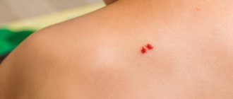
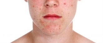
Vascular formations of this type are, as a rule, congenital pathologies. They can be smooth or protruding above the surface of the skin. The color of the new growths can be scarlet, burgundy, sometimes with a bluish tint. Vascular angioma grows on any part of the body, but mainly on the upper part of the body. The formation of dilated capillaries reaches a size of 10 cm in diameter. The anomaly is not dangerous, but its presence on the skin can lead to emotional imbalance. After all, the stain greatly spoils the appearance and becomes the reason for the close attention of others and their ridicule. Capillary hemangioma can especially ruin life when it is located on the face, neck, or arms.
Cavernous angiomas
A benign formation of this type looks like a pulsating purple spot. The structure of the pathology is characteristic. Blood enters the cavernous chambers through narrow arteries and is drained from the formation through wide venous channels. The spot is soft on palpation, and after squeezing it quickly restores its shape and appearance.
Branched angioma
The pathology often affects the limbs, less often appears on the scalp. The structure of the formation looks like intertwined dilated vessels. The skin over such a pathology is often affected by simple angioma. This is why capillary hemangioma must be deeply investigated. After all, a branched angioma may be hidden under it.
Intraosseous angiomas
These formations develop on the bones and are detected when they spread to nearby tissues. Bleeding becomes a symptom of the pathology. Accurate diagnosis is possible after obtaining radiographic images. The photo clearly shows the boundaries of the pathology.
Of all the listed types of angiomas, it is simple capillary formations that affect the skin.
BENIGN TUMORS OF MUSCLE TISSUE
Tumors of muscle tissue are divided into smooth muscle tumors - leiomyomas, and striated muscle tumors - rhabdomyomas. Tumors are quite rare.
Leiomyoma
Mature benign tumor. Occurs at any age in both sexes. It is often multiple. The tumor can become malignant. Treatment is surgical.
Leiomyoma, developing from the muscular wall of small vessels - small, often multiple, poorly demarcated and slowly growing nodes, often with ulcerated skin, is clinically very similar to Kaposi's sarcoma.
Genital leiomyoma is formed from the muscular lining of the scrotum, labia majora, perineum, and nipples of the mammary gland. May be multiple. Cellular polymorphism is often observed in the tumor. Hormone dependent. Treatment is surgical.
Angioleiomyoma from the trailing arteries
Clinically, a sharply painful tumor that can change size under external influences or emotions. The sizes are usually small, more often found in older people, on the limbs, near the joints. It is characterized by slow growth and a benign course.
Rhabdomyoma
A rare mature benign tumor based on striated muscle tissue. Affects the heart and soft tissues. It is a moderately dense node with clear boundaries, encapsulated. Metastases of rhabdomyoma have not been described. Relapses are extremely rare. Microscopically, 3 subtypes are distinguished - myxoid, fetal cell and adult. Rhabdomyoma of the female genitalia is also distinguished. The adult type mainly recurs.
Angiomas: causes of appearance
Until now, scientists cannot name the exact reasons for the appearance of vascular formations. Doctors agree that vascular pathology begins during embryonic development. Infectious diseases suffered by the mother can affect this. Toxicosis, anemia of a woman during pregnancy, and serious hormonal fluctuations also contribute to the appearance of abnormalities. Acquired angiomas occur due to sunburn, liver disease, skin injuries, and increased fragility of blood vessels.
Angiomas in children
Angioma in children is usually diagnosed at birth or during the first weeks of life. Girls get sick twice as often as boys. Angioma in a child goes through several stages. In the first few months, the formation actively grows, the size of the spot increases. At the next stage, growth stops. By the age of 9, in 90% of cases, self-healing occurs - spontaneous involution of the pathology. The stain disappears, leaving no traces in its place.
Unfortunately, in some cases, benign formation is accompanied by the following complications:
- ulceration and bleeding of the skin as a result of trauma, for example, due to rubbing with a diaper when the formation is located on the leg or buttock;
- formation of scars at the site of healing ulcers after ulceration of the skin at the site of formation;
- dysfunction of internal organs due to the germination of the formation or compression of them by its boundaries;
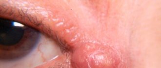
- When the formation is located on the forehead, ophthalmological diseases develop.
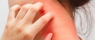
Important: Very often, angioma in a child is located on the face, neck, or chest. This leads to a pronounced cosmetic defect.
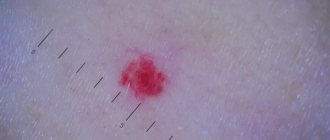
It is recommended to observe uncomplicated disease in children. To do this, during the appointment, photographs of the formation are taken using a dermatoscope. At subsequent appointments, images can be compared to analyze the behavior of the formation.
Diagnostics
Angiomas located on the surface of the skin are diagnosed during examination and palpation. The characteristic coloring, ability to contract and increase in size when pressed and strained allow the doctor to easily make the correct diagnosis.
If the localization of the lesion is more complex (internal organs, bones, brain), a number of diagnostic measures are required:
Skeletal hemangiomas are identified using x-rays of the spine, ribs, bones, skull and pelvis.
To identify angiomas of internal organs, antiography (contrast x-ray examination of blood vessels) of the kidneys, brain, lungs, etc. is performed.
Pharyngeal angiomas are examined by an otolaryngologist.
Ultrasound diagnostics allows you to determine the exact depth of growth of the angioma, its structure and location features. If a vascular angioma is suspected, the doctor may additionally prescribe a puncture. The resulting yellowish liquid is examined in the laboratory. This makes it possible to differentiate angioma from lymphadenitis, lipoma, cyst, hernia.
Angiomas: treatment
Angioma in adults and children is easily diagnosed. The doctor performs a dermatoscopy to examine the formation. A digital dermatoscope repeatedly magnifies the skin, allowing you to determine the depth, nature, and boundaries of the pathology. In some cases, angiography, blood flow analysis, and histology may be prescribed.
Important: Many patients are interested in why angiomas are removed if the formation is benign and non-aggressive. The main reasons have already been mentioned above. A large angioma on the face disfigures the appearance and there is a risk of complications. In this case, it is enough to observe congenital small spots, since there is a high probability of their disappearance without a trace.
When an angioma on the body or face is perceived as a serious aesthetic defect, if the risk of complications is high, in case of bleeding the formation must be removed. Today, several methods of treating vascular formations are practiced: radio wave therapy, cold treatment (cryotherapy), electrocoagulation, surgery and laser treatment. Each method has its own advantages. But there are also disadvantages. For example, removing part of the skin with a scalpel leaves scars and does not allow you to work with pathology accurately. Cold treatment can negatively affect adjacent tissues. And electrocoagulation is not suitable for neoplasms of significant depth and large area.
The most effective treatment for pathology of any complexity is laser removal of angioma. The laser beam removes abnormally dilated capillaries without affecting adjacent tissue. The doctor controls the intensity and depth of the impact, so he works with high precision. It is important that the treatment is bloodless, painless, and safe. The beam also cauterizes blood vessels, preventing infection. There are no scars left on the skin.
Important: Many people try to get rid of senile moles and red spots on their legs, arms, and face with dubious means at home. Aggressive agents and various ointments, including those containing hormones, are used. Doctors strongly do not recommend doing this, since you most likely will not be able to get rid of the pathology. But serious harm to health can be caused. Moreover, it is not difficult to treat skin lesions professionally today.
In each case, the dermatologist practices an individual approach to the patient. This allows you to get optimal results.
Important: If the formation does not bother you and is located in an inconspicuous place, you can replace treatment with a wait-and-see approach. Because there is a high chance that it will disappear.
BENIGN TUMORS OF PERIPHERAL NERVES
Traumatic or amputation neuroma
Arises as a result of post-traumatic hyperregeneration of the nerve. It is a small painful node.
Neurofibroma
A single, slowly growing benign tumor of the mesenchymal sheath of the nerve trunk of any location, but most often develops on the sciatic nerve and intercostal nerves. Occurs in people of any age. Clinically defined as a small, densely elastic consistency with a smooth surface of the tumor node, upon palpation of which the pain radiates along the nerve. Some tumors can reach large sizes. Tumor growth can occur both to the periphery of the nerve and in the thickness of the nerve trunk, which is revealed during its morphological examination.
Treatment is surgical. The prognosis is good. A special disease is multiple neurofibromatosis (Recklinghausen's disease), which belongs to the group of dysplastic processes. Cases of malignancy of one of the multiple neurofibromas in this disease have been described.
Advantages of treating angiomas at the Lazersvit clinic
Our specialists have extensive experience in treating skin pathologies and vascular formations of the skin. Highly qualified dermatologists, the use of international protocols, and the use of the latest laser equipment in treatment ensure high cosmetic and therapeutic results. In the vast majority of cases, we completely remove vascular anomalies without consequences or complications. In the case of large and deep pathology, several sessions may be required at intervals of 2-3 weeks.
Important: Laser removal of angiomas does not require complex preparation and does not require a long rehabilitation period. The procedure is performed on an outpatient basis. In one session you can completely get rid of a small formation. Immediately after the intervention, the patient returns to his usual lifestyle.
You can learn more about the work of the Mole Diagnostic Center and directly about the treatment of angiomas during a face-to-face consultation with a doctor. You can make an appointment by phone.
Soon after the birth of a child, parents may notice a formation on the skin of blue, red, pink color, which can quickly grow, capturing new territories on the baby’s tiny body. Most likely, this is a benign vascular tumor, a hemangioma, with all the inherent signs of a benign tumor.
A feature of the course of vascular neoplasms - hemangiomas in young children is the unpredictability of their “behavior”. A punctate hemangioma of the cheek in a one-month-old baby can turn into an extensive and deep tumor with a complex anatomical localization and without a tendency to stop growth within 2-3 weeks. This leads to disruption of the functions of various important organs responsible for vision, hearing, breathing, etc. Due to the unpredictability of the development of hemangiomas in children under one year of age, hoping for independent resorption of large and deep hemangiomas using expectant management means voluntarily taking a conscious risk of the health and life of the baby. It is necessary to distinguish between a vascular tumor and other formations: medial spot, nevi Unna, rosacea, etc.
Therefore, if you find any spots, spots, or blood vessels on the baby’s body, immediately contact pediatric surgeons and oncologists.
Why does hemangioma appear?
It is believed that a tumor as a disease occurs as a result of the body’s reaction to harmful external and internal influences. Therefore, to prevent vascular tumors during pregnancy, simple rules must be followed: contact with harmful carcinogenic substances should be completely avoided; You should not smoke, you should not be in a room where people smoke or have smoked; Uncontrolled use of any medications is strictly prohibited. There is an opinion that the appearance of hemangiomas is associated with complex disorders of intrauterine development of the fetus, especially in the first trimester of pregnancy, when the vascular system of the unborn baby is formed. During this period, emotional and chemical stress, low-frequency electromagnetic influences, injuries, infections, hormonal disorders and any unfavorable factors of a physical or chemical nature that have embryotoxic properties are especially dangerous. Particularly dangerous factors of a physical nature include ultraviolet radiation (solariums!), ionizing radiation, mechanical injuries and elevated temperature in a pregnant woman with long-term exposure. Among the biological factors include viral and bacterial diseases, decreased immunity, and parasitic infections of the mother during pregnancy. Sometimes it is enough to dye your hair in the early stages of pregnancy and the child develops a “stork’s kiss,” as hemangiomas are often called.
Experts are still debating whether hemangiomas are true tumors or a congenital malformation of the vascular system. These tumors have the ability to self-resorb and malignant degeneration. Nowadays, hemangiomas occur in every tenth newborn. In girls it is 2-3 times more common than in boys. If a child already has one hemangioma, then during the first six months of life, 75% of such children may develop hemangiomas in other parts of the body. Recently, the number of children with multiple hemangiomas has increased to 43%. Hemangiomas can grow unevenly. Along with rapid growth, often leading to severe irreversible damage, sometimes there is a complete lack of growth, and in some rare cases even reverse development.
What complications can occur with hemangiomas?
The vast majority of hemangiomas do not have complications. However, in some cases, ulcers and ulcerations occur on them; some hemangiomas are complicated by bleeding. A feature of the course of hemangiomas in the perineum and external genitalia is their tendency to frequent ulceration. Contamination of these areas with urine and feces, friction with diapers or clothing can lead to the formation of ulcers, drip bleeding, and infection. In cases of internal hemangiomas, for example, the larynx or the inside of the nasal passage, the breathing process is complicated. Vascular tumors of the eye can cause occlusion and amblyopia. Hemangiomas of the oral mucosa and lip area disrupt the processes of sucking and digestion. With very large tumors, cardiac dysfunction may occur due to the large amount of blood that must be pumped into the excess blood vessels of the hemangioma. Angiomas adjacent to the bone can also lead to bone erosion. Children with hemangiomas may develop thrombocytopenia due to the death of platelets in the vascular tumor. There are gigantic hemangiomas of skeletal muscles, consisting of thin-walled capillaries infiltrating the entire limb.
How to assess the condition of hemangioma in a child yourself?
Signs of growth, stabilization and regression:
1.Color - a fast-growing tumor is bright red, tense, bulging, the surface is shiny or peeling
2.rapid increase in tumor size: dynamic measurement of the tumor is necessary. You can transfer the contours to cellophane and get a stencil that reflects its size and area. Mark on the stencil the date and overall height of the child at the time of measuring the tumor or the length and circumference of the body part at the measurement site. After 3-4 weeks, take a repeat measurement to create a repeat stencil, observing the same position of the child’s body (standing, lying, sitting). Re-enter the date and information about the child’s height. The assessment is carried out by simple mathematical comparison. If the increase in the size of the hemangioma area in millimeters is equal to or exceeds in digital value the number of centimeters by which the child has grown, then this is an indicator of the progressive development of the neoplasm itself. Such a growing hemangioma requires immediate treatment.
A sign of stabilization of growth or the beginning of regression is its pallor, a decrease in volume, the appearance of areas of whitish coloring in the mass of the tumor - a symptom of “fly agaric”.
If there is a hemangioma, in the interests of the child, taking into account the unpredictability of its development, it is necessary to carry out therapeutic or preventive measures, and the sooner the better.
What treatment methods for hemangiomas exist?
Currently, more than 60 methods of treating hemangiomas are known: surgical, sclerosing, electrocoagulation, radiation, hormonal, laser, cryogenic, medicinal, etc. Each of the methods has certain advantages and disadvantages, and the question is which one is the most rational with in terms of simplicity, accessibility, convenience for the patient, effectiveness in terms of cosmetic and functional results. The choice of treatment method should be based on a comprehensive assessment of each case, taking into account the type, location, clinical form, size, nature of the hemangioma, and the age of the patient. Conservative and invasive methods are used to treat hemangiomas. Of the conservative methods, the most commonly used are hormone therapy, treatment with propranolol, radiation methods, and photochromotherapy. Among the invasive methods, the most popular are surgical excision, cryotherapy (cold treatment), electrocoagulation, laser therapy, sclerotherapy, and embolization of regional vessels. Unfortunately, each of these methods does not provide a guaranteed cure, but has a number of side effects. At the Oncology Research Institute in Rostov-on-Don, a non-invasive, painless method of treating hemangiomas in children was developed and patented: photochromotherapy (a treatment method in which a certain modified red light). This medical technology is protected by Russian Patent No. 2240159 for the invention “Method for treating skin tumors in children under one year of age” and has been widely and successfully used for more than 10 years.
Photochromotherapy - the use of the method is based on the ability of monochrome red light to induce photobioadaptive processes in the child’s body, leading to the activation of humoral factors regulating local blood flow and induction of reparative tissue reactions, which contributes to the stabilization of growth and regression of vascular neoplasms of the skin. This technique is non-invasive and allows it to be used for both therapeutic and preventive purposes. The developed method for treating skin tumors is suitable for children of any age - both up to one year of age and older.
Contraindications:
individual intolerance to light therapy, severe neurological symptoms, acute infectious diseases of unknown origin. Considering the simplicity, harmlessness, painlessness and high efficiency of this method, it can be actively recommended for the treatment of hemangiomas in children.
Errors in the treatment of hemangiomas
The traditional mistake is to wait and see
in the hope of spontaneous resorption. It is not possible to hope for spontaneous regression of large and deep hemangiomas due to their rapid growth or other complications.
Another common mistake is prescribing troxevasin
to the area of hemangiomas. Monitoring of children after the administration of troxevasin in the early stages showed stimulation of tumor growth in 98% of children. This also applies to Lyoton and other heparin-containing drugs. One of the erroneous prescriptions is to apply ice; after such an effect, a phase of sharp vasodilation begins, and the hemangioma grows. There is no point in using Vishnevsky's liniment, and the use of decoctions of St. John's wort, primrose, oats, bee products, acetic and sulfuric acids can significantly worsen the process, lead to dangerous bleeding and tumor growth. The obvious harm is lost time, a neglected process.
What should children with hemangiomas be wary of?
1. Injuries . Danger of bleeding in case of injury - call an ambulance if there is bleeding. It is necessary to apply a hemostatic sponge and press firmly. Cold compress - ice cubes, preferably made from a decoction of oak bark, but you can keep it for no more than 10-15 minutes, then remove it for 15 minutes and repeat the procedure. Hydrogen peroxide 1-2% and antiseptic disinfectants (miramistin or chlorhexidine) are used.
2. Sun. _ You should be wary of direct sunlight. You should take a walk in the summer until 10 a.m. and from 6 p.m. From 11 a.m. to 5 p.m., direct sunlight causes irreparable damage to the skin. Before a walk in the sun, give your baby salted water or tea with lemon. Use sunscreen.
Sunscreens for babies: AVEN: sunscreen for babies spf 50+, Garnier AMBRE SOLER: sunscreen milk-screen baby spf 50, spray, sunscreen color, control for babies, spf30, Vichy Capital Soleil, baby sunscreen spray, spf 50, sunscreen hypoallergenic baby cream, spf 30. La Roche Pose antiheliosdermoKIDs: milk (for babies), spf 50. Mustella sunscreen milk for babies spf 50+, sunscreen spray for children, spf 30. Uriage baby sunscreen milk for hypersensitive skin spf50 spf 30. Floresan, sunscreen milk for babies spf25. Bübchen sunscreen spray for children spf 30, sunscreen milk for children spf 20, sunscreen foam for children spf 15 and 25. Nivea sun sunscreen spray color for children spf 30, children's sunscreen lotion spf 50+. Oriflame children's sunscreen spf 40, spray spf30."Our Mother" Russia children's sunscreen cream spf 15, children's sunscreen mousse with a water-repellent effect, spf 20, children's sunscreen milk spf15."Our Sun", Russia children's sunscreen cream spf 20.1. You need to choose the right protection product: creams are useful for children For those with dry skin, sprays are most convenient.2. Check safety: apply the cream behind the ear, on the elbow and knee bends, observe the skin and the child’s reaction to the smell for 24 hours. 3. Apply the product half an hour before going outside. 4. Distribute correctly over the entire surface of the skin - colored sprays are convenient. 5.Storage sunscreen in the refrigerator for a maximum of six months.
3. Bathing . It is not recommended to bathe in hot water up to 37 degrees, do not bathe in a manganese solution, use neutral soap on the hemangioma area once every 10 days.
4. Before recovery, it is not recommended to do routine preventive vaccinations, since the possibility of provoking the rapid growth of hemangiomas in children after vaccination cannot be ruled out.
Photochromotherapy.
This is a type of treatment with visible electromagnetic radiation (light quanta) with a certain wavelength. The clinical use of red light with a wavelength of 0.67 microns allows for a therapeutic effect due to the photolytic breakdown of biomolecules, which leads to the growth of free forms with high biological activity. This medical technology is protected by RF patent No. 2240159 for the invention “Method for treating skin tumors in children under one year of age.” The technique is non-invasive, which allows it to be used in the treatment of early infants with hemangiomas of critical locations (head and neck, genitals, mammary glands, places of natural skin folds, articular surfaces. The high efficiency of the FCT method lies not only in achieving a local therapeutic result (photo-induced regression of the vascular tumor itself), but also in increasing the overall protective and compensatory potential of the child, achieving an immunostimulating and anti-stress effect on his body.
Prevention of angioma
It is impossible to prevent the occurrence of congenital vascular pathologies, so effective prevention does not exist. There are several ways to reduce the risk of abnormalities such as red spots. Women are advised to carefully plan their pregnancy, and while carrying a child, follow all the recommendations of the obstetrician-gynecologist.
As for senile vascular anomalies, their occurrence can be prevented by avoiding prolonged exposure to the sun and cold, and by building a healthy diet rich in minerals and vitamins. It is also recommended that older people lead a healthy lifestyle and do not ignore physical activity agreed upon with their family doctor.