Skin weighs up to 15% of body weight.
It is the largest organ on which many neoplasms arise and disappear. People are especially familiar with nevi. This is what doctors call them, and the name mole has taken root in colloquial speech.
What it is?
A nevus is a benign formation of melanocyte cells, usually dark and smooth. Most people have between 10 and 40 moles nestled on their skin. Some appear at birth. The rest - throughout life. During puberty, old nevi enlarge and darken, and they also grow due to exposure to the sun. The more often a person sunbathes, the more moles decorate him. Or it doesn’t decorate, depending on your luck.
Nevi often change. For example, they turn into flabby and soft new growths, or vice versa - into dense and colorless ones.
There are many types of moles: complex, transitional, borderline, non-pigmented, epidermal. Some are harmless. Others are more dangerous and can develop into malignant tumors. A doctor should regularly examine complex nevi to prevent their transformation.
Make an appointment with a dermatologist
Other types of moles also have their own characteristics.
Types of nevi
Doctors classify moles by location, pigmentation and other characteristics.
Main types of nevi:
- Congenital
These are moles you were born with. They come in small, medium or giant sizes with different colors and shapes. Some nevi in newborns cover large areas of skin and often develop into cancerous formations.
- Regular
Smooth one-color moles. Sometimes they appear at birth, but mostly grow out during adolescence. Common colors are brown and pink. The surface is flat or dome-shaped.
- Dysplastic or atypical
Non-standard moles, of which there are not very many on the skin - fewer than ordinary nevi . May develop into melanoma . The skin on moles can be multi-colored, and the edges of the new growths are asymmetrical with unclear boundaries. Dysplastic nevi often degenerate into melanoma. The more there are, the higher the risk.
- Blue
Blue moles can be congenital or acquired. The shade of the new growths ranges from gray to black, but they are always clearly distinguishable due to their blue color. Most cases of these nevi occur in people of Asian descent. For others - rarely.
- Miescher's nevi
Brown or flesh-colored growths on the neck or face. Smooth and hard dome-shaped moles from which hair often grows.
- Nevi of Unna
Soft brown moles similar to Miescher nevi. They appear mainly on the arms and neck, and their texture resembles raspberries.
- Meyerson's nevi
Eczema surrounds the moles, which causes a red rash. Such tumors grow even in patients without eczema. Mainly found in men. In women - 3 times less often. Most Meyerson nevi form in people over 30 years of age.
- Halo nevi
Moles are surrounded by a pale ring. They do not remain until the end of their lives - first they acquire a pink color, then completely disappear. Sometimes new white rings appear around the growths.
- Spitz nevi
Raised dome-shaped moles. Such pink neoplasms mainly grow at a young age - up to 20 years. Sometimes they have a different color. Some nevi bleed and are difficult to distinguish from malignant tumors.
- Reed nevi
Black or dark brown raised growths that grow very quickly. Therefore, doctors often confuse them with melanoma, but these moles are not dangerous. Occurs mainly in women.
- Agminated
Clusters of similar moles on a small area of skin. They are not the same - one group may consist of smooth nevi, the other of dome-shaped ones.
As you can see, dermatologists have managed to identify many types of moles. We haven't listed everything, just the main ones. Doctors perform dermatoscopy of the nevus to determine its species. A typical example is papillomatous nevus.
Pigment formations
All pigmented formations on the skin can be divided into two groups: non-dangerous (melanoma-non-dangerous) and dangerous (melanoma-dangerous) pigmented nevi (Table).
Table 1. Classification of skin tumors of melanocytic origin
| Skin tumors of melanocytic origin | |
Melanomic nevi
| Melanoma-dangerous nevi
|
What is papillomatous nevus
Not all skin growths are moles. There are also warts, condylomas and papillomas, which are sometimes difficult to distinguish from nevi. It is especially difficult with papillomatous pigmented nevus .
It looks like a regular papilloma.
But in fact, this is a convex, bumpy mole - a benign formation that never turns into cancer. Usually a nevus is flesh-colored, brown or brown. Black is rare.
Most interdermal pigmented nevi grow hair and form on the head or neck. In rare cases - in other areas.
Such moles appear at any period of life: at birth, in adolescence, in old age. At first they are invisible. However, nevi continually grow and soon begin to interfere. If they appear among the hair on the head, then combing can damage them. Inflammation will begin. Moles constantly hurt, and the skin around them turns red.
A papillomatous nevus on the face is a serious cosmetic defect, because it constantly grows and is striking.
People with such moles often want to have them removed. But we recommend making a decision only after consulting a dermatologist and checking the tumor for the risk of degeneration. This nevus is similar to papillomas. Without the help of a doctor, you will not be able to determine how dangerous a mole is.
Usually, a dermatologist prescribes surgery if the nevus is constantly injured and inflamed or spoils the patient’s appearance. Before surgery, the doctor must rule out the possibility of melanoma. He performs a dermatoscopy of the mole.
Papillomatous nevus is removed using different methods:
- Laser
- Cryodestruction
- Radio wave surgery
- Electrocoagulation
- Surgical excision
A common method for removing tumors on the face and neck is laser.
Surgical operations
Large papillomatous nevus undergoes surgical excision. The application of small cosmetic stitches after the removal operation guarantees minimal cosmetic defects. The possibility of relapse is excluded, since along with the nevus, the doctor also captures part of the healthy tissue when removing it. Giant nevi are also treated surgically. They are rare, but the risk of developing cancer in them is very high. Treatment of giant pigmented nevus is carried out in stages, with the involvement of plastic surgeons for further cosmetic operations.
Currently, there is no method that can prevent the appearance of a nevus. The only thing that can be avoided is the transformation of a nevus into melanoma. To do this, you just need to make sure that the nevus is not injured. Under no circumstances should you engage in self-extraction and self-diagnosis of nevi, as this may lead to undesirable consequences. Do not neglect your health, and if a large mole appears or damages an existing one, consult a doctor immediately.
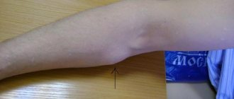
Intradermal nevus
This is another name for common moles - dome-shaped nevi.
We have already mentioned that such neoplasms rarely grow in children - only in every hundred, while in adults they pop up in dozens.
Why are they called that?
Intradermal - moles under the top layer of skin. Therefore, their color does not differ from the color of the surrounding skin and they are noticeable due to their height.
Intradermal nevi grow everywhere. Most often they appear on the neck, head, upper arms and even on the eyelids.
Most moles are small - from 5 mm to 1 cm, and are hardly noticeable in children. As you grow older, nevi change. They become more convex and darker. In people over 70 years of age, intradermal nevi, on the contrary, become discolored and are almost invisible.
Why do common moles appear?
Such neoplasms arise for 3 main reasons:
- Heredity
If parents have more than 50 moles, then the child is likely to get many nevi.
- Sun exposure
When the sun damages skin, especially light skin, moles often appear.
- Immunosuppressive treatments
Some drugs reduce immunity, which causes new nevi to form.
Should intradermal moles be checked?
We recommend checking any nevi regularly. At least once a year. See a dermatologist outside of your regular schedule if the growths have changed color, shape, or size.
The main reasons for removing a mole:
- Degenerates into melanoma
- Gets damaged on clothes every day
- Looks disgusting
The nevus is easily removed. Doctors even remove tumors on the face or neck with laser or other methods.
Epidermal nevus
This type of neoplasm grows from cells in the outer layer of the skin. Up to half of epidermal moles appear at birth, the rest - in the first year of life. After that they don't change.
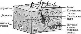
Varieties of epidermal nevus differ in the type of cells they are composed of. The outer layer of the skin, or epidermis, is made up of keratinocyte cells.
A separate group of moles is keratinocyte or non-organoid. It includes neoplasms that consist of keratinocytes and grow on the trunk or limbs. Never on the head. Nevi can be flat or raised. As a person gets older, moles thicken and darken.
Another group of epidermal nevi is organoid. These moles are not only made up of keratinocytes, but also cells that form sweat glands. Yellow-orange neoplasms. They grow exclusively on the face and head, making it appear that a person is bald. Like non-organoid nevi, these moles gradually become denser. A quarter of nevi cause the development of malignant tumors.
Typically, people develop one or more moles of varying sizes and shapes. Less often - more. According to studies, out of 1000 people, no more than 3 will become carriers of epidermal nevus.
What is an apigmented nevus?
Most moles have a characteristic color: from pink to black.
There are exceptions.
Pigmentless nevi have almost no pigment cells and appear as light or whitish spots of irregular shape. Moles appear in childhood and remain with a person for life. However, not everyone has them. Mostly such neoplasms settle on the skin of representatives of the Caucasian race.
The size of the nevus does not exceed 2 cm. In most cases, only moles on the face are removed if they spoil the appearance. This happens quite often.
That's why.
Unlike other neoplasms, non-pigmented nevi do not darken due to the sun. Against the background of tanned skin, they are striking, which for many people is a good reason for removing moles.
However, the chance of their appearance is small - only 1%.
Causes and risk factors
There is no single cause of melanoma, either pigmented or non-pigmented. In 5-12% of cases, the disease occurs under the influence of hereditary predisposition. Then one of the genes that has undergone mutation is passed on to the descendants. The main ones are:
- CDK4, responsible for the production of an enzyme that suppresses tumor development;
- CDKN2A, essential for normal cell division.
The risk of developing melanoma in relatives is 8-14% if:
- amelanoma has been diagnosed in two or more of the person's siblings, especially if they are twins;
- malignant melanocytic formation was present in both parents;
- Two or more of the person's children have been diagnosed with melanoma.
Melanoma genes can “accumulate” and be passed on in families living in regions with a high incidence of this tumor. In this case, the risk of developing such a neoplasm is 5-10%. In approximately every 12-50th case, the formation of non-pigmented neoplasia will occur.
The majority (about 90%) of cases of melanoma development are sporadic, that is, not associated with hereditary transmission, cases. In this case, the cause of the tumor is considered to be ultraviolet radiation - rays of types B and A.
Risk factors for malignant neoplasms from melanocyte cells are:
- fair skin (Fitzpatrick phenotype I and II), especially in combination with red hair and freckles;
- received sunburn (including in early childhood);
- use of PUVA therapy (for the treatment of psoriasis, vitiligo);
- large congenital nevus - when it occupies an area of more than 5% (5 palms) of the body area;
- immunodeficiency - congenital, caused by taking glucocorticoid drugs or cytostatics for the treatment of rheumatoid arthritis, autoimmune diseases, to prevent transplant rejection.
In 70% of melanomas do not develop on “clean” skin, but as a result of the degeneration of such benign formations as:
- blue nevus;
- nevus of Ott;
- dysplastic nevus;
- Dubreuil's melanosis;
- complex pigmented nevus;
- Pick's melanosis.
The process of melanoma formation is triggered by injury, frequent exposure of pigment formation to ultraviolet rays. A change in hormonal balance may also be a trigger factor. The risk of developing non-pigmented melanoma is 1.8-8.1%.
Borderline nevus
A type of mole that often turns into a malignant formation. They need to be checked regularly.
A borderline nevus grows under the stratum corneum of the skin and looks like a uniform brown spot. It has different shades: from light to dark. Not much different from a regular mole - the same round or oval shape, clear boundaries.
The following signs indicate degeneration:
- The tumor increases in size
- Nevus darkens or brightens
- Pigment spots appear near the mole
- The neoplasm becomes denser
- There is itching or pain in the mole
Contact a dermatologist if any of these symptoms appear - he will examine the moles and, if necessary, refer you to an oncologist.
You also need to be careful about giant tumors.
What to do with huge nevi
We are accustomed to the fact that most moles are small formations and are almost invisible on the skin. Giant nevi do not live up to expectations.
As the name suggests, they are very large - with a diameter of 20 cm. Such moles cover a significant part of the arms, legs and face.
Congenital nevi of this type grow in people of any race - they spoil the lives of 2% of the world's population. But it is not their size that is dangerous.
A giant mole is likely to degenerate into melanoma, especially when the nevus grows near the spinal column.
The surface of the new growth is rough, and its color is gray, brown or black. Gradually the nevus grows. The skin on it thickens, and pigmentation intensifies. Very often, hair grows in certain areas of the mole.
The neoplasm looks especially terrifying after the birth of a child - on a tiny body it looks simply gigantic. The baby grows faster than the nevus. Therefore, the mole appears to shrink over time, but in fact it continues to grow.
In 30% of cases, a giant nevus degenerates into melanoma. Transforms at any age. Some people face this threat in their youth, others live with it into old age. It is noteworthy that large moles do not disappear even in old people.
When reborn they:
- Are increasing
- Change colors and shapes
- Bleeding
- Covered with a crust
Doctors recommend removing these nevi. Usually a surgical operation is performed - the doctor cuts off the mole with a scalpel. This method is not always available if the tumor is located in a complex location or for other reasons.
Instead it is assigned:
- Curettage
- Dermabrasion
- Ablative laser therapy
These procedures do not completely remove the nevus, but eliminate its external imperfections: hair, strong pigmentation.
Large moles on the face are especially difficult to deal with.
Nevi on the head
One of the main reasons why people remove moles is cosmetic. Growths on the chest and legs can also look unsightly, but they are almost always hidden under clothing.
With nevi on the head everything is more complicated.
For some people, these moles emphasize beauty, for others they spoil the whole impression. Many people want to get rid of even harmless tumors that will never develop into malignant tumors.
Nevi on the neck are removed for similar reasons.
Moles in this place constantly touch clothing and become damaged. Of course, not all. Flat growths are usually invisible and do not rub against tight clothing. High nevi are another matter. Dermatologists recommend removing such moles if they become damaged and inflamed. In general, they cause a lot of trouble without possibly turning into deadly melanoma.
The face and neck are not the only places where nevi appear. They also appear in the hair, and it is much more difficult to detect them. And it's easier to damage.
In this part of the body, nevi of the sebaceous glands are often found - a warty yellow-pink plaque without hair. Has an oval or irregular shape.
This mole grows due to:
- Human papillomavirus
- Genetic predisposition
Nevus of the sebaceous glands appears both among the hair and near the ears, on the neck or face. In most cases, it is formed during fetal development, but it can also grow in children.
How to identify such a mole?
Doctors usually notice it immediately after the baby is born. They perform examination and dermatoscopy. Sometimes a histological examination is necessary - dermatologists examine a sample of the nevus under a microscope.
Do I need to remove a mole right away?
No. She is reborn only at puberty. Therefore, nevi of the sebaceous glands are removed in adolescents, and not in infants.
Moles of different sizes are removed.
Large ones are eliminated surgically, small ones - with laser, cryodestruction or electrocoagulation.
Before surgery, the child should have the nevus checked regularly by a dermatologist.
Treatment of the disease
At Euroonko, non-pigmented melanoma is treated according to modern protocols, taking into account the stage of the process:
- In stage I of the disease, surgical removal of the tumor and tissue around it is performed.
- In stage II, the tumor is removed and the lymph nodes are additionally checked for cancer cells. If mutated melanocytes are found in the lymph nodes, the nodes are removed. Treatment may include photodynamic therapy and radiation therapy. Here, if there is a mutation in the BRAF gene, treatment is carried out with BRAF inhibitors.
- Stage III is treated by removing the tumor and all nearby lymph nodes. Treatment is complemented by chemotherapy and radiotherapy. Immunotherapy and the introduction of BRAF inhibitors are involved. Therapy is required to improve the condition and relieve symptoms.
- At stage IV, large tumors and metastases are removed, if necessary. Chemotherapy, radiotherapy and palliative treatment are prescribed. Immunotherapy is mandatory.
Modern treatment of melanoma includes such a new and very promising direction as immunotherapy. And if previously only chemotherapy was carried out, which was successful only in 10% of cases, now the concept has changed greatly for the better. Immunotherapy involves the administration of special drugs that bind to certain proteins on the membranes of lymphocytes and “show” them that there are cancer cells in the body. Then the lymphocytes start working again and destroy them - throughout the body, and not locally.
Immunotherapy helps the body fight cancer. Even with advanced forms of malignant melanocytic tumor, patients can count on successful treatment.
For amelanotic melanoma containing a BRAF gene mutation, Euroonko provides targeted therapy with BRAF inhibitor drugs. These drugs suppress tumor progression and enhance immune activation. BRAF inhibitors are used starting from stages IIC-III. They increase long-term survival and are recommended for use even with an inoperable tumor or the presence of distant metastases.
Therapy with BRAF inhibitors is carried out for a long time - 6-9 months.
Out of sight, out of sight. How to get rid of moles
Not an easy question. Many factors influence the ability to remove tumors. Unfortunately, no pills have yet been invented that will relieve you of annoying or dangerous moles.
Yes, treatment of small nevi usually does not cause problems.
Successful removal depends on:
- Doctor's qualifications
- Mole size
- Nevus locations
The first option is surgery. The doctor cuts off the mole, some adjacent skin, and then stitches the wound. After removal, the nevus heals quite quickly, but a noticeable mark remains.
Whether this is good is up to you to decide.
What about laser surgery?
Yes, modern technologies do offer new treatments, but they do not work miracles, as some people believe. Some skin diseases are treated with lasers. But not all nevi.
The laser beam vaporizes the outer layer of skin and cells of the mole. The deeper it is rooted, the more difficult it is to destroy it completely - sometimes the surface layer is burned out, but the base remains intact. Then the nevus grows back.
The operation has to be repeated again and again, and there is no guarantee of results. So the surgical procedure is sometimes more reliable than laser surgery.
However, in some situations, a high-tech procedure accomplishes the task. Removes small nevi. Working with large tumors is much more difficult.
We recommend choosing laser nevus removal only after consulting a dermatologist and checking the tumor.
Another important point is the choice of doctor. Many doctors have accumulated vast experience in removing small moles. Everyone has them. Therefore, doctors work with them very often and know well how to quickly remove nevi.
But what about large moles?
They are not common and therefore it is difficult to find a doctor who regularly operates on them. Read reviews about nevus removal on the websites of different clinics. This is useful. You will learn what operations the doctors of these medical institutions performed and the patients’ impressions of their work.
Gather more information.
When you see your dermatologist, ask about available methods for removing moles. Perhaps in your case, surgery and laser therapy are not the best options. Harmless nevi do not need to be removed at all.
Many medical institutions remove moles. But where to go? We would like to recommend one of these clinics.
Complications and consequences of the disease
Pigmentless melanoma can be complicated by screenings of daughter tumors, metastases, and internal organs. This leads to a deterioration in their performance:
- metastases in the lungs impair blood oxygen saturation;
- brain metastases cause seizures, personality changes, and impaired movement and/or sensation in the limbs. Daughter tumors in the brain stem lead to disturbances in breathing, heartbeat, and maintaining normal blood pressure levels;
- metastases in the bones cause severe pain, bone destruction, difficulty in movements - up to their complete impossibility;
- metastases to the liver lead to indigestion, yellowing and itching of the skin, and deterioration of brain function;
- metastases to the skin cause pain.
When neoplasia is detected by the body's immune system, intoxication develops. It manifests itself as weakness, loss of appetite, drowsiness, and nausea. A person stops eating normally, and the body does not receive the nutrients it needs for life and to fight the tumor.
Over time, cancer cachexia develops - exhaustion. It is manifested by weight loss, constant weakness, and lack of appetite. Cachexia further worsens a person’s condition, disrupting metabolism and the functioning of all his internal organs.
Removal of nevi at the Lasersvit clinic in 100 seconds or less
Nowadays, beauty salons easily remove nevi of various types. And the procedure is inexpensive. But the quality and result of the operation are not always guaranteed.
Only have moles removed in well-equipped clinics.
And that's why.
The basis of a successful operation is accurate diagnosis of tumors. Sometimes simply examining a mole is not enough and dermatoscopy is needed. That is, the use of a dermatoscope.
The Lasersvit clinic uses a Delta 20 T dermatoscope, which magnifies the image of the skin 16 times. With its help, our doctors can easily determine the type of nevus and promptly notice signs of degeneration. Helps quickly detect melanoma.
Our doctors have accumulated enormous experience in diagnosing moles - they have examined over 100,000 patients. They are familiar with cutting-edge research in the field of neoplasms because they studied abroad. Even dermatologists of the second category.
Lazersvit doctors can easily remove nevi from an adult or child. Benefits of the procedure:
- Rapidity
On average, one small mole is removed in 1-2 minutes. Therefore, sometimes doctors remove many nevi at once, one after another - for 20 moles it will take from 30 to 40 minutes.
- Painless
The laser pulses are very short - they last a fraction of a second, and the skin is constantly cooled. Patients do not have time to feel anything. In long operations on large moles, local anesthesia is used.
- Bloodlessness
The laser beam destroys nevi, seals blood vessels and prevents bleeding.
- Easy healing
A protective crust grows at the site of the removed mole, which prevents infection. It disappears within a week. Skin redness disappears within 3 weeks.
- Tracelessness
The laser destroys only nevus cells and when they are destroyed, the skin is restored. The new skin is smooth and without scars.
- Efficiency
Evaporated tumors are not rebuilt, as doctors burn out all their cells.
What about the price of nevus removal?
It depends on the type of tumor, its size and location. Moles in hard-to-reach places are more difficult to remove, which means the cost of the procedure increases. Sometimes additional surgery is needed.
Large nevi cannot be removed immediately and doctors treat them in several stages. Talk to your dermatologist after your examination and he will explain in detail how much it costs to remove a specific mole and how long the procedure will take.
Come to Lasersvit for quick and comfortable removal of dangerous and inconvenient nevi.
Diagnostics
At the slightest suspicion of melanoma, you should consult an oncologist or dermato-oncologist. With the help of an examination, which at the first stage is simple and painless, the doctor will be able to distinguish non-pigmented melanoma from similar diseases: seborrheic keratosis, pyogenic granuloma, Spitz nevus, basal cell skin cancer, hemangioma.
The first thing Euroonco doctors begin diagnosing melanoma with is an examination of the formation. The dermato-oncologist needs to evaluate the appearance, consistency, symmetry, bleeding of the tumor, and palpate the nearest lymph nodes.
Next, he collects an anamnesis - he finds out from the patient when the non-pigmented neoplasm appeared, whether a diagnosis of melanoma was made in one of the closest relatives, whether there were frequent injuries to the tumor, whether it was exposed to excessive sun exposure.
At the same stage, the doctor performs digital dermatoscopy - examination of the tumor using a special device. A dermatoscope magnifies the tumor and allows you to analyze the structure of its deep layers, assess the location and course of blood vessels. This is very important for diagnosing amelanoma.
If, according to dermatoscopy, malignancy of the tumor cannot be excluded, a second diagnostic stage is prescribed. This is a biopsy - obtaining tumor tissue for research. It is performed under local anesthesia and is excisional. This means that both the tumor and the surrounding tissue are completely removed.
The area obtained by biopsy is examined under a microscope. Immunohistochemical studies (IHC) are also performed. It involves detecting certain types of proteins in a tissue sample (biopsy) to determine the type of melanoma and its sensitivity to chemotherapy drugs.
When the diagnosis of non-pigmented melanoma is confirmed, the third diagnostic stage is carried out - determining the extent of the process (tumor stage). To do this, metastases are detected in the lymph nodes and internal organs:
- Lymph node metastasis is determined by sentinel lymph node biopsy
The procedure is not mandatory. Euroonko doctors recommend undergoing it if the thickness of the removed melanoma is more than 1 mm. This is due to the fact that in this situation the risk of metastases to regional lymph nodes increases. Thus, even with a tumor thickness of 0.75-1 mm, micrometastases in the “sentinel” node are observed in 4-12% of patients.
A sentinel lymph node biopsy is performed as follows. A special drug is injected into the area of the removed melanoma. Next, the doctor observes which lymph node the drug entered first and removes it. This happens under local anesthesia.
After this, the lymph node is carefully examined under a microscope, using also the IHC method. If no melanoma cells are detected, it means that the tumor has not sent daughter cells beyond the occupied area.
- Metastasis to internal organs
It is determined by magnetic resonance imaging (MRI) and computed tomography (CT) methods.