The structure of the skin
The skin is the largest integral multifunctional organ, interconnected with all other organs and systems of the body. In direct contact with the external environment, it performs a barrier-protective function. On the surface of the skin there is a complex pattern in the form of triangular and rhombic fields, formed by numerous grooves. Coarser grooves form folds in the palms, soles, scrotum, and facial wrinkles.
Histologically, three layers of skin are distinguished (Fig. 1):
- epidermis;
- derm (dermis);
- subcutaneous fatty tissue (subcutis), or hypodermis (hypodermis).
Rice. 1. Skin structure
The epidermis is the epithelial part of the skin, and the dermis and hypodermis are connective tissue. The border zone between the epidermis and dermis has the appearance of a wavy line due to the presence of outgrowths in the dermis - papillae, which cause the formation of ridges and furrows on the surface of the skin, forming a skin pattern. The connective tissue part of the skin (dermis and hypodermis) contains nerves, blood and lymphatic vessels, and muscles. In addition, the skin has its own appendage structures, which include hair, sebaceous and sweat glands, as well as nails.
Table of contents
- Etiology and pathogenesis
- Clinical manifestations
- Treatment methods
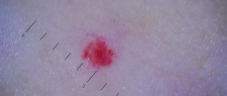
Port-wine stains, also called capillary angiodysplasia or flammeus , are one of the most common vascular malformations. They are a network of pathologically altered capillaries located superficially or deep under the skin and visible through it.
In our company you can purchase the following equipment for the treatment of wine stains:
- M22 (Lumenis)
According to world statistics, port-wine stains are registered on average in 0.1–2.0% of newborns. Most often they occur in representatives of the Caucasian race, with equal probability in both boys and girls.
Pigment formations
All pigmented formations on the skin can be divided into two groups: non-dangerous (melanoma-non-dangerous) and dangerous (melanoma-dangerous) pigmented nevi (Table).
Table 1. Classification of skin tumors of melanocytic origin
| Skin tumors of melanocytic origin | |
Melanomic nevi
| Melanoma-dangerous nevi
|
Etiology and pathogenesis of port-wine stains
In most cases, port-wine stains (nevus flaminga) are a birth defect . They are present in the baby immediately after birth and increase in size in proportion to the further growth of the child. An important feature of port-wine stains is the lack of spontaneous regression - they form for a lifetime and, as a rule, have a negative impact on a person’s psychological state.
In rare cases, port-wine stains may appear on apparently healthy skin in adulthood - this is the so-called acquired capillary angiodysplasia . Sometimes port-wine stains may not be an independent pathology, but signs of severe syndromes: Sturge-Weber (encephalotrigeminal angiomatosis with damage to the pia mater) or Klippel-Trenaunay (vein malformations with hypertrophy of bones and soft tissues).
The International Society for the Study of Vascular Anomalies (ISSVA) classifies capillary angiodysplasia according to the predominant vessel type—arterial, venous, lymphatic, capillary, or complex.
Melanomic nevi
Congenital melanocytic nevus
All congenital nevi are harmless. Among them there are small, medium and giant formations:
1. Congenital small melanocytic nevus (Fig. 2).
Elements of the rash. A spot or plaque raised above the skin measuring up to 1.5 cm. The shape of the nevus is round or oval, the boundaries are clear or blurred. The surface of the nevus is smooth or wrinkled, lumpy, folded, lobed.
Color. Light and dark brown. In rare cases, a depigmented rim is noted.
Localization. Any.
Course and prognosis. The risk of developing melanoma before puberty is virtually non-existent; in later life it ranges from 1 to 5%.
Rice. 2. Congenital small melanocytic nevus
2. Congenital medium melanocytic nevus (Fig. 3).
Elements of the rash. A round or oval plaque raised above the skin from 1.5 to 20 cm. The surface of the formation is smooth or wrinkled, lumpy, folded, lobulated, covered with papillae or polyps.
Color. Light or dark brown, there may be small dark inclusions on a lighter background.
Localization. Any.
Course and prognosis. There is virtually no risk of developing melanoma before puberty. Average congenital nevi change slightly throughout life. Due to the growth of the child, there is a proportional increase in education. It is advisable to remove the nevus before reaching puberty.
Rice. 3. Congenital medium melanocytic nevus
3. Congenital giant melanocytic nevus (Fig. 4).
Elements of the rash. A plaque raised above the skin level measuring more than 20 cm in diameter. There may be satellite lesions along the periphery of the main lesion. A disorder of the skin pattern is characteristic. On the surface of the formation there are nodules, papules and, as a rule, coarse dark hair. The boundaries can be either smooth or uneven.
Color. Darkly pigmented formation.
Localization. Any.
Course and prognosis. According to various sources, the risk of transformation of congenital giant melanocytic nevus into malignant melanoma reaches 5%. Surgical excision of the formation followed by plastic correction should be carried out as early as possible, however, often the operation is not possible due to the size or location of the formation.
Rice. 4. Congenital giant melanocytic nevus
Acquired melanocytic nevus
Acquired melanocytic nevus can be represented by a borderline (transitional) or complex (mixed) nevus. The transition from borderline nevus to complex nevus to intrademal nevus over time demonstrates the normal evolution of neogenesis.
1. Borderline nevus
Elements of the rash. A round or oval spot, sometimes slightly raised above the surface of the skin, up to 1 cm in size, with clear, even boundaries.
Color. Homogeneous (various shades of brown).
Localization. Any.
Course and prognosis. Borderline nevus, which arose in early childhood, becomes mixed as a result of the proliferation of nevus cells and their advancement into the dermis. This usually occurs during puberty. After the disappearance of the borderline component, the nevus becomes intradermal. This natural transformation usually occurs before the age of 30. In some cases, common borderline nevi remain unchanged throughout a person's life.
Rice. 5. Borderline nevus
2. Complex nevus
Elements of the rash. A formation in the form of a papule or node, usually up to 1 cm in size. The surface is smooth, less often warty, often with the growth of bristly hair. In shape, complex nevi are predominantly formations that rise evenly above the skin.
Color. Generally uniform: dark brown.
Localization. Any.
Rice. 6. Complex nevus
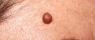
Dermal (intradermal, “resting”) nevus
Elements of the rash. A round, dome-shaped formation, usually up to 1 cm in size, rising above the surface of the skin. Over time, the nevus may develop a stalk and may take on the appearance of a warty (papillomatous) nevus. This phenomenon is most typical for formations localized in the torso area.
Color. Yellow-brown, brown or with brown spots, telangiectasias may be observed.
Localization. The most common are the face and neck. Formations of this group on the trunk and limbs are less common.
Course and prognosis. In most cases, intradermal nevi are not treated. Indications for removal of the formation are: localization in which there is permanent injury to the lesion.
Rice. 7. Dermal nevus
Prevention of the occurrence of acquired nevomelanocytic nevi is to reduce exposure to sunlight (especially in the early period of a person’s life). This is achieved by reducing the time spent in the sun (especially during the peak solstice period from 11 a.m. to 4 p.m.) and using sunglasses.
Blue nevus
Blue nevus can be either congenital or acquired; it most often appears in childhood or adolescence. These skin lesions are much less common than nevi of epidermal melanocytic origin. A blue nevus develops from ectopic melanocytes of the dermis. The coloring characteristic of a blue nevus is due to the Tyndall phenomenon - the refraction of light by the pigment of tumor cells located deep in the dermis.
Currently, several types of dermal melanocytic nevi are considered in the nosological form of “blue nevus”: simple blue nevus, cellular, cellular non-pigmented, combined and deeply penetrating.
Rice. 8. Blue nevus
1. Simple blue nevus (blue nevus, Jadassohn–Tiche nevus
Elements of the rash. The nodule, usually up to 1 cm in size, is sharply demarcated from the surrounding skin, round in shape, hemispherically protruding above the skin level, with a smooth surface.
Color. Blue, light blue, dark blue, gray, bluish - black.
Localization. Any type is possible, but the typical location for blue nevi is the dorsum of the hands and feet. A rare arrangement – soft and hard palate.
Course and prognosis. This formation grows quite slowly, often remaining unnoticed for several years. Over time, a blue nevus may acquire a flatter shape and lose pigment. Transformation of a blue nevus into melanoma is extremely rare.
2. Blue cell nevus (proliferating)
Elements of the rash. The development of a nevus begins with the formation of a spot or compaction in the dermis, which then transforms into a node or plaque up to 2.5 cm in diameter.
Color. Blue.
Localization. Any is possible. Quite often the localization of nevus in this group is the gluteal, lumbosacral region, less often on the dorsum of the hands and feet. Rare tumor localizations have been described - the conjunctiva and the scalp.
The diagnosis is made on the basis of clinical and histological signs; immunohistochemical studies are necessary.
Course and prognosis. Depending on the clinical picture, pathomorphological signs, course and prognosis of the process, two forms of blue cell nevus are distinguished: typical (classical) and atypical with uncertain biological potential. Malignant transformation usually occurs over a long period of time in mature or elderly people and is manifested by rapid tumor growth, ulceration and discoloration. With a cellular blue nevus, cells similar to this formation may be observed in the regional lymph nodes. This process is called “benign metastasis.” Treatment is surgical excision with pathological examination.
Spitz nevus (atypical spindle cell, juvenile, benign juvenile melanoma)
Elements of the rash. A small single dome-shaped hairless node measuring up to 1 cm in diameter. Education, as a rule, is singular.
Color. Pink-red with abundant vascularization, yellow-brown, dark brown, uneven coloring possible.
Localization. Any is possible. In children and adolescents, the most common location is the scalp and face.
The diagnosis is made on the basis of histological examination. A quickly growing dome-shaped formation in a child allows one to suspect a Spitz nevus.
Course and prognosis. Spitz nevus is characterized by a sudden appearance. From the moment it appears, the formation is characterized by rapid growth, then it flattens and remains unchanged for years. Some nevi may undergo morphological transformation into dermal melanocytic nevi, and transformation into melanoma is also possible. The risk of malignant tumor degeneration occurs during puberty. Malignant degeneration is rare, but such formations require careful monitoring. This formation must be removed before the end of puberty. Then, in the postoperative period, dynamic observation is advisable.
Rice. 9. Spitz nevus
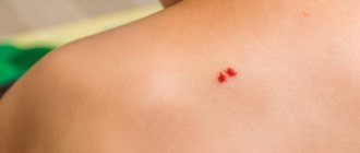
Halo nevus (Sutton's nevus)
Elements of the rash. A slightly elevated, slightly infiltrated reddish-brown nodule of round or oval shape, with an average diameter of 4-5 mm. The halo nevus is surrounded by a halo of depigmentation. The diameter of the rim, as a rule, is 2-3 times larger than the size of the hyperpigmented nodule. Characterized by multiple skin lesions.
Localization. Any type is possible, but the most common is the back.
Course and prognosis. There are several stages in the course and resolution of halo-nevus:
- melanocytic nevus with a surrounding rim,
- the central element loses pigmentation and acquires a pink color,
- disappearance of the central element,
- complete repigmentation of the entire nevus over several months or years.
No treatment required.
Rice. 10. Halo nevus
Introduction
Every child has melanocytic nevi; they can be single, multiple, exist at birth (congenital) or appear over time (acquired).
The tactics for managing patients with melanocytic formations in childhood can be implemented in three ways: surgical excision of the element, dynamic observation of the formation, and zero-intervention tactics, when further observation or surgical intervention is not required. The doctor makes a decision based on an analysis of all factors characterizing the nevus: the child’s age, morphology, location, size and melanoma danger of the element.
The predominance of aggressive surgical tactics in relation to nevi in children forced us to turn to scientific literature data reflecting the state of this problem in the world.
The purpose of the work is to analyze literature data over the past 10 years on the pathogenesis, clinical picture, diagnosis and management tactics of congenital and acquired nevi in children.
Melanoma-dangerous nevi
Phenotypically, these nevi do not reveal clinical signs of malignancy, but are distinguished by melanocyte dysplasia and a tendency to malignancy, thus giving reason to consider them as premalignant or borderline formations. These are the only tumors that require mandatory prophylactic removal with morphological verification of the pathological process. If it is multiple in nature, it is advisable to classify this group of patients as a risk group and subject them to mandatory clinical examination with dynamic monitoring by an oncologist.
Dysplastic nevus
Elements of the rash. A spot with a separate slightly raised area, usually in the center, above the skin level. The shape is round, oval or irregular with “ragged” edges. The boundaries are irregular and blurred.
Color. Various shades of black, brown, reddish, light red.
Localization. The most common are the torso, arms, buttocks, dorsum of the feet, and less commonly, the face.
Course and prognosis. A dysplastic nevus may not undergo any transformations throughout a person’s life, but may transform into a superficial melanoma; Complete regression of education is also possible. Against the background of a dysplastic nevus, melanoma develops in 9% of cases. It differs in clinical, histological, and biological manifestations from the known forms of melanoma, and is considered by some authors as “minimal melanoma.” The appearance of these nevi in young patients is not an alarming syndrome; they, as a rule, do not transform into melanoma.
With dysplastic nevus syndrome, the prognosis is unfavorable, and the risk of developing malignant melanoma increases significantly.
Treatment. Not all dysplastic nevi require immediate removal. Dynamic observation with photography and measurement of the size of the formation is necessary. Changing, suspicious, and traumatizing nevi are subject to excision.
Rice. 11. Dysplastic nevus
Treatment Methods for Port Port Stains
The simplest method of not so much treating as masking port-wine stains is the use of cosmetics . Properly selected products help hide the tumor and allow such patients to feel quite comfortable in public places. A more radical option, which is also not aimed at treating a port-wine stain, is to tattoo over the tumor.
Drug therapy for capillary angiodysplasia has not demonstrated significant effectiveness in treating port-wine stains. However, there is evidence of positive results from topical use of the immunomodulator imiquimod 5 times a week under occlusion with plastic film for 4 weeks.
Before the invention of lasers, the radical treatment for port-wine stains was skin grafting . This operation, like other invasive interventions, has limitations and carries certain risks.
Today, light and laser therapy - IPL and Nd:YAG lasers - are actively used to treat port-wine stains. The principle of such therapy is the selective destruction of pathological vessels due to their heating when hemoglobin absorbs light or laser energy. This type of therapy is more suitable for situations where the vascular defect is completely formed and there is no way to influence it with other types of therapy. Destruction of blood vessels leads to a pronounced and persistent aesthetic defect. Laser and light therapy procedures are carried out without compromising the integrity of the skin, are well tolerated and do not require long-term rehabilitation.
Diagnostics
Typically, diagnostic measures are limited to an examination by a dermatologist or oncologist, since the specific appearance of a nevus allows one to immediately identify the disease. But if you suspect its possible malignancy, the doctor may send you for additional examinations:
- siascopy (assessment of the structure of education);
- dermatoscopy (analysis of the structure, clarification of the boundaries and depth of the nevus);
- morphological examination of biopsy specimens or material obtained during excision of a nevus (many melanocytes located in the basal layer of the epidermis, along with their absence in the papillary layer of the dermis);
- determination of tumor markers specific for melanoma (SU100, TA90).