This is an acute purulent inflammation of soft tissues and/or internal organs (mediastinum, abdominal cavity, retroperitoneal space, pelvis, etc.).
The name comes from a Greek word meaning inflammation or swelling. Cellulitis can affect internal organs such as the appendix, kidneys, liver, spleen, intestinal vessels, prostate gland, pancreas, or anywhere under the skin. It tends to spread quickly and in some cases can be life-threatening.
What causes phlegmon, reasons
The reason for the development of phlegmon is the penetration, active development and spread of infection. As a result, a purulent melting of tissues (fat, muscle, etc.) is formed with damage to nearby organs (vessel walls, intestines, kidneys, etc.). Cellulitis is usually caused by bacteria: more often aerobic, such as streptococci, Staphylococcus aureus, etc., less often by anaerobes (forming spores).
Other factors that influence the development of phlegmon:
- the presence of chronic intoxication due to chronic inflammation, poisoning, chemotherapy, radiation sickness,
- extensive severe injuries,
- patient's immune status,
- virulence of bacteria (ability to cause disease).
Bacteria can enter through a scratch, insect bite, or injury to form a cellulitis just under the skin. They can also move with the blood flow from an inflamed organ (abscess of the kidney, lung, liver, appendiceal abscess, perirectal abscess, etc.).
The bacteria can also attach to the wall of an internal organ, such as the stomach wall or appendix, and form a cellulitis.
People with weakened immune systems may be especially vulnerable to cellulitis formation.
Symptoms and manifestations of phlegmon
Vary depending on the location and severity of the infection. The main symptoms are redness of the skin and swelling at the site of inflammation.
Soft tissue phlegmon against the background of a progressive abscess
The affected part of the body is sensitive to touch and hurts. Fever is later added to this symptom. Swollen associated lymph nodes may occur.
Manifestations of skin phlegmon
- skin redness
- inflammation
- pale swelling of tissues
- swollen lymph nodes
- elevated local temperature
- fatigue
- fever
- headache
Internal organs
Cellulitis can affect any internal organ. Symptoms may vary depending on the organ and the specific bacteria.
- organ dysfunction
- abdominal pain
- fever
- chills
- nausea
- vomit
- diarrhea
- intestinal obstruction
- elevated body temperature
- loss of appetite
- increased thirst
- sweating
- increase in white blood cells (leukocytosis)
Purulent diseases of the hand
The occurrence of felon is almost always associated with a previous violation of the integrity of the skin, most often in the form of various kinds of microtraumas, among which the most dangerous are small puncture wounds and splinters. The edges of these wounds quickly stick together, and sometimes epithelization of the skin defect occurs, but the source of infection remains in the wound canal. According to most studies, fingers I, II, and III of the right hand are more often affected (56.8-64.5%), which is explained by their greater functional load and frequency of trauma. A comparative study of the microflora of purulent diseases of the hand over 20 years (1960-1980) conducted by B. V. Stombreky (1984) suggests that Staphylococcus aureus is the dominant infection, although the number of staphylococcal infections is decreasing and non-staphylococcal infections are increasing. The role of gram-negative flora, as well as aerobic-anaerobic associations, has increased. Classification of panaritiums : I. Superficial forms of felon: • Skin felon. • Paranychia. • Subungual felon. • Subcutaneous panaritium. • Furuncle (carbuncle) on the back of the finger. II. Deep forms of felon: • Bone felon. a) Spicy. b) Chronic (fistula form) • Tendon panaritium. • Articular felon. • Osteoarticular panaritium. • Pandactylitis. Skin felon Exudate is located under the epidermis and exfoliates in the form of a bubble, the contents of which are serous, purulent or hemorrhagic in nature. Subcutaneous panaritium When examining a finger, attention is drawn to the direction of the tissues, sometimes the smoothness of the interphalangeal flexion groove located near the inflammatory focus. Connective tissue cords that penetrate the fatty tissue of the finger and connect the skin itself with the periosteum prevent the spread of edema to the periphery. The tension of these bridges causes intense pain in the finger. With subcutaneous panaritium, pus tends to spread deeper. Indication for surgery is “first sleepless night syndrome.” Paranychia is accompanied by painful swelling of the periungual fold and hyperemia of the surrounding tissues. Upon examination, attention is drawn to the overhang of the affected periungual fold over the nail plate, peeling it off along its entire length, i.e. subungual panaritium occurs. Subungual felon Accumulating under the nail plate, purulent exudate slightly raises it. Its fixation to the bed is lost. Surgical removal of the nail plate creates the necessary prerequisites for recovery. Articular panaritium Occurs after injury to the interphalangeal or phalangeal areas of the finger from their dorsal surface, where the joints are covered only by a thin layer of soft tissue. The inflamed joint takes on a fusiform shape, and the dorsal interphalangeal grooves are smoothed out. An attempt at flexion-extension movements of the finger leads to a sharp increase in pain in the affected joint. When the ligamentous, cartilaginous and bone apparatus of the finger is involved in the inflammatory process, pathological mobility and a feeling of crepitus of the rough parts of the articular surfaces occur. Such “looseness” of the joint indicates a significant change in the osteochondral apparatus of the finger. Bone panaritium It develops, as a rule, when the pathological process transfers from the soft tissues of the finger to the bone, i.e. the process is secondary. The bones of the fingers are primarily affected by the inflammatory process extremely rarely (when the infection is transferred by blood flow from distant inflammatory foci). Basically, bone felon develops from neglected or non-radically cured subcutaneous felon. X-ray examination of bone healing is determined only by the end of the 2nd to the beginning of the 3rd week. Tendon felon Subcutaneous felon in some cases is the cause of tendovaginitis. If the therapy does not create conditions for the successful elimination of inflammation, then it becomes possible for the infection to spread to deeper tissues and, above all, to the tendon sheaths and finger flexor tendons. Deterioration of the general condition, the appearance of twitching, throbbing pain throughout the finger, uniform swelling of the tissues with smoothness of the interphalangeal grooves is a symptom of tendon panaritium. The finger takes on the appearance of a sausage. Pandactylitis This is a purulent inflammation of all tissues of the finger. With pondactylitis, there is no predominance of one of the forms of acute inflammation that were listed earlier. The clinical picture of the disease consists of a combination of all types of purulent laceration of the finger. The disease develops gradually and is severe, accompanied by severe intoxication, regional lymphangitis, cubital and axillary lymphadenitis. General principles of treatment of panaritium and the postoperative period The key to success in the treatment of purulent infection of the fingers and hand is timely and adequate surgical intervention, the main point of which is a complete necrectomy. • The incision should provide a complete revision and sanitation of the purulent focus and at the same time be gentle, ultimately allowing for a good functional and cosmetic effect. • After evacuation of pus, it is necessary to perform a full necrectomy, focusing on the color and structure of the tissue. Surgical treatment of a purulent focus should be carried out taking into account the important anatomical structures located nearby in order to avoid their damage. • For more radical removal of a purulent-necrotic focus during surgery, it is rational to evacuate the wound, treat it with low-frequency ultrasound, a pulsating jet of antiseptic, a defocused laser beam, etc. • A purulent wound after thorough surgical treatment must be drained. • After complete surgical treatment of the abscess, in the absence of pronounced perifocal inflammation of the surrounding tissues, mobility of the wound edges and its active drainage, primary delayed sutures can be applied to the wound. Immobilization of the hand is necessary during the conservative treatment of felon and in the postoperative period until the wound is cleansed of purulent-necrotic tissue. Improving the results of surgical treatment can be achieved by rational antibiotic therapy, the main condition for which is the creation of a high concentration of the antibiotic and its long stay at the site of inflammation. It should be noted that antibiotics do not penetrate avascularized necrotic tissue and do not affect the processes of their rejection. In this regard, proteolytic enzymes are used. In case of congestion in soft tissues, electrical stimulation is performed to prevent contractures and muscle paralysis. In order to resolve scars and adhesions, iodine electrophoresis and Trilon B phonophoresis are used. In the postoperative period, various physiotherapeutic procedures are used for the purpose of rapid rehabilitation. Therapeutic baths with 1% Lysol solution, 0.1% potassium permanganate solution, furacillin, chloramine, furagin, hypertonic sodium chloride solution. Local oxygenation and ozone-air mixture are successfully used.
Phlegmon of the hand - This is a diffuse purulent lesion of the cellular spaces of the hand. Classification • Interdigital phlegmon. • Cellulitis of the thenar region. • Phlegmon of the hypothenar region • Supraneural phlegmon of the median palmar space. • Subgaleal phlegmon of the median palmar space: a) superficial. b) deep. • Phlegmon of the dorsum of the hand. • Crossed (U-shaped) phlegmon of the hand with damage to the Pirogov-Paron space. • Combined phlegmon of the hand. Interdigital phlegmon of the hand Usually develops secondarily. The inflammatory focus is formed, as a rule, in the commissural spaces of the II - IV fingers. The openings of the palmar aponeurosis contribute to the spread of infection from superficial abscesses deeper. Phlegmon of the thenar area Develops when puncture wounds of the thenar area become infected, as a complication of subcutaneous panaritium or purulent tendivaginitis of the first finger. It is possible that pus may spread to the thenar area through the canal of the lumbrical muscles of the second finger or from the median palmar space. Phlegmon of the hypothenar region The purulent process is limited to the fascial bed of the hypothenar muscles. Phlegmons of the median palmar space Supraneuratic (subcutaneous) phlegmon is a restriction no deeper than the median palmar aponeurosis, limited laterally by the deep fascia of the thenar and hypothenar. Subgaleal phlegmon of the median palmar space can be superficial (supratendinous), when the purulent process is localized between the palmar aponeurosis and the free tendons of the II-III fingers; and deep (subtendinous) - when the purulent process is localized between the fascia lining the interosseous muscles on the palmar side and the posterior surface of the long flexor tendons. Phlegmon of the dorsum of the hand is characterized by peeling of the skin from deep-lying tissues with the subsequent spread of the process along the plane, the formation of areas of skin necrosis. Crossed (U-shaped) phlegmons of the hand
It is a combination of purulent tendovaginitis and tendobursitis of the 1st and 5th fingers. It develops when purulent inflammation spreads from the radial sinovesal bursa to the ulnar bursa or vice versa. General principles of treatment of phlegmons of the hand: • Treatment of patients with phlegmons of the hand should be carried out in a surgical hospital. • It is necessary to determine as accurately as possible which cellular space of the hand is affected, which is important for choosing adequate access. • Surgery should be early and carried out with strict adherence to aseptic rules. • Optimal pain relief and precise bleeding of the hand is necessary. • Necrectomy and drainage of the abscess are the most important aspects of the operation. • Adequate antibiotic therapy taking into account the sensitivity of the microflora. • The use of various types of local therapy after surgery, depending on the location of the inflammation. • Immobilization. • Rehabilitation of patients, prevention of complications.
Diagnosis of phlegmon
- symptoms, complaints, when they started and how long they last,
- physical examination of the patient,
- the patient's previous medical history, medications taken,
- palpation on physical examination.
Cutaneous cellulitis is always visible, while internal cellulitis is more difficult to diagnose.
Anaerobic phlegmon of the back
Examinations may include:
- blood analysis
- Analysis of urine
- Ultrasound
- x-ray
- MRI (magnetic resonance imaging)
- CT (computed tomography)
Classification of panaritiums
Taking into account the location and nature of the affected tissues, the following types of panaritium are distinguished:
- Cutaneous panaritium is the mildest form: an abscess forms in the thickness of the skin.
- Periungual panaritium (paronychia) - inflammation is localized in the area of the periungual fold.
- Subungual felon - develops under the nail plate.
- Subcutaneous felon - occurs in the subcutaneous tissue of the palmar surface of the fingers.
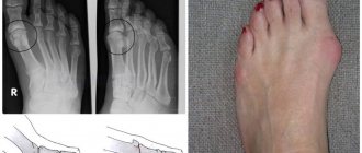
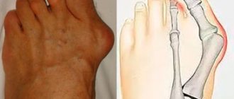
- Bone panaritium - a distinctive feature is the involvement of bone in the purulent process.
- Articular felon - develops in the interphalangeal or metacarpophalangeal joints.
- Osteoarticular felon - usually occurs with the progression of articular felon, when inflammation spreads to the articular ends of the phalangeal bones.
- Tendon felon - localized in the tendon area.
How is phlegmon treated?
Treatment is only surgical. The extent of the operation directly depends on the location, depth and area of the lesion.
The main goal of treatment is:
- relief of purulent melting of tissues,
- drainage of purulent discharge, which eliminates the absorption of decay products and poisoning of the body with decay products,
- elimination of the focus of phlegmon development, which ensures the elimination of the cause of the phlegmon process so that a complete recovery can be achieved and relapse can be prevented.
Conservative treatment is possible only in two cases:
- when phlegmon is at the very beginning of its development and there is no clear focus of suppuration (this is extremely rare),
- as the second stage of treatment after a full autopsy.
In severe cases, surgery is required to drain the cellulitis, remove dead tissue and prevent further spread of infection.
Drainage of phlegmon of soft tissues of the back
The operation is performed under general anesthesia, where the cavity in which the phlegmonous process is located is cleaned with antibiotics and antiseptic solutions, and dead tissue is removed. Intravenous antibiotics are prescribed to the patient in the postoperative period.
How the treatment method works
After processing the surgical field, the surgeon makes an incision in the place of greatest fluctuation (fluctuation), where the maximum accumulation of pus is usually observed. After opening and emptying the purulent cavity, it is washed with antiseptic solutions. Before this, the pus must be cultured for microflora.
After cleansing, the surrounding tissues are examined. All areas of questionable viability must be removed; if there are open interosseous joints, then such fingers must be removed.
If the infection spreads along the tendon, it is necessary to make an additional incision higher up the tendon, away from its purulent lesion. After this, the tendon is divided and its affected end is removed through the wound on the foot.
During an operation to open phlegmon, surgeons are guided by the principle of not leaving anything doubtful. The foot wound will heal if the infection is stopped and blood flow is restored.
Complications of cellulitis
Purulent-necrotic phlegmon of the leg in the process of cleansing the wound surface and forming granulations.
The main ones are:
- gangrene,
- sepsis,
- thrombosis of arteries and veins of the lower extremities, mesenteric vessels,
- abscesses of the liver, kidneys, spleen, respiratory organs, lungs, brain,
- thromboembolism of small and large branches of the pulmonary artery,
- multiple organ failure.
Purulent-necrotic phlegmon of the back
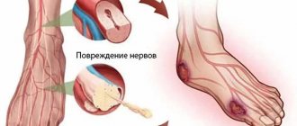
With a progressive disease, pus accumulates in the deeper layers of the skin, causing the development of inflammation of the surrounding tissues, affecting the blood and lymphatic vessels and nerves.
Cellulitis of the posterior thigh
How does the inflammation process start?
Initially, how the body’s immune system is activated:
• Blood flow to the damaged area increases sharply.
• As a result, the local temperature increases.
• There is a release of the liquid part of the blood and white blood cells from the capillaries into the intercellular space to fight the foreign agent (bacteria).
• Hyperemia in the area of damage (redness).
• Soft tissue swelling causes unbearable pain.
These four signs of hand cellulitis - fever, swelling, redness, and pain - characterize inflammation.
As inflammation of the hand develops, pus begins to form in the tissues. The purulent process with phlegmon spreads quite quickly through the cellular and fascial spaces. Finding the path of least resistance and literally melting soft tissue.
That's why emergency surgery is needed. And no amount of antibiotics or anti-inflammatory drugs can suppress this process that has already begun.
Severe intoxication of the body begins, body temperature rises, and chills appear.
Aseptic phlegmon occurs more mildly, without the formation of pus. But lymph is released from the vascular bed in huge quantities, which causes a sharp increase in pressure in the interfascial spaces, which entails intolerable pain, compression of blood vessels, nerves, even necrosis of the fingers.
In such cases, an emergency operation is also necessary to decompress the cellular and fascial spaces - fasciotomy.
Difference between cellulitis and abscess
With an abscess, the purulent focus is clearly demarcated from the surrounding tissues without a tendency to spread.
Post-injection abscess An abscess of the leg that developed after a cat bite
With phlegmon, there is no clear demarcation, and the lesion has a “creeping” character and very quickly spreads to healthy tissue.
Phlegmon of the back of the leg
But sometimes there are cases when it is not easy to distinguish an abscess from a phlegmon, especially in the initial stage of its formation, at the very period when the contents of the purulent cavity (ichor, pus, fibrin) get out of control and just begin to spread throughout the surrounding tissues.
Abscess complicated by soft tissue phlegmon
Forecast and prospects
Depends on the severity of the infection and the area that is infected. With timely initiation of complex treatment (full surgery and adequate conservative therapy), the prospects and prognosis are favorable. In advanced cases and with inadequate treatment, the prospects and prognosis are much worse, even negative due to extremely rapidly developing severe complications (sepsis, renal-hepatic and cardiopulmonary multiple organ failure).
Prevention of the development of phlegmon
It consists of timely and complete treatment of any wounds and any suppuration of the skin, which will prevent infection from penetrating into the soft tissues. With the development of any suppuration (infected wound, boil, hidradenitis, abscess, paraproctitis, etc.), a complete and timely opening of the abscess + adequate sanitation and drainage of the purulent cavity is necessary.
Prognosis after treatment method
Predicting the outcome of treatment for diabetic phlegmon is quite difficult. This outcome depends on many factors - general health, the state of the immune system, the ability to restore blood flow and close wounds.
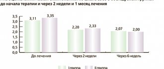
Our practice shows that with timely surgery to open the phlegmon and restore blood flow, it is possible to save the supporting limb in 75% of patients. Mortality is about 5% and is associated with worsening concomitant diseases.
Predisposing factors and reasons for the development of panaritium
The direct cause of panaritium is most often Staphylococcus aureus , which penetrates the tissue through wounds, abrasions, injections, cracks, splinters or hangnails, which most often go unnoticed, because look so insignificant that the patient simply does not pay attention to them. Less commonly, panaritium is caused by gram-negative and gram-positive bacilli , streptococcus , Escherichia coli , Proteus , as well as anaerobic non-clostridial microflora and pathogens of putrefactive infections .
External factors contributing to the development of felon include:
- systematic cooling,
- hydration,
- vibration,
- maceration,
- contamination or exposure to irritants.
Internal factors that increase the likelihood of felon occurring are:
- endocrine diseases,
- hypovitaminosis,
- metabolic disorders,
- decreased immunity.