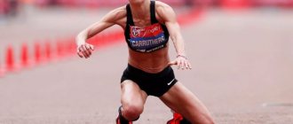
Leg pain is the third most common complaint among our patients. The reason for this is the high prevalence of varicose veins, arthritis and the seeming insignificance of such a problem as pain in the legs. At the same time, if your legs hurt from the knee to the foot, you cannot ignore this symptom as unimportant, since it may be the beginning of a more serious disease.
Pain below the knee in the front tends to occur in a few common situations. These include varicose veins, chronic venous insufficiency, myofascial pain syndrome (in which the pain is accompanied by cramps) and, less commonly, deep vein thrombosis, which is essentially an emergency condition.
At the appointment, we explain to our patients that pain in the legs must be treated and all necessary measures must be taken to eliminate it, since it deprives a person of the main factor of health - his mobility.
Pain below the knee with varicose veins
A typical situation in which a person is bothered by heaviness in the calves, swelling, “roaring in the legs,” and sometimes cramps. This picture is due to poor outflow of venous blood from the calf muscles, resulting in swelling, fluid accumulation, heaviness and pain. Superficial veins swell, blue venous nodes appear, and the skin of the lower leg takes on a brownish tint (pigmentation). The causes and treatment of varicose veins are described in detail in our article Varicose veins: causes, treatment, symptoms. Osteopathy helps not only to get rid of the symptoms of this disease, but also to prevent its further development, and often, the need for surgery.
Myofascial pain syndrome
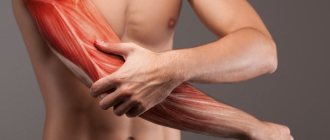
Overexertion of the lower leg muscles is the main cause of myofascial pain in the legs. Even in a healthy person, the deep muscles of the lower leg, as a rule, are in spasm, and if you run your hand over them, an acute characteristic pain appears. It is distinguished from venous pain by its greater severity, localization in one or several points, and irradiation into the foot or deep into the lower leg. Often muscle overstrain turns into spasms and cramps. In this case, neither taking calcium tablets nor increasing the dose of vitamins and microelements helps.
Myofascial pain is eliminated using osteopathic methods in 2–3 sessions, and from the first appointment the patient already feels significantly better. But you will have to be patient a little: trigger points on the legs are quite painful when touched, and their relaxation is associated with pressing on the spasmed area of the muscle. However, the positive effect quickly and completely covers the unpleasant sensations from the manipulations.
How to treat pain in the calf of the left or right leg
If you feel pain in your calf muscles, contact the Yusupov Hospital. Doctors will prescribe anti-edematous and anti-inflammatory treatment. After a comprehensive examination using x-rays, computed tomography and magnetic resonance imaging, blood and urine tests, a collegial decision will be made on treatment for the syndrome that causes pain in the calf muscles.
Severe cases of diseases that cause pain in the lower leg muscles are discussed by professors and doctors of the highest category at the Yusupov Hospital at a meeting of the expert council. Leading neurologists, rheumatologists, orthopedists, and rehabilitation specialists establish an accurate diagnosis and jointly develop tactics for further patient management. A comprehensive treatment is carried out aimed at eliminating the cause of pain, the mechanisms of development of the pathological process and relieving the pain syndrome. Therapy is carried out with effective drugs that have a minimal range of side effects.
After treatment, patients are offered a comprehensive rehabilitation program. It allows you to get all the necessary procedures at a stable price and save money. If you experience pain in the calf muscle, call the Yusupov Hospital, where the call center is open every day 24 hours a day, without days off or lunch breaks. Doctors provide emergency medical care around the clock.
Acute thrombosis of the veins of the leg
An emergency condition that requires immediate hospital treatment. Hospitalization cannot be delayed; a blood clot can completely block the outflow of venous blood and lead to gangrene of the limb. In addition, a detached blood clot can clog the blood vessels of the lungs and cause thromboembolism. If a patient comes to us with symptoms of acute thrombosis, we immediately send him to the hospital. It will be possible to continue osteopathic treatment after discharge - eliminate the causes that led to the development of thrombosis, improve venous outflow from the lower extremities, eliminate blood stagnation at the pelvic level, etc.
Diagnostics
Patients with pain in the lower leg most often initially turn to orthopedic traumatologists. If indicated, patients are referred to surgeons, neurologists, and other specialists. The examination plan includes:
- Survey
. The doctor finds out when and under what circumstances the pain first appeared, establishes the connection between the pain syndrome and external factors, identifies other complaints, and studies the patient’s life history. - Physical examination
. The specialist assesses the condition of the limb, determines swelling, hyperemia, and other pathological changes. If vascular disease is suspected, pulsation in the arteries of the foot is examined; if neurological symptoms are present, a neurological examination is required. - X-ray of the lower leg.
It is a basic study for lesions of solid structures. For some soft tissue pathologies, it is prescribed for differential diagnosis. Shows fractures, changes in bone structure, periossal growths, and other changes. - and MRI.
They are carried out at the final stages of the diagnostic search in case of ambiguous X-ray results, to clarify the conservative or surgical treatment plan. They allow you to accurately localize the pathological focus, determine its size, structure, and configuration. - Ultrasound.
For vascular diseases, Doppler sonography and duplex scanning are performed. The techniques make it possible to assess the condition of the vascular bed, the speed of blood flow, and detect places of obliteration or dilation of blood vessels. - Electrophysiological studies
. For pain of neurological origin, electromyography, electroneurography, and electroneuromyography are performed to determine the level of damage to the nerve trunk and study the condition of the nerves and muscles. - Lab tests
. They are used to assess the severity of inflammatory processes and study the state of the body in systemic pathologies.
Plaster splint for shin
LUMBOSACROSIS OSTEOCHONDROSIS
Shooting pain spreading along the back of the leg from the buttocks to the heels is characteristic of lumbosacral osteochondrosis. Many diseases come from a problematic back - tension, poor lifestyle, and little physical activity aggravate the poor condition of the spine and lead to pain in different parts of the body, including the legs from the lower leg to the foot.
Neurologists, vertebrologists and osteopaths deal with osteochondrosis: you can always contact our doctors for advice and help, which will not only relieve pain, but also eliminate the original cause of its occurrence.
Osteopathy is one of the most effective methods of treating osteochondrosis, relieving pain literally after the first appointment and helping, in addition, to eliminate the original cause of the painful condition.
Causes of shin pain
Traumatic injuries
A shin bruise usually occurs after a blow, less often it results from a fall.
It manifests itself as short-term severe acute pain, which quickly subsides, becomes dull, aching, weak or moderate. Swelling is detected in the area of the bruise, and bruising is possible. Support is preserved, movements are somewhat limited due to pain. Sometimes there is lameness. According to the mechanism of occurrence and clinical manifestations, a hematoma resembles a bruise, but the pain is of a pressing, bursting nature, which is caused by the accumulation of blood in the soft tissues. The difference from a bruise is a dense, limited swelling or area of fluctuation that does not disappear for a long time. Bruising is usually found on the skin.
Damage to the Achilles tendon is manifested by sharp pain, reminiscent of a blow or cut, on the back of the lower leg just above the ankle joint. Upon examination, swelling, pain on palpation, and retraction at the site of the Achilles are revealed. Plantar flexion of the foot is impossible with a complete rupture, but with a partial rupture it is limited. Support is significantly difficult.
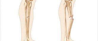
Fractures of the lower leg bones are the result of a high-energy impact: a fall from a height, being hit by a car bumper, or an industrial accident. They manifest as explosive pain, which subsequently decreases somewhat, but remains very intense. At the time of injury, a crunching sound may be heard. The following types of damage are possible:
- Fracture of the tibia.
Develops as a result of impact, twisting, falling. Usually associated with a fracture of the fibula. Accompanied by unbearable pain, displacement of fragments, significant swelling, deformation of the limb, widespread bruising, crepitus, and pathological mobility. Support is impossible, movements are sharply painful. - Isolated fracture of the fibula.
Formed by direct impact. Local swelling and bruising at the site of injury are detected. The pain is moderate, sharply intensifies with palpation of the fracture zone. When the fracture line is located in an area not covered by muscles, a step is detected. The supporting function of the limb suffers slightly.
With a pathological fracture, the clinical manifestations are smoothed out, the pain syndrome is moderate. Bone fragments are often pressed into each other, so there is no crepitus or pathological mobility. Signs that allow one to suspect a violation of the integrity of the bone are the prolonged persistence of symptoms, previous disease of the bone structures, and periodic pain in the area of this segment of the limb.
Inflammatory diseases
Myositis of the lower leg muscles develops against the background of previous overloads: intense training, long walks. It manifests itself as aching pain that spreads throughout a muscle or group of muscles. The pain increases with tension and palpation of the muscle. Sometimes a slight diffuse thickening of muscle tissue, mild swelling, and hyperemia are detected.
Pain in the back of the leg above the ankle joint is caused by inflammation of the Achilles tendon. With tendinitis, the zone of maximum pain is located 3-6 cm above the Achilles insertion point, with peritendinitis it spreads throughout the tendon. At first, the pain syndrome appears only in the first minutes of exercise, then the pain intensifies, becomes prolonged, increases rather than decreases with continued movements, and occurs at rest, at night.
The tibia is often affected by osteitis deformans. The pain is deep, localized in the area of the affected bone, dull, aching, constant, and persists for many months. Intensifies at rest, after rest. They may be accompanied by curvature of the limb, pathological fractures, and when the lesion is located near the joint, the development of osteoarthritis is noted.
Shin pain
Bone infections
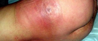
Aseptic periostitis usually develops along the anterior surface of the tibia, in an area with little soft tissue volume. It occurs after a bruise and is characterized by moderate pain, aggravated by palpation of the bone, and slight swelling. Sometimes serous inflammation is observed with the formation of a significant amount of fluid, manifested by bursting pain and the formation of a “bump” in the affected area.
Purulent periostitis becomes a consequence of infectious diseases of soft tissues and is formed in the projection of the inflammatory focus or in the area of swelling. The pain is rapidly increasing, twitching, pulsating, sharply intensifying when palpated. They appear against a background of increased temperature, chills, weakness, fatigue, and significant swelling of the limb. Within a few days they become unbearable and deprive you of night sleep.
Hematogenous osteomyelitis often affects the tibia. It develops in children, in half of the cases – after minor trauma or a general infectious disease. Manifests with severe hyperthermia, chills, fever, against which a pain syndrome appears after a few hours or 1-2 days. The pain is deep, extremely intense, pulsating, tearing. The slightest movement causes an explosion of pain, so patients try to lie still.
In some cases, a favorable course of hematogenous osteomyelitis is observed with a predominance of local symptoms and moderate pain. A hurricane development of the disease with delirium and severe impairment of general condition is also possible. In the case of postoperative and post-traumatic osteomyelitis, a clinical picture similar to the hematogenous type of the disease is observed, but the symptoms are not so pronounced, and the progression of the pathology is longer.
Soft tissue infections
In case of infectious lesions of soft tissues, the pain in the first hours is pressing, and when touched it is stabbing. They grow quickly, become twitching, pulsating, constant, and exhausting. They intensify when palpating, lowering the limb. The formation of an abscess usually occurs against the background of a sleepless night. In the affected area, swelling, hyperemia, purple-bluish coloration of the skin, and local hyperthermia are detected.
The degree of disturbance of the general condition depends on the prevalence of the purulent process, ranging from minor malaise to severe fever, severe intoxication syndrome. Local pain in the lower leg is observed with boils. Intense pain syndrome, spreading over a significant part of the segment, is observed with carbuncles, abscesses and phlegmon.
Arterial diseases
The cause of pain in the lower leg is sometimes arterial disease. With obliterating endarteritis, pain initially appears only when walking long distances (over 1 km). The pain is localized in the calf muscles, provokes intermittent claudication, and forces the patient to stop while moving. In the future, the distance to the onset of pain is reduced. In the later stages, pain is observed at rest, trophic ulcers form, and gangrene develops.
In terms of the nature and conditions of the onset of pain, obliterating atherosclerosis resembles obliterating endarteritis, but is detected in older men rather than in young men, and has a more favorable course. Only 14% experience rapid development of pathology, leading to pain at rest and severe trophic disorders; in other cases, pain remains at the same level for a long time or intensifies during periods of seasonal exacerbations. Similar symptoms are observed with Mönckeberg arteriosclerosis, which is diagnosed in the second half of life and occurs equally often in both sexes.
Diseases of veins and lymphatic vessels
A common vascular cause of pain is varicose veins. At first, the pain is minor, dull, local, short-term, appears in the evening, after a long stay in an upright position, and is combined with a feeling of heaviness. Subsequently, the pain syndrome becomes long-lasting, occurs after minor exertion, and is accompanied by noticeable pastiness of the limbs. At the final stage, the pain is prolonged, supplemented by night cramps, swelling, hyperpigmentation, and trophic disorders.
Acute phlebitis of the superficial veins of the leg often develops with varicose veins and is manifested by rapidly progressing pain, the appearance of a strip of hyperemia along the vein. The vein is compacted, its palpation is painful. With chronic phlebitis, the symptoms are smoothed out, the pain is recurrent. When deep veins are affected, there is no red stripe on the skin, the pain is localized deep in the tissues, combined with severe swelling and general hyperthermia.
With thrombophlebitis, as a rule, varicose veins of the upper third of the segment are affected. There is a nagging sharp pain in the projection of the vein, which intensifies when walking, the formation of a red stripe, a dense painful cord, and a disturbance in the general condition. With deep vein thrombosis, the pain is deep, bursting, aggravated by palpation, and can be combined with swelling, pallor of the limb, and swelling of the superficial veins.
With lymphedema, pain in the legs is constant, dull, bursting, complemented by heaviness in the legs and significant dense swelling of the limb. Primary lymphedema is characterized by a gradual spread of pain and swelling in the peripheral direction (from the thigh to the lower leg), while secondary lymphedema occurs in the central direction (from the foot to the lower leg). Palpation of the limb segment is painless.
Other skeletal diseases
In Schlatter's disease, local tenderness occurs in the area of the tibial tuberosity, just below the knee joint. The pain syndrome is associated with physical activity, appears when squatting, walking up stairs, and disappears with rest. At first the pain is mild, then it becomes significant, and attacks of acute cutting pain are possible. Palpation is painful; a prominence of bone density is detected in the area of the tuberosity.
Pain in the legs can occur with congenital anomalies of the lower extremities, Blount's disease, X-shaped legs, gonarthrosis and arthrosis of the ankle joint. Usually dull, aching, intermittent, associated with physical activity. Caused by disruption of the normal mechanisms of standing and walking, constant overload of the limb.
Oncological pathologies
Benign tumors of the tibia are characterized by slow growth, low-intensity, intermittent pain with unclear localization. The exceptions are osteoid osteoma and osteoblastoma, in which the pain is sharp, acute, and intense. Chondromas most often form on the tibia. Osteochondromas usually affect the fibula, the pain syndrome is localized in the upper third of the leg along the outer surface.
With malignant neoplasia, the pain is initially unclear and dull. They progress quickly, become permanent, extremely intense, painful. They can only be eliminated with narcotic analgesics. There is a deterioration in the general condition, swelling, deformation, and dilation of veins in the area of the neoplasm. With osteogenic sarcomas, pain occurs closer to the knee or ankle joint, with chondrosarcomas - in the upper part of the leg.
Neurological causes
Sometimes painful sensations are provoked by neurological pathologies. The pain is burning, shooting, piercing, spreading throughout the segment or the entire leg, combined with sensitivity disorders, weakening of the strength of the limb. The cause of pain is:
- Radicular syndrome.
Occurs after spinal injuries, in many degenerative diseases. The affected area depends on the root involved. Pain in the leg is combined with pain in the back, which is provoked by sudden movements, laughing, coughing, sneezing. - Femoral nerve neuropathy.
The pain is localized along the anterior inner surface of the lower leg and intensifies with extension of the knee joint. - Peroneal nerve neuropathy.
Pain occurs in the outer part of the lower leg (mainly in the lower third of the segment), and intensifies with squats. - Tibial nerve neuropathy.
With pathology of traumatic origin, patients complain of burning causal pain along the back, partially outer surfaces of the leg. With tarsal tunnel syndrome, there is pain in the foot, radiating to the calf muscle. - Sciatic nerve neuropathy.
The pain spreads along the buttock to the back of the thigh and lower leg to the foot. Very sharp, reminiscent of a lumbago, a dagger strike.
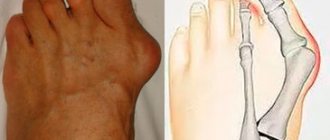
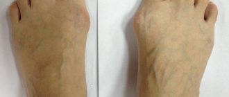
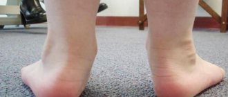
FLAT FOOT
The cause of the painful condition of the legs from the feet to the legs can be “normal” flat feet. This pain is characterized by an increase in the evening and aggravation during long walking and other physical activities; it is aching, tiring, and spreads throughout the lower part of the limbs. The feet may become visually larger, and the bone often begins to protrude.
Flat feet are “treated” by choosing shoes after consultation with a doctor, physical therapy complexes, and osteopathy also helps to cope with it. Osteopaths work on the foot, improving blood circulation, eliminating congestion, and stimulating the production of synovial fluid, which works as a lubricant for the joints. After just a few techniques, even an adult who has suffered from flat feet all his life becomes easier and more comfortable to shift.
Treatment
Help before diagnosis
For minor injuries and non-traumatic lesions, it is recommended to ensure rest and an elevated position of the limb. For tibia fractures, temporary immobilization using splints or improvised materials is required. The leg is fixed from the foot to the upper third of the thigh. The victim is given an analgesic. For pain of non-traumatic origin without signs of severe inflammation, it is possible to use local anesthetics. In case of intense pain or a violation of the general condition, an immediate examination by a specialist is necessary.
Conservative therapy
For patients with tibial fractures, a blockade is performed, followed by fixation using skeletal traction. For other diseases and injuries, depending on the severity of the pathology, a plaster splint is applied, rest or a gentle regimen of physical activity, and the use of orthopedic devices are recommended. The following methods are used:
- Drug therapy
. The list of drugs is determined by the etiology and symptoms of the disease. For intense pain, analgesics are prescribed. For purulent lesions, antibiotics are necessary. For vascular pathologies, antiplatelet agents, anticoagulants, and antispasmodics are indicated. - Exercise therapy
. Therapeutic exercise is a mandatory part of rehabilitation measures. Allows you to maintain muscle strength and joint mobility, prevent the development of complications, and improve limb function. - Physiotherapy
. Physiotherapeutic procedures reduce pain and inflammation, activate blood circulation, and stimulate recovery processes. Widely used techniques include medicinal electrophoresis, UHF, and magnetic therapy. Electrical stimulation has been successfully used for some diseases.
Patients are prescribed massage. According to indications, manual therapy is performed. For a number of pathologies, kinesio taping is used. Patients are referred to sanatorium-resort treatment.
LEGS HURT RIGHT NOW! WHAT TO DO?
1. Relax. Lie down with your ankles on a bolster to elevate your legs above heart level. The blood will drain, the swelling will decrease, and the muscles will relax.
2. Apply a cold compress. For example, a thin towel with ice or some package of semi-finished products taken out of the freezer. Keep the compress for up to 20 minutes, repeat several times a day.
3. Take an over-the-counter pain reliever such as ibuprofen. But take only those medications that you are sure of, and if the pain does not go away, be sure to visit a doctor.
4. Get a massage. This is true if your legs have cramps, or if they “hum” after a long period of exercise. You can stretch the muscles yourself, or you can consult a specialist, including an osteopath.
How to relieve pain in calf muscles
In order to relieve pain in the calf muscle after an injury, it is necessary to provide rest to the lower limb, place an ice pack on the back of the shin, apply an elastic bandage and elevate the leg. 72 hours after the injury to the calf muscle, a heating pad should be applied to the lower leg.
As soon as the swelling subsides, you need to rub the skin over the calf muscle with a pain-relieving balm (Bengay, Finalgon) and wrap it with an elastic bandage. If there is severe pain in the calf muscle, you should take a tablet of a non-steroidal anti-inflammatory drug – ibuprofen, ketanov – orally. If the pain does not go away after 7 days, you should consult a doctor.
If you have pain in the calf due to an injury, you need to reduce the load on the heel. Heel pads can reduce stress on the tendons and calf muscles. You can make them yourself. To do this, just cut out pieces of cork 6 cm thick and put them in your shoes. It is necessary to avoid activities that involve additional stress on the calf muscles. No need to walk to the upper floors or ride a bicycle. Wearing comfortable shoes reduces pain in the calf muscle. Traumatologists recommend wearing sneakers and avoiding stiletto heels during illness.
If the cause of pain in the calf muscles is varicose veins of the lower extremities, it is useful to use compression stockings. Arch supports, which are installed in shoes under the instep of the foot, help correct imbalances that affect gait and cause pain in the calf muscles.
If acute pain suddenly appears in the calf muscles, you must stop, lie down on a bench, place a cushion under your feet, and call an ambulance. In this case, the patient needs to consult a phlebologist and provide emergency care. Sharp pain in the calves, reminiscent of a whiplash, goes away after resting for a few minutes. If it appears again, you should contact an angiosurgeon.
The benefits of osteopathy in the treatment of foot diseases
- Osteopathy restores not only the function of the damaged area, but also the biomechanics of your entire body.
- We find the reason that led to your dysfunction and eliminate it.
- We do not give you injections into the joint cavity, we do not use hormonal drugs, which means you have no risk of infection or other complications.
- We provide all patients with a full osteopathic examination and examination, regardless of complaints. This allows for correction at a higher level, including the body’s reserves and directing them towards self-healing.