Infectious mononucleosis - a disease also known as glandular fever, Filatov’s disease, monocytic tonsillitis, Pfeiffer’s disease, “kissing disease”, etc. This is an acute manifest form of Epstein-Barr virus infection, occurring with prolonged fever, generalized lymphadenopathy, tonsillitis, pharyngitis, enlargement of the liver and spleen, characteristic changes in the hemogram in the form of leukocytosis, absolute lymphomonocytosis with the appearance of atypical mononuclear cells in the peripheral blood.
- Causes and causative agent
- Epidemiology
- Pathogenesis
- Pathomorphology
- Symptoms and classification
- Atypical forms
- Outcomes of infectious mononucleosis
Causes and causative agent
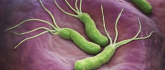
The causative agent of MI is the Epstein-Barr virus (EBV) - a large enveloped DNA virus of type 4 from the family Herpesviridae, subfamily of Herpesviridae, genus Lymphocryptovirus.
Based on differences in the structure of the genome and the ability to cause a blast transformation reaction of B-lymphocytes, 2 strains of EBV are distinguished: the most active type 1(A) in the reaction and the less active type 2(B). Type 1(A) is widespread, type 2(B) is found mainly on the African continent and several countries in North America.
EBV has an oval shape with a diameter of 150-200 nm. The virus genome is represented by two DNA molecules enclosed in an icosahedral capsid consisting of 162 capsomers. The complex of virus capsid proteins (p150, p18, p23) is represented by its capsid antigen - VCA (Viral capsid antigen). The immunodominant proteins in this complex are p18 and p23.
At the earliest stages of EBV replication, a highly immunogenic complex of viral proteins (p54 and p138), called the early EA antigen, appears in the body of an infected person.
The targets for EBV are cells that have the CD21 receptor on their surface. These are epithelial cells of the mucous membranes of the nasopharynx and oropharynx, excretory ducts of the salivary glands; B-lymphocytes (primarily located in the follicular structures of the Pirogov-Langans lymphoid ring and peripheral lymphoid tissue); dendritic cells; epithelium of the cervix.
To learn more
|
The immunosuppressive effect of EBV, on the one hand, causes long-term (lifelong) persistence of the virus in the body with the possibility of periodic exacerbations of the disease, on the other hand, it explains the possible role of the virus in the development of immuno- and oncopathology. The virus is unstable to environmental factors and quickly dies under the influence of disinfectants when dried at room temperature.
EBV is extremely thermolabile - it “dies” at a temperature of 50-52°C for 30 minutes, at a temperature of 37.5°C for 20 hours, and is stable at a temperature of minus 70°C; tolerates lyophilization well and is stored in tissues for a long time in a 50% glycerol solution. On metal surfaces (coins, door handles, water taps) EBV survives for 2 hours, on plastic and wood - up to 3 hours, in damp medical cotton wool and gauze - until they dry at room temperature - up to 6 hours.
Methods for diagnosing mononucleosis
Symptoms of mononucleosis are not specific. Therefore, diagnosis is based primarily on laboratory data. The necessary studies are combined in the profile “Diagnostics of infectious mononucleosis”.
General blood analysis
A complete blood count for mononucleosis usually shows an increase in the number of leukocytes and ESR. Characterized by a significant increase in lymphocytes and monocytes, together these varieties can account for up to 80-90% of the total number of leukocytes. The main symptom of mononucleosis is an increase in the number of specific cells - atypical mononuclear cells (which are altered lymphocytes) to a level of over 10%. However, they are detected no earlier than on the 5th day from the onset of the disease.
More information about the diagnostic method
Blood chemistry
Mononucleosis can manifest itself in the results of a biochemical blood test in the form of a two- or three-fold increase in the activity of ALT and AST. When the skin turns yellow (jaundice), bilirubin increases.
More information about the diagnostic method
Serological blood test
Using serological analysis, specific antibodies to the causative agent of mononucleosis, the Epstein-Barr virus, can be detected.
More information about the diagnostic method
Sign up for diagnostics To accurately diagnose the disease, make an appointment with specialists from the Family Doctor network.
Epidemiology
The source of infection is a person with acute and chronic manifest or latent forms of EBVI. Infected individuals actively shed the virus from the last days of incubation and for 6-18 months after the initial infection. More than 90% of asymptomatic seropositive individuals harbor the virus in oropharyngeal secretions.
The main transmission mechanism is airborne, the transmission route is aerosol. EBV is mainly transmitted through direct close contact through saliva (kissing, toys with contaminated saliva) containing oropharyngeal epithelial cells. Infection is possible if sanitary and hygienic rules for handling utensils in the public catering system are not followed, as well as when using “shared” glasses, bottles, etc. Parenteral (during organ transplantation and blood transfusions) and sexual transmission are also possible. The possibility of vertical transmission of the pathogen with the development of congenital EBVI has been established. EBV is transmitted through blood transfusions and transplants. The entire non-immune population is susceptible to infection, regardless of age.
Pathogenesis
Incubation period (7-21 days). Introduction of EBV and its replication in epithelial cells of the nasopharyngeal mucosa (primary foci of infection). The entry of newly formed virus generation from lysed epithelial cells into the intercellular environment every 18-24 hours, “horizontal” spread of infection in the area of the entrance gate. Release of the virus into the environment.
Entry of EBV into the follicular lymphatic structures of the oropharyngeal mucosa, into regional lymph nodes, its introduction into B-lymphocytes, their transformation into lymphoblastoid cells. The presentation of virus antigens by macrophages and DCs to immunocompetent cells is the beginning of the formation of humoral and cellular immunity. Lymphogenous and hematogenous dissemination of EBV.
Initial period (from several hours to 1 week). Main manifestations: sore throat, tonsillitis, pharyngitis, adenoiditis, enlarged regional lymph nodes. Intoxication syndrome is manifested by fever, chills, sweating, and weakness in the body.
The peak period (from 1 to 3 or more weeks). There is an increase in specific immunity, necrotic changes in the mucous membranes at the entrance gate. Bacterial flora (follicular, lacunar, necrotic tonsillitis) may join or become more active. The virus continues to be released through saliva and nasopharyngeal and oropharyngeal secretions.
The period of convalescence (from 1 to 3-4 months). Peak formation of specific immunity. The affected organs and tissues are restored. In most cases, clinical recovery occurs. The virus will remain in the body of most people who have been ill for the rest of their lives.
Pathomorphology
Due to the low mortality rate, the pathomorphology of MI is presented mainly based on the results of studies of biopsy material.
A pathological examination reveals a macroscopic increase in all groups of lymph nodes, tonsils and other lymphoid formations of the pharynx, spleen, and liver. Histological examination reveals proliferation of lymphoid and reticular cells, and in the liver periportal infiltration with lymphoid elements. In severe cases, focal necrosis in the lymphoid organs and the appearance of lymphoid infiltrates in the lungs, kidneys, central nervous system and other organs are possible. Rare deaths are most often caused by splenic rupture, hematological complications (hemolysis, thrombocytopenic purpura) or central nervous system damage.
Complications after illness
Complications after infectious mononucleosis are not very common, but quite serious:
- hematological complications (autoimmune hemolytic anemia, thrombocytopenia, granulocytopenia);
- splenic rupture;
- neurological complications (encephalitis, paralysis of facial muscles, polyneuritis, transverse myelitis, psychosis);
- hepatitis;
- cardiac complications (pericarditis, myocarditis);
- complications of the respiratory system (interstitial pneumonia, airway obstruction).
1 Consultation with an otolaryngologist in MedicCity
2 Consultation with an otolaryngologist in MedicCity
3 Consultation with an otolaryngologist in MedicCity
Symptoms and classification
By type, infectious mononucleosis comes in the following forms:
- typical,
- atypical (erased, asymptomatic)
Based on severity, the following forms of the disease are distinguished:
- light,
- medium-heavy,
- heavy.
According to the course, MI can be uncomplicated or complicated.
In ICD-10 (1995) it is designated as follows: B27.0 “Mononucleosis caused by the gammaherpetic virus.”
Below is the most typical clinical picture of MI , observed in moderate cases of the disease.
The incubation period is from 8 to 21 days (maximum 6-8 weeks).
Initial period (up to 4-5 days). The onset of the disease can be either acute or gradual. Febrile fever is accompanied by progressive weakness, headache, decreased appetite and other symptoms of intoxication. Hyperplasia of the third pharyngeal tonsil, located on the vault of the nasopharynx, and impaired lymphatic drainage are clinically realized by the appearance of signs of adenoiditis: nasal congestion, “nasal” voice, night snoring (even in small children), swelling of the eyelids, pastiness of the upper half of the face.
Patients complain of moderate sore throat. Objectively - catarrhal phenomena on the tonsils and mucous membrane of the oropharynx. Noteworthy is the discrepancy between moderate catarrhal symptoms in the oropharynx and the severity of the intoxication syndrome. Already now, the contours of the neck can change due to a significant increase in the cervical, occipital, and submandibular lymph nodes. The lymph nodes are moderately painful on palpation, are not fused to each other or to the surrounding tissues, are clearly contoured, and the surrounding subcutaneous tissue is not swollen.
During the peak period (from 1 to 3 or more weeks), the patient’s well-being worsens: the fever can reach 40″C, accompanied by chills and increased headache. Sore throat gets worse when swallowing. The symptoms of acute tonsillopharyngitis are objectively revealed, which can occur in catarrhal, follicular, membranous or ulcerative-necrotic forms. The most common picture of lacunar tonsillitis is observed: hypertrophy of the tonsils to the 2-3rd degree, the presence of loose whitish deposits on them in the form of islands, “pillars”. In some cases, plaques may resemble diphtheria - difficult to remove films, tightly bound to the underlying tissue.
Multiple petechiae may appear on the mucous membrane of the soft and hard palate; the posterior wall of the pharynx is sharply hyperemic, loose, granular, with multiple small hyperplastic follicles. The duration of a sore throat reaches 2-3 weeks. The phenomena of tonsillitis do not have a strict correlation with the severity of intoxication syndrome.
Lymphadenopathy becomes generalized. The size of the lymph nodes varies from a pea to a walnut (0.5-3 cm), palpated in the form of single lymph nodes or a chain. Already from the 6th day of illness, most patients experience an enlargement of the liver and spleen. It is worth noting that the spleen enlarges more than the liver.
On days 8-11 of illness, an exanthema of a maculopapular nature may appear. The rashes do not have a specific localization and quickly disappear without treatment. Exanthema is of an immunocomplex nature and is the result of polyclonal activation of B lymphocytes under the influence of a virus, including the formation of epitopes capable of binding ampicillin. Subsequently, in those who have recovered from myocardial infarction, sensitization to penicillins is leveled out.
The period of convalescence (from 1 to 3-4 months).
The patient’s well-being gradually improves, body temperature normalizes, and the clinical symptoms of acute tonsillopharyngitis disappear. Lymphadenopathy regresses slowly - over 1.5 months or more. Hepatolienal syndrome can be determined within 3-6 months. The duration of the convalescence period varies from person to person; sometimes low-grade body temperature persists for several weeks. During the period of convalescence, asthenic syndrome is expressed - weakness and malaise may persist for several months. If the patient has normal immunity (the body's defenses), he recovers in 3-4 months.
The severity criteria for MI are:
- severity of intoxication syndrome,
- the severity of changes in lymphoid tissue,
- hemogram changes,
- presence of complications.
In a mild form, the patient’s general condition changes slightly, the temperature rises to 37-37.5˚ C, and catarrhal changes in the nasopharynx and oropharynx of an inflammatory nature are observed. Lymph nodes are slightly enlarged. Hepatosplenomegaly is mild. Reversal of symptoms occurs quickly (by the end of the 2nd week).
In severe forms , high fever is recorded (body temperature above 39.5°C), severe intoxication (lethargy, adynamia, vomiting, headache, anorexia) persists for more than 3 weeks. Hyperplasia of the lymphoid tissue located on the vault of the nasopharynx manifests itself in impaired nasal breathing, up to its absence, and the characteristic appearance of the patient (puffy “adenoid face”). Damage to the palatine tonsils is characterized by severe purulent-necrotic, sometimes fibrinous inflammation. Generalized lymphadenopathy and severe hepatosplenomegaly are observed. High probability of complications. Reversal of symptoms occurs slowly, over 4-5 weeks.
In patients with infectious mononuclear disease, there is a parallelism between the severity of the disease and the number of atypical mononuclear cells in the peripheral blood.
Complications of infectious mononucleosis in children
With timely diagnosis and treatment, complications of infectious mononucleosis do not occur so often. However, the most common among them is liver inflammation , which can cause jaundice in children. In very rare cases, rupture of the spleen occurs - this is the most dangerous consequence of mononucleosis, which sometimes turns fatal.
When a child has had mononucleosis, his immunity remains weakened for about a year. Therefore, during this period it will be effective to do routine vaccinations after an illness and, if possible, limit the child’s contact with a large number of people, as well as refrain from traveling to places with an unusual climate.
The most insidious feature of mononucleosis is that the mononucleosis virus is oncogenic , i.e. it can have a stimulating effect on the development of cancer. Therefore, it is very important to re-take a blood test after recovery in order to track how the blood composition is being restored.
To prevent these complications, if there are obvious symptoms of mononucleosis in a child, you need to consult a doctor, who, in the case of an acute form of the disease, will ensure hospitalization at the SM-Clinic hospital. Our doctors will quickly diagnose, prescribe treatment and make every effort to ensure that the child recovers as quickly as possible.
Sources:
- A. V. Razgulyaeva, O. P. Ukhanova, S. M. Bezrodnova. Modern ideas about the etiology, pathogenesis, clinic and treatment of infectious mononucleosis in children // Bulletin of the Stavropol State University, 2012, No. 78(1).
- I. A. Zaitseva, S. A. Khmilevskaya, I. A. Berezhnova. Infectious mononucleosis in children // Children's infections, 2004, No. 3.
The information in this article is provided for reference purposes and does not replace advice from a qualified professional. Don't self-medicate! At the first signs of illness, you should consult a doctor.
Atypical forms
Erased - with an incomplete, weakly expressed, quickly passing symptom complex.
Asymptomatic - there are no clinical signs of the disease. Diagnosis in this case is possible based on laboratory data, taking into account the epidemic situation.
Complications . Specific:
- asphyxia (due to supraglottic stenosis associated with a significant increase in the lymphoid formations of the Pirogov-Waldeyer pharyngeal ring);
- rupture of the spleen, which can occur spontaneously or from physical influences (for example, during palpation);
- lesions of the nervous system (encephalitis, meningitis, cranial nerve palsies, Guillain-Barré syndrome);
- hemolytic anemia, thrombocytopenia, neutropenia.
- In rare cases, bilateral interstitial pneumonia with severe hypoxia is noted.
The most common nonspecific complication is the addition of bacterial infections (otitis, sinusitis, pneumonia, etc.) caused by Staphylococcus aureus, streptococci, etc. Complications of the disease can occur at different periods, including during the period of long-term convalescence.