Helicobacter bacterium: symptoms and causes
Abdominal pain, nausea, belching of air - all this may indicate that a dangerous, harmful Helicobacter bacterium has settled in the body, the treatment of which must be serious and thorough.
Helicobacter pylori is a very dangerous pathogenic microorganism that can lead to gastric and duodenal ulcers, gastritis and other diseases dangerous to the digestive system. Helicobacter was first discovered only 30 years ago. Medical research conducted since that time has proven that gastritis can have an infectious etiology. Also, according to studies of this bacterium, scientists have proven that, according to statistics, 75% of cases of stomach cancer in developed countries are caused by Helicobacter. In developing countries, this figure is even more frightening: 90% of patients with stomach cancer contracted the disease due to Helicobacter pylori.
Thus, it is worth pointing out the special role of early diagnosis of gastritis and gastric ulcers. It is a timely visit to a doctor that can save health and life.
Bismuth preparations
Bismuth was one of the first drugs to eradicate H. pylori. There is evidence that bismuth has a direct bactericidal effect, although its minimum inhibitory concentration (MIC - the smallest amount of drug that inhibits the growth of a pathogen) against H. pylori is too high. Like other heavy metals such as zinc and nickel, bismuth compounds reduce the activity of the enzyme urease, which is involved in the life cycle of H. pylori. In addition, bismuth preparations have local antimicrobial activity, acting directly on the bacterial cell wall and disrupting its integrity.
What is the Helicobacter bacterium?
Helicobacter is a special type of pathogenic microorganism. It is a dangerous bacterium that infects the duodenum and stomach of humans. The microorganism itself is a parasite that produces toxic substances that damage the mucous membranes of organs. It is damage to the mucous membrane that leads to peptic ulcers, gastritis and other dangerous ailments.
It has long been believed that no organism is able to survive in the acidic environment of the stomach. But this does not apply to Helicobacter. The bacterium, on the contrary, thrives in an acidic environment, which is preferable for it to any other habitat. Helicobacter pylori has a spiral shape and flagella. This structure of the microorganism allows it to move through the mucous membranes of internal organs and cause irreparable harm to their integrity.
Helicobacter can adapt to almost any habitat. This is explained by the fact that this organism practically does not require oxygen. And another distinctive feature of Helicobacter pylori is its ability to change its own shape and become oval or round.
Russian scientists have come to the decision to introduce a special term - helicobacteriosis. It denotes all the processes that begin to occur in the body immediately after the introduction of this pathogenic microorganism into it. But Western scientists conducted studies of patients’ charts and came to the conclusion that about 60-65% of the population of the entire Earth are susceptible to infection. Thus, Helicobacter is the most common infectious disease of humanity after herpes, which is a viral disease.
Metronidazole
H. pylori is generally very sensitive to metronidazole, the effectiveness of which is independent of pH. After oral or infusion use, high concentrations of the drug are achieved in the gastric juice, which makes it possible to achieve the maximum therapeutic effect. Metronidazole is a prodrug that undergoes activation by bacterial nitroreductase during metabolism. Metronidazole causes H. pylori to lose its helical DNA structure, causing DNA damage and killing the bacterium.
NB!
The result of treatment is considered positive if the results of the test for H. pylori, carried out no earlier than 4 weeks after the course of treatment, are negative. Performing the test before 4 weeks after eradication therapy significantly increases the risk of false negative results. It is preferable to stop taking PPIs two weeks before diagnosis. Helicobacter pylori eradication therapy: scheme
What happens in the body
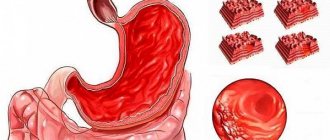
As soon as Helicobacter enters the human body, it immediately descends into the stomach, where it lives for the rest of the time. In order not to die from gastric juice, the composition of which is quite aggressive, Helicobacter secretes special enzymes that surround it with a shell and neutralize the acid. The spiral-shaped antennae that Helicobacter has allow it to drill through the mucous layer and get to the cells of the parietal tissues, which are usually hidden from acid.
It is the parietal cells of tissues that become the main food for Helicobacter. The pathogenic microorganism eats them and poisons the environment with the products of its vital activity. At the same time, those blood cells that react to Helicobacter and strive to destroy them (neutrophils) wake up in the body. However, by destroying the pathogenic microorganism, neutrophils also destroy mucosal cells that have already been touched by Helicobacter.
After the layer of the mucous membrane is damaged, hydrochloric acid begins to actively affect the tissue. This is how the inflammatory process develops and an ulcer occurs. The localization of ulcers is most often similar. This is explained by the fact that Helicobacter itself is very fond of two parts of the stomach - the bulb and the pylorus.
Proton pump inhibitors (PPIs)
PPI therapy has proven effective in various clinical studies. Although PPIs have a direct antibacterial effect on H. pylori in vitro, they do not play an important role in eradicating the infection.
The mechanism by which PPIs synergize when combined with antimicrobials to enhance the clinical efficacy of eradication therapy has not been fully established. It is assumed that antisecretory drugs of the PPI group may help increase the concentration of antimicrobial agents, in particular metronidazole and clarithromycin, in the gastric lumen. PPIs reduce the volume of gastric juice, as a result of which the leaching of antibiotics from the surface of the mucosa decreases, and the concentration, accordingly, increases. In addition, reducing the volume of hydrochloric acid maintains the stability of antimicrobials.
Causes of Helicobacter pylori infection
Helicobacter bacteria cannot exist in air; with active air intake, these pathogenic organisms die. They are transmitted mainly through human mucus and saliva. Thus, most often infection occurs in the following ways:
- Sharing utensils;
- Using only personal hygiene products;
- Kisses;
- From mother to child.
Thus, friends, family and cohabitants of the patient may be at risk.
In general, infection is facilitated by a low standard of living and neglect of hygiene rules. Very often, helicobacteriosis occurs in people living in communal apartments and dormitories, orphanages, as well as in medical workers. It is worth noting that in third world countries the disease occurs much more often than in developed countries. In Russia, in recent years, the incidence of gastritis and ulcers due to the influence of Helicobacter has begun to be noted among people from wealthy segments of the population.
You can protect yourself, and it is better to take care of prevention in advance than to suffer from an illness and look for ways to fight Helicobacter urgently.
Eradication therapy
Despite the use of combination treatment regimens, 10–20% of patients infected with H. pylori fail to achieve elimination of the pathogen. The best strategy is to select the most effective treatment regimen, but the possibility of using two or even more sequential regimens should not be ruled out if the treatment of choice is insufficiently effective.
If the first attempt at eradication of H. pylori fails, it is recommended to immediately proceed to second-line therapy. Antibiotic sensitivity testing and switching to salvage therapy regimens are indicated only for those patients in whom second-line therapy will also not lead to eradication of the pathogen.
One of the most effective “rescue regimens” is a combination of PPI, rifabutin and amoxicillin (or levofloxacin 500 mg) for 7 days. An Italian study led by Fabrizio Perri and published in Alimentary Pharmacology & Therapeutics in 2000 confirmed that the rifabutin regimen was effective against strains of H. pylori resistant to clarithromycin or metronidazole. However, the high price of rifabutin limits its widespread use.
NB! To avoid the development of resistance to both metronidazole and clarithromycin, these drugs are never combined in the same regimen. The effectiveness of such a combination is very high, but patients who do not respond to therapy usually develop resistance to both drugs at once (a study by German scientists led by Ulrich Peitz, published in Alimentary Pharmacology & Therapeutics in 2002). And further selection of therapy causes serious difficulties.
Research data confirm that a 10-day salvage therapy regimen, including rabeprazole, amoxicillin and levofloxacin, is much more effective than standard second-line eradication therapy (a study by Italian scientists led by Enrico C Nista, published in Alimentary Pharmacology & Therapeutics in 2003 year).
How to get infected?
Infection with this microorganism is becoming a real problem for modern medicine, because almost every second adult is a carrier of the bacterium
Both adults and children can become infected with Helicobacter pylori. The main route of transmission is fecal-oral, oral-oral. Thus, you can become infected by kissing, sharing utensils, cups, bottles, toothbrushes or lipsticks of other people, through dirty hands, etc.
That is why this bacterium is often called a family problem.
- if it appears in one person, with a high probability expect it to appear in his loved ones.
References
- Ivashkin V.T., Maev I.V., Abdulganieva D.I., Alekseenko S.A., Ivashkina N.Yu., Korochanskaya N.V., Mammaev S.N., Poluektova E.A., Trukhmanov A. S., Uspensky Yu.P., Tsukanov V.V., Shifrin O.S., Zolnikova O.Yu., Ivashkin K.V., Lapina T.L., Maslennikov R.V., Ulyanin A.I. . Practical recommendations of the Scientific Community for Promoting the Clinical Study of the Human Microbiome (NSOM) and the Russian Gastroenterological Association (RGA) on the use of probiotics for the treatment and prevention of gastroenterological diseases in adults. Russian Journal of Gastroenterology, Hepatology, Coloproctology. 2020
- GuevaraB, CogdillAG. Helicobacter pylori: A Review of Current Diagnosticand ManagementStrategies. DigDisSci. 2020
- Choi IJ, Kim CG, Lee JY. Family History of Gastric Cancer and Helicobacter pylori Treatment. N Engl J Med. 2020
Guarantees of effectiveness - in the hands of the pharmacist?
The effectiveness of eradication therapy has been calculated and sorted out. First-line therapy is successful in more than 75% of cases, and treatment using an alternative regimen (second line) brings recovery to 93% of patients. The unsatisfactory result of treatment is explained simply: bacteria quickly adapt to antibiotics and new, resistant strains appear. To defeat H. pylori, you must strictly follow the treatment regimen. And an undisciplined patient trying to replace, cancel or delay taking one or even several components of eradication therapy should be aware of the danger that awaits him.
A pharmacist and pharmacist can tell a doubting client how many additional problems may befall him when trying to “improve” the anti-ulcer complex in his own way. Don't skimp on colors when your customer wants to turn off the straight road to recovery towards traditional medicine or homeopathy. Paint a picture of resistant Helicobacter pylori infection brightly and on a grand scale, and another good deed will appear on your account.
Long courses of treatment with NSAID drugs
There are 2 methods of treating Helicobacter pylori infection.
Treatment is carried out comprehensively. According to the WHO methodology, any drug regimen must meet the following criteria:
- Efficiency and speed
- Safety for the patient
- Convenience – use long-acting drugs, short course of treatment
- Interchangeability - any drug must be interchangeable with a complete analogue or generic
Currently, 2 methods have been adopted for the treatment of Helicobacter pylori infection. It is not recommended to use them simultaneously. If 1 scheme does not give a positive result, then the second one is used and vice versa. This prevents Helicobacter from developing immunity to drugs. Treatment regimens:
- Three-component method - 2 antibacterial drugs and 1 agent to reduce the acidity of gastric juice
- Four-component method - 2 antibacterial drugs, 1 - to reduce the secretion of hydrochloric acid, 1 - bismuth compounds
There is a 3rd treatment regimen for combating the microorganism. It is used when the first 2 did not have the desired effect. In this case, they speak of a resistant Helicobacter strain.
In this case, a preliminary endoscopic examination is carried out with the collection of material for a biopsy. In the laboratory, drugs for the treatment of Helicobacter pylori are individually selected. And only after that the doctor develops an individual course.
Blood and stool tests
20–30 days before stool examinations, you should stop taking antibiotics, and three days before, you should stop using coloring products. Blood is donated in the morning on an empty stomach. One day before, you need to eliminate fatty foods from your diet.
The results of stool tests are released the next day. An enzyme-linked immunosorbent test is just as quick, but a Western blot test, which requires more time and more complex equipment, can take up to six days.
A stool test only shows the presence or absence of infection. Blood tests provide more accurate results and reveal the presence or possibility of developing peptic ulcers.
Helicobacter: diagnostic measures
To diagnose Helicobacter, you need to undergo tests.
Diagnostic procedures begin with interviewing and examining the patient. Then special studies are carried out to confirm or refute the preliminary diagnosis. Tests for Helicobacter pylori:
- Non-invasive procedures - blood for specific antibodies, breath test, stool and saliva analysis
- Invasive techniques - endoscopy with collection of material for histological examination
- To determine the microorganism in biological media, PCR analysis is carried out.
- For breath tests, the patient takes a solution of urea with labeled carbon atoms. The microorganism breaks down urea, and the labeled atoms are found in the air exhaled by a person. Additionally, an analysis is carried out to determine the concentration of ammonia in exhaled air.
The most accurate results are provided only by invasive examination methods.


