Lichen planus is a dermatosis characterized by its chronic course. Its only element is a papule.
Symptoms include lesions of the skin, nails, and mucous membranes of the body. There are many clinical forms of lichen planus, which is due to the process of localization and grouping of papules. If a specialist discovers a complex form of lichen planus, a biopsy will be used to diagnose it.
This is a fairly rare disease in humans. Lichen planus occurs in only 2.5% of all dermatological cases diagnosed by specialists. However, in the situation of diseases of the oral mucosa, this percentage increases significantly, and, according to some data, here it is 35%. Today, experts note an increasing number of diseases with lichen planus, with women getting sick more often than men.
Reasons for development
The true causes of lichen planus in humans are not clear, as are all the links in the complex pathogenesis of this disease. There are a number of theories, but none of them have become generally accepted:
- Viral concept. It was assumed that the cause of the disease was the penetration of a virus into the skin, weakening the patient’s immunity and causing characteristic symptoms. But there is no objective data confirming this theory.
- Neurogenic concept. In patients with lichen planus, increased excitability of the autonomic nervous system, changes in the electroencephalogram indicating excitation in the brain, and an increase in the concentration of adrenaline and dopamine, stress hormones, were detected. In addition, the appearance of rashes is often preceded by severe emotional trauma or mental illness.
- Hereditary concept. There is no data indicating a direct influence of genetic information on the disease. Only in 11% of cases did the patients' relatives suffer from some kind of dermatosis. But in the presence of a relationship, the symptoms of the disease appear earlier, and the frequency and duration of relapses are higher than in patients without a hereditary nature of the process.
- Liver and gastrointestinal dysfunction. In diseases of these organs, special substances are produced that attack skin cells. This can lead to symptoms of shingles.
- Diabetes. In patients with diabetes mellitus, the likelihood of developing any skin diseases increases due to microcirculatory disorders.
- Immunological concept. Immune complexes have been identified in the skin of patients that increase the activity of lymphocytes and force them to attack the body’s own cells. This indicates the role of immune system disorders in the pathogenesis of the disease.
Reasons for appearance
The disease was first identified in 1860, but since then doctors have not been able to determine exactly what the prerequisites for its appearance are.
Today, doctors do not have a common opinion about the origin of lichen. This is partly why there are no therapeutic agents that eliminate the cause of this disease and cure it completely. Scientists can identify only a certain group of factors that contribute to the occurrence of the disease:
- Genetic predisposition (genetically predetermined characteristics of immunity).
- Internal disorders (immune problems, neurological problems).
- External influences (the influence of viruses, allergens, side effects of using certain drugs).
On the surface of the body there are characteristic places that are most susceptible to lichen planus - the joint area of the wrist, forearm, lower leg, sacrum, skin of the penis. Speaking of mucous membranes, plaques appear in the mouth and genitals.
Is lichen ruber contagious?
There is no definite answer to this question, since all theories about the occurrence of this dermatosis are currently unproven. However, there are known cases of lichen planus occurring in members of the same family, including both spouses. In addition, an episode of infection of a doctor who took tissue from a patient’s lesion (biopsy) for further research is described. A month after the manipulation, his first single lesion appeared, and three weeks later, new numerous rashes, identical to those that his patient had.
Therefore, we should not forget that infection is probably still possible, but most likely through close contact. And if you live with a sick person, then make it a rule: do not share scissors, a razor, a toothbrush cup, underwear, a towel, clothes or shoes with him.
Etiology
The etiology of the disease is unknown, but there are many pathogenetic factors that can provoke the development of the disease. There is often a combined manifestation of LP with autoimmune diseases, diseases associated with metabolic disorders (diabetes, hypercholesterolemia), and genetic predisposition.
Pathogenetic factors:
1. Neuropsychological factors and autonomic disorders. Chronic stress, increased excitability, fatigue and other psychogenic disorders often precede the onset of the disease and influence its course.
2. Infectious, toxic and immunological disorders. The viral etiology of LP is being considered, although at present it has not been proven, however, it cannot be completely excluded, bearing in mind the manifestations of some viral diseases.
3. A large group of medications, such as antimalarials, heavy metals, penicillin, salicylates, etc. can provoke manifestations of LP. This provocation is accompanied by immunological reactions (usually 4 types of immune reactions according to the Gall-Coombs classification) and blocking the activity of small salivary glands. The presence of immunoglobulins and complement noted in the area of the epidermal-dermal border of the skin confirms the role of immune mechanisms of damage in LP.
4. Hereditary predisposition. Often the disease occurs in members of the same family.
5. Local trauma. A positive isomorphic phenomenon, when a nonspecific stimulus causes lesion elements characteristic of LP (Kabner reaction). Studies have shown that local trauma to the mucous membrane contributes to the manifestation of the disease, as rashes appear in the area of damage and in response to it.
Manifestations of LLP on the skin:
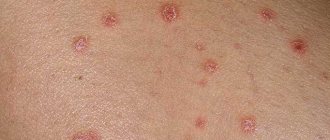
Small, polygonal, flat papules. They can merge into plaques, the surface of which is covered with thin, shiny scales. Papules are sharply limited from the surrounding skin. At the beginning of the disease, the papules are red, but soon acquire a purple tint, later a brownish tint appears. The center of the papule has an umbilical depression.
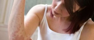
Papules can occur on any part of the skin, but usually occur symmetrically on the extensor surface of the forearms, hands, lower extremities and trunk. The primary manifestation of LLP on the skin is severe itching, often intolerable, but scratching only occasionally leads to erosions and excoriations.
Wickham grid
The surface of the papule has very thin grayish-white lines, which are called Wickham's stripes (Wickham's grid, which can be clearly seen on each papule after smearing the surface with vegetable oil).
Manifestations in the oral cavity
In the oral cavity, LLP appears differently from the skin and is classically characterized by the formation of white or gray miliary papules grouped in lines, circles and networks, forming a characteristic mucosal pattern resembling lace, rings and stripes on the buccal mucosa, and to a lesser extent on the tongue, gums, hard palate and lips.
Symptoms of red lichen
In humans, the symptoms of lichen planus (see photo) are varied, however, they all boil down to the formation of a monomorphic rash, which consists of small flat papules with a diameter of up to 0.5 cm.
- The nodules can be red-violet or crimson-red. The center of the papules is retracted, their surface is shiny. The nodules are especially visible in side lighting.
- Peeling, as a rule, is not too pronounced. The scales can be separated with difficulty. Sometimes the peeling resembles the picture of psoriasis. In this case, we are talking about the psoriasiform variety of the disease.
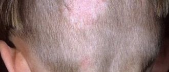
- The scalp, palms, soles and face are not attractive places for inflammation caused by lichen planus to develop. However, in approximately 10% of people, the pathology affects the head with its scalp and face. In this case, the lichen looks like a dark red lump that is very itchy. As the pathology progresses, hair begins to fall out of the head.
- In 25% of people whose lichen has formed on the mucous membranes, papules do not appear on the dermis. They affect the oral cavity, the head of the penis, and the entrance to the vagina. They can be grouped into rings, circles, or a grid. On the mucous membranes, the nodules have a grayish tint. If the tongue is involved in the pathological process, then flat plaques with jagged edges form on it. If the lips are affected by the disease, then purple plaques with mild peeling at the top are visible on them.
- Multiple rashes predominate when the nodules are located in groups. They can be combined into garlands, stripes, circles, or merge to form plaques. Elements of the rash form again around these fusions. After the nodules resolve, an area with persistent red pigmentation remains on the affected area.
- All rashes cause anxiety to the patient in the form of severe itching. This often leads to problems with night rest, ultimately provoking neurotic disorders. The ones that itch the most are the ones that are localized on the fingers. Therefore, many people mistake lichen for scabies. (
Lichen planus (LP) is an inflammatory dermatosis with diverse clinical manifestations, involving the skin, its appendages (hair, nails) and mucous membranes. The prevalence of LP is unknown, but according to statistics, patients with LP at an outpatient appointment with a dermatologist in a large clinic account for 1.3-2.4% [1]. The incidence of LP varies depending on the population studied, with a particularly high incidence in India and only 1% in the United States. LLP most often affects middle-aged people, but cases of this disease have also been described in children. Women get sick just as often as men [2]. In more than 50% of cases in patients with the skin form of the disease, resolution of the rash occurs within 6 months; in 85% of cases, regression of efflorescence occurs within 18 months. However, according to an epidemiological study, resolution of the process can last from 1 month to 7 years [2]. Thanks to the use of a number of local and systemic drugs, improvement and regression of the manifestations of LP were observed.
It is currently believed that LP is a specific delayed-type hypersensitivity reaction in response to an unidentified epidermal neoantigen [3].
The initiating moment in the pathogenetic chain of LP is the presentation of an unknown antigen by cytosolic (using the major histocompatibility complex type I - MHC I, located on the surface of keratinocytes of the basal layer) and endosomal (using MHC type II, located on the surface of Langerhans cells of the epidermis) pathways. Both mechanisms are involved in the activation of CD8+ cytotoxic lymphocytes.
In the first case, there is a direct interaction of MHC I with the T-cell receptor of CD8+ lymphocytes, in the second - the interaction of MHC II with the T-cell receptor of CD4+ lymphocytes and their subsequent differentiation into type I T-helpers, which produce a number of cytokines, including IL-2 and interferon-γ. Cytokines interact only with MHC I activated CD8+ lymphocytes, induce their proliferation and are an additional confirmatory factor that triggers cytotoxic mechanisms. The main mechanism of cell death in LP is apoptosis, induced by the interaction of tumor necrosis factor family cytokines produced by CD8+ lymphocytes, such as TNF-alpha and FasL, with the corresponding receptors on the surface of keratinocytes (Fig. 1)
.
Figure 1. Scheme of the pathogenesis of lichen planus [4].
Despite the dominant role of cytotoxic CD8+ lymphocytes in the pathogenesis of LP, CD4+ lymphocytes predominate in the inflammatory infiltrate; CD8+ lymphocytes are located in the epidermis and along the dermis-epidermal junction.
Morphological changes in LP are a consequence of a combination of processes of damage and regeneration of keratinocytes (Fig. 2)
.
Figure 2. Vacuolization of the cytoplasm of cells of the basal layer;
apoptosis of keratinocytes, melanophages in the dermis. Hematoxylin and eosin staining. Uv. 400 (provided by M.A. Bobrov). A fully formed element of a skin rash is characterized by: - in the epidermis
— orthohyperkeratosis, wedge-shaped hypergranulosis in the zone of intraepidermal components of sweat glands and hair follicles (clinically represented by Wickham’s grid), acanthosis in the form of “saw teeth”
(Fig. 3)
, vacuolization of the cytoplasm and apoptosis of cells of the basal layer;
Figure 3. Epidermis with orthohyperkeratosis, hypergranulosis, acanthosis in the form of “saw teeth”.
In the upper parts of the dermis there is a band-like lymphohistiocytic infiltrate. Hematoxylin and eosin staining. Uv. 100 (provided by M.A. Bobrov). - in the upper dermis
- strip-like lymphohistiocytic infiltrate, melanophages, colloidal bodies
(see Fig. 2)
.
Characteristic signs of LLP are polygonal, flat-topped, purple papules, prone to crowded arrangement, grouping with the formation of rings, garlands, lines ( lichen ruber anularis, marginatus, striatus
and etc.).
lichen ruber corymbiformis
appear , which is explained by the “push-like” nature of the appearance of rashes in this dermatosis
(Fig. 4)
.
Figure 4. Typical shape and location of lichen planus. The elements are located symmetrically on the skin of the flexor surfaces of the wrist joints and forearms, the anterior surface of the legs, torso, genitals, and quite often on the mucous membrane of the oral cavity. Rarely the palms, soles and face are affected. Unilateral skin lesions (zosteriform LP) may also be observed (Fig. 5)
.
Figure 5. Zosteriform localization of lichen planus (photo courtesy of M.A. Kochetkov, Ph.D.). The number of rashes varies: from single ones (in the mouth, on the genitals) to multiple ones, covering large areas of the body so that the impression of a complete skin lesion can be created, sometimes like erythroderma. Subjectively, patients are bothered by itching [5].
On the surface of the formed relatively large (0.5 mm in diameter) nodules, one can find the Wickham grid, pathognomonic for the disease, characterized by opalescent white or grayish dots and stripes. Its formation is explained by uneven granulosis. The mesh becomes more visible if the surface of the papules is moistened with water or oil during dermatoscopy [6].
In the progressive stage of the disease, a positive Koebner phenomenon is observed (the appearance of rashes in the area of even minor skin trauma). Patients are bothered by intense itching (Fig. 6)
[7].
Figure 6. Koebner phenomenon in lichen planus.
When the process regresses, secondary hyperpigmentation usually remains in place of the papule. Changes in the nails are often observed, especially when multiple rashes occur. Changes in the nails may precede skin rashes and be the only manifestation of dermatosis. The rash usually lasts a long time, the process can worsen, but over time it regresses, usually without a trace. With a long course, especially with irrational treatment, spinalioma can develop in the foci (mainly in erosive-ulcerative and verrucous forms).
In addition to the described typical form of LP, there are a number of varieties.
Clinical variants of lichen planus
The classification of clinical variants of LP is based on the characteristics of morphology and localization of rashes. Some systems take into account the etiological factor [5].
Anular (ring-shaped) lichen planus (lichen ruber anularis)
(Fig. 7)
is a common variant of damage not only to the skin, but also to the mucous membranes [8].
Figure 7. Anular form of lichen planus (photo courtesy of M.A. Kochetkov, Ph.D.).
The most typical localization of rashes is the skin of the trunk, limbs, genitals (scrotum) and mammary glands [9]. The color of the plaques varies from pink to purple. When their central part is reabsorbed or due to the peripheral growth of individual nodules, elements in the form of rings are formed. After regression of the elements, an area of atrophy is noted in the center [10]. Even after regression of the rash, brownish pigmentation remains, which is also found in subtropical LP, found in the Middle East. Hypertrophic lichen planus (lichen ruber hypertrophicus, s. verrucosus)
(Fig.
also known as verrucous or hyperkeratotic form.
Figure 8. Hypertrophic form of lichen planus.
Mostly, the rashes are localized on the skin of the anterior surface of the legs, rarely on the skin of the upper extremities and torso [11]. Rashes in the form of red, gray-yellow, dark red infiltrated hyperkeratotic papules or plaques are formed as a result of the fusion of individual small plaques and nodules [12]. The rash is hard on palpation. Most rashes are intensely itchy. Individual isolated hyperkeratotic lesions resemble senile keratosis or basal cell carcinoma. It is possible to develop squamous cell carcinoma at the site of long-existing rashes [13, 14]. Atrophic lichen planus (lichen planus atrophicus)
(Fig. 9)
is a rare form localized on the skin of the lower extremities [11].
Figure 9. Atrophic form of lichen planus (photo courtesy of M.A. Kochetkov, Ph.D.).
Clinically, the rashes are presented as round or oval, with a depression in the center, brown or purple papules and plaques [9, 15]. In place of the regressing nodular elements, atrophy develops. Ulcerative lichen planus (lichen ruber ulcerosus)
(syn.: erosive) is most often located on the feet, namely in the interdigital spaces
(Fig. 10)
[16-18].
Figure 10. Ulcerative form of lichen planus.
Only a few cases of localization of this form on the trunk and in the perianal region have been described [9, 19]. Single or multiple rashes are sharply demarcated, with a bizarre configuration, forming small ulcers and erosions with a raised edge or mesh areas of leukoplakia. As a rule, there is a pain syndrome that seriously impairs walking to the point of impossibility. They are often preceded by bullous rashes. Dystrophic changes in the nails are expressed, up to their rejection; paronychia, which, like bullous rashes, may be the initial manifestation of the disease. Bullous lichen planus (lichen ruber planus bullosus)
(Fig. 11)
is characterized by typical blisters.
Figure 11. Bullous form of lichen planus.
The skin of the lower extremities is affected in the form of numerous small, sometimes large grouped blisters with a tense tire; some blisters are cellular and contain serous or serous-yellow fluid [20-22]. In lichen planus pemphigoides
(Fig. 12),
the clinical manifestations, histopathology and immunofluorescence are similar to those of bullous pemphigoid [23].
Figure 12. Pemphigoid form of lichen planus.
Characteristic of this form is the development of vesicles and blisters not only on existing rashes, but also on unaltered skin [24]. In the latter case, the possibility of developing bullous pemphigoid cannot be excluded. Along the periphery of the rash there are tense flat multi-chamber blisters. The rash predominates on the extremities, most rarely on the torso; located in groups or disseminated [25]. This form of LP has not yet been classified pathophysiologically. When the elements are localized on the scalp, cicatricial alopecia may develop. In some cases, this form is a manifestation of toxicoderma or paraneoplasia. Atrophic changes such as anetoderma and hyperpigmentation may remain on the skin. Pigmented spotted or papular elements are characteristic of pigmented lichen planus (lichen planus pigmentosus)
(Fig. 13, 14)
.
Figure 13. Pigmented form of lichen planus (photo courtesy of M.A. Kochetkov, Ph.D.).
Figure 14. Pigmented form of lichen planus.
In addition to the widespread perifolicular linear or encircling nature of the location, rashes can be localized along Blaschko's lines, along the vessels of the lower extremities and on the skin of the extensor surfaces of the extremities. Intertriginous areas are also most characteristic [26]. Rashes have also been repeatedly described in the lumbar region. Pigmentation ranges from grey-brown, violet to dark brown. Patients with fair skin are most susceptible to this form of LP [26]. Pigmentation may also appear during the period of regression of typical nodular elements, and sometimes changes similar to poikilodermic ones develop. It is possible that one of the variants of pigmented LP is erythema dyschromicum perstans
. It is also assumed that the formation of foci of pigmentation may precede the appearance of a common papular rash; in some cases, pigmentation on the skin may be combined with typical LP rashes in the mouth.
Erythrodermic variant (erythrodermic lichen planus)
is the rarest [11]. Morphologically, red or purple papules and extensive infiltrated erythematous plaques with or without a tendency to grow are detected. In addition to erythroderma, typical elements of LP, blisters or erosions may be detected. The rash is accompanied by intense itching, which reduces overall well-being.
Inverse lichen planus (lichen planus inversus)
(Fig. 15)
typically affects the skin of intertriginous areas - the armpits, inguinal folds, flexor surfaces of the extremities and the submammary region.
Figure 15. Inverse form of lichen planus.
Clinically, the rashes are localized, erythematous, with blurred boundaries and areas of lichenification. In addition, keratotic (keratinized) papules and erosions with bizarre outlines can be detected. Pigmentation is also typical for this form [27]. Linear form (lichen ruber planus linearis)
most common in children and adolescents. Its clinical manifestations are stripes that can be limited to several centimeters or segmentally, have a girdling character, or follow Blaschko's lines. Individual rashes may look like typical flat papules, but they are itchy; rashes can also be cellular, hyperkeratotic or ring-shaped [28].
Follicular or genital lichen planus (lichen planopilaris)
(Fig. 16)
is localized on the skin of the scalp, armpits, groin, scrotum and flexor surfaces of the extremities.
Figure 16. Follicular form of lichen planus (Little-Lassueur syndrome). Clinically, they are grouped or disseminated, follicular raised or hemispherical erythematous papules with a horny plug [15]. On the surface of individual nodules there are horny spines ( lichen ruber spinulosis
).
If they are localized on the scalp, the process often ends with atrophy and baldness ( lichen ruber follicularis decalvans
), which in combination with alopecia in the armpits and pubic area, spiny horny papules on the trunk and limbs are designated as
Little-Lassuer
. The onset of the disease may be onychodystrophy.
Actinic or subtropical lichen planus (lichen planus subtropicus)
(Fig. 17)
typically appears in autumn or summer on open areas of the skin exposed to sunlight (face, arms, shoulders).
Figure 17. Actinic form of lichen planus.
Children and adolescents with dark skin living in the tropics and subtropics are most susceptible to the disease. Clinical variants of the course are possible. Lichenoid or annular hyperpigmented papules and erythematous infiltrated plaques with varying boundaries and distribution are noted [20]. Cases of a combination of signs of LP and discoid lupus have been described. In this case, the lesions are located on the distal parts of the extremities, have a bluish-red or purple color, with hypo- and hyperpigmentation, telangiectasia, atrophy, and sometimes with warty, bullous-ulcerative changes. In other areas, including on the oral mucosa, typical LP rashes or atrophic lilac-reddish plaques may be detected. Pronounced dystrophic changes in the nails up to anonychia are also observed; Some patients develop cicatricial alopecia. Histological and immunofluorescence (direct reaction) studies also reveal signs of both diseases. One of the reasons for this combination is the commonality of pathogenetic mechanisms. Apparently, for accurate diagnosis, in addition to a complex of histological, serological, clinical, immunofluorescence parameters, the indirect immunofluorescence reaction proposed by R. Olsen et al. may be useful. (1984) to identify the specific antigen of LLP. This antigen was found in 80% of patients with LP and in no patients with lupus erythematosus.
Differential diagnosis.
LLP should be distinguished from papular syphilide, toxicoderma, amyloid and myxedematous lichen, lichenoid and warty tuberculosis of the skin, small nodular form of sarcoid, nodular prurigo, chromomycosis, diffuse and limited neurodermatitis, pityriasis rubra pilaris, callus, granuloma annulare, bowenoid nogo papulosis, sclerotic white lichen , pemphigus, toxic melasma, Darier's disease, psoriasis, lichenoid parapsoriasis, flat juvenile warts, Kaposi's sarcoma, lichen planus, Fox-Fordyce disease, Kirle disease, papular acrodermatitis, malignant atrophying papulosis, vulvar kraurosis, pretibial myxedema.
Differential diagnosis of common forms of LP is usually not difficult.
Lichen planus of mucous membranes
The most common localization of LP is the oral mucosa (Fig. 18)
.
Figure 18. Damage to the oral mucosa in LLP (photo courtesy of A.V. Tereshchenko, Ph.D.). Almost half of the patients have single lesions; multiple lesions are very rare. The topography of the rashes is very diverse, and the morphology of the rashes is even more diverse. There are six morphological types of lesions of the oral mucosa: reticular, plaque-like, atrophic, papular, erosive and bullous (Fig. 19)
.
Figure 19. Damage to the oral mucosa in LLP. Erosive-ulcerative form (photo courtesy of A.V. Tereshchenko, Ph.D.).
Reticular and erosive forms are the most common [15, 20]. The combination of individual morphological forms with a specific localization on the oral mucosa has not been described, with the exception of the plaque form with damage to the apex and lateral part of the tongue.
The erosive or atrophic forms are often associated with burning pain, which can be triggered by eating sour, spicy or hot foods.
In other forms, these symptoms do not appear or are less pronounced. Damage to the oral mucosa is an optional precancerous lesion (according to various sources, 0.4-5.3% of cases). A recent review describes a case of esophageal involvement. Moreover, damage to the esophagus was described without characteristic lesions of the oral mucosa.
The most common location for lesions of the genital organs is the glans penis in the form of ring-shaped rashes (Fig. 20)
.
Figure 20. Lesions of the skin of the glans penis in LP. In women, the vulva is most often affected compared to the vagina (Fig. 21)
.
Figure 21. LLP of the vulva (photo courtesy of M.A. Kochetkov, Ph.D.). The risk of developing squamous cell carcinoma cannot be reliably estimated. Estimates range from "low" with a frequency of up to 2.4%. Penile malignancy is very rare [29].
In 80% of women, a syndrome occurs that combines damage to the mucous membranes of the oral cavity, vulva and vagina, and HLA DQB1*0201
a gene indicating a predisposition to this syndrome. There is a risk of developing scars and strictures. Damage to the mucous membrane of the urethra, nasopharynx and esophagus is also possible [30].
Lichen planus of the nail plates
LLP affects the nail matrix, which can lead to a variety of morphological changes in the nail plate that are not pathognomonic (Table 1)
.
The first sign of isolated nail damage is thinning of the nail plate and longitudinal striations [31].
Characteristic features of LP are dorsal pterygium and trachonychia. Pterygium develops due to the adhesion of the eponychium and matrix, which leads to cracking of the nails and, sometimes, complete loss of the nail plate. Trachonychia is characterized by roughness of the nail plate, loss of transparency and gray color.
Characteristic changes usually develop simultaneously on several nails.
It is not uncommon for all 20 nails to be affected. This may be preceded by rashes on the skin or mucous membranes [31]. With isolated nail lesions, the diagnosis must be confirmed by pathological examination.
Treatment
Phototherapy.
Disseminated rashes with an acute onset and severe itching require effective and timely treatment
(Table 2)
.
Phototherapy can be considered as the therapy of choice.
Thus, UVB (narrowband 311 nm and broadband 280-320 nm) and PUVA therapy (psoralen + UVA 320-400 nm) have proven to be highly effective in the treatment of common forms of LP. The duration of PUVA therapy is on average 10-12 weeks, for narrowband phototherapy - 8-9 weeks. However, LP may recur in approximately half of patients treated with phototherapy. The long-term results of PUVA therapy are comparable to the results of narrow-band phototherapy. In most cases, a combination of phototherapy with topical corticosteroids (prednicarbate or mometasone furoate) for 4 weeks is recommended. It is the treatment of choice for the treatment of common forms of LP.
First-line systemic therapy.
Systemic therapy is considered if the above phototherapy methods have failed. There are a number of drugs with a high evidence base for the treatment of patients with LP, which have their own advantages and disadvantages. Therefore, an individual and personalized approach to the choice of therapy is important.
Systemic retinoids
— Acitretin is prescribed at a dose of 0.5-0.7 mg/kg per day until remission is achieved, then at 0.3-0.5 mg/kg per day as monotherapy or in combination with topical or systemic corticosteroids. This method is most indicated in cases of acute onset of the disease with severe inflammation, intense itching or bullous/erosive-ulcerative rashes.
— Isotretinoin at a dose of 0.3-0.5 mg/kg per day for up to 2 months. Effective in the treatment of LP of the oral mucosa and cutaneous forms, it causes fewer side effects compared to acitretin.
The combination of PUVA therapy with retinoids (re-PUVA) improves efficacy and reduces side effects in severe cases by reducing the cumulative radiation dose. However, there is a high risk of hyperpigmentation, especially in patients with dark skin. The combination of narrowband phototherapy with retinoids may be an alternative to re-PUVA therapy with comparable efficacy and a reduced risk of side effects.
— Methotrexate is prescribed at a dose of 15-20 mg once a week for 10 weeks, while most patients achieve complete remission within a month after starting treatment, and only 5% of patients experience a relapse during the observation period (6 months). Low doses of methotrexate (10 mg subcutaneously once a week) are most effective and safe compared to high doses.
Systemic corticosteroids.
Although systemic corticosteroids have not been thoroughly tested in clinical studies in traditional LP therapy, their undisputed effectiveness has been described in long-term follow-up. Durant corticosteroid injections given at 2-week intervals resulted in complete remission in 61 (83.6%) of 73 patients with the generalized form. An oral corticosteroid (prednisolone) at a dose of 0.5 mg/kg per day was as effective as its injectable form. The course is usually 2-4 weeks.
Hydroxychloroquine (Plaquenil)
prescribed 200 mg or 6.5 mg/kg per day in rounds: 5 days with a break of 2-5 days; up to 5 rounds per course. It is possible to prescribe hydroxychlorine daily at a dose of 200 mg/day for up to 2 months. This is a safe and effective method in the treatment of the generalized form as monotherapy or in combination with other drugs.
Dapsone
Prescribe 50-150 mg per day for 1-2 months. It should be noted that this drug was one of the first systemic agents in the treatment of torpid forms of LP. Many studies indicate the high effectiveness of dapsone, especially in severe cases in children and in the treatment of bullous form of LP. However, side effects such as dose-dependent hemolysis and methemoglobinemia limit the use of this drug in elderly patients (over 65 years of age) and in patients with anemia or heart disease.
Second-line systemic drugs
used for torpid course of the disease. Quite high effectiveness in the treatment of severe generalized forms of LP was recorded with the use of cyclosporine (2.5-5 mg/kg body weight per day for up to 2 months). Their combination with phototherapy is not recommended, as there is a high risk of skin cancer.
Alternative methods used in treatment include:
— azathioprine
(up to 2.5 mg/kg per day) in combination with topical corticosteroids can be used for a long time (up to 6 months);
— mycophenolate mofetil
use 2 g per day, or in small dosages (500 mg/day), combination with plaquenil is possible. Usually the course is 1-2 months;
— pulse therapy with dexamethasone
at a dose of 100 mg 3 days every 4 weeks. There are 4-6 rounds per course.
Other drugs:
- griseofulvin 7.5-10 mg/kg for 3-6 months;
— itraconazole 200 mg/day for 1 week of each month; course - 3 months;
— metronidazole 500 mg/day for 20-60 days, both as monotherapy and in combination with PUVA therapy;
— thalidomide 150 mg/day for 4 months. Due to possible side effects, including irreversible neuropathy, thalidomide should be used with caution.
Complications
Lichen planus is especially unpleasant in the genital area, in the vagina. The disease leads to pain and disrupts sex life. Scars may remain in place of the elements. Sexual dysfunction may subsequently persist for a long time.
There is evidence that lichen planus, although to a small extent, may increase the risk of squamous cell carcinoma. Your dermatologist may recommend that you be screened for cancer cells in the lesions.
Skin pigmentation in places where nodules and plaques were located persists for a long time.
Causes
There is no definite data regarding the appearance of lichen planus in the human body. However, many scientists and doctors are inclined to certain theories.
Immunoallergic factor
Studies show that T-helpers and T-suppressors (groups of immune cells) were found in the blood of patients with lichen red. The tests also show the presence of antibodies - originating from immune complexes circulating in the blood vessels.
The theory is the main one, unifying all other versions. They lean towards it most often.
This analysis, aimed at detecting immune cells, reflects the fact that the body does not recognize its own cellular structure, and periodically sends antibodies and white blood cells to fight its components. This explains the fact that inflammatory processes often predominate in the affected area.
Virus/infection penetration
If we refer to this theory, then the provocateur of the rash is a virus that has penetrated into the lower layer of the epidermis. Due to certain reasons, it begins to negatively affect skin cells, and the immune system becomes unable to act adequately and only directs its actions to destroy its own cells.
Heredity
The theory is that a person has a gene inside him that he inherited from close relatives. If a certain type of provocateur appears, then it begins to manifest itself, resulting in lichen planus.
Studies have shown that 1.5% of patients actually had relatives who had the disease. As a rule, rashes appeared in childhood and were not eliminated for a long time, becoming recurrent in nature.
Neurogenic factor
It was found that most of the patients suffering from lichen planus had diseases of a nervous nature - neurosis, neurasthenia, etc. This gave reason to assume that dermatosis of this type may have a neurogenic origin.
Intoxication
The provocateurs of the disease, according to the founders of this theory, are medications containing chemicals, which, in turn, provoke rashes. Also, if you believe the same version, toxic substances that enter the body can also provoke formations.
Diagnostics
In most cases, a doctor can easily make a diagnosis during an examination. Difficulties may arise if the skin is not affected, and the elements are only on the mucous membrane.
Tests for lichen planus that a dermatologist may prescribe:
- Biopsy. The doctor obtains a piece of tissue in the area of the nodules and plaques and sends it to the laboratory for analysis. Most often this is done using scrapings. When microscopying samples, you can see cells characteristic of lichen planus.
- Blood test for hepatitis C. This viral disease often provokes the occurrence of lichen planus. If during the conversation the doctor decides that you had a risk of contracting hepatitis, he will order this test.
- Allergy tests. Allergies can also trigger the occurrence of lichen planus. Your dermatologist may refer you to an allergist who can perform an allergy test. Solutions containing different potential allergens will be applied to your skin. A positive reaction will be indicated by the appearance of redness and a blister in the place where the “culprit” allergen was applied.
Not everyone needs these studies. They are prescribed if there is a suspicion of a particular disease in cases where the diagnosis is difficult to establish.
Answers to popular questions:
- Is lichen planus contagious? Is it transmitted from one person to another? Lichen planus is not a contagious disease; it is not transmitted from person to person. However, hepatitis C may be a factor influencing the development of lichen. In turn, hepatitis C is a contagious disease, so if you have symptoms of dermatological diseases, it is necessary to consult a doctor.
- How and how long does it take for lichen planus to go away? Could it be chronic? Can it develop again? After the first appearance on the skin, lichen planus can remain on them from six months to 2 years. After this time, the lichen goes away. However, the risk of relapse cannot be excluded. When the oral mucosa is affected, the disease has a longer course and can exist for 5-20 years with periods of calm and exacerbation.
- Is it possible to sunbathe at sea or in a solarium if I have lichen planus? Lichen planus is not a contraindication for swimming in the sea or spending time in the sun. Only one extremely rare form of lichen is dependent on ultraviolet rays - radial lichen ruber. But this variety is found only among residents of India, Africa and the Middle East. However, doctors still do not recommend excessive tanning, since in this case there is a risk of developing skin cancer (not associated with lichen).
- Is it possible to shower or take a bath if you have lichen planus? You can shower and take a bath without any worries. These water procedures do not affect the development of the disease.
- How can lichen planus affect the development of pregnancy? There are no data regarding the effect of lichen planus on pregnancy. However, if there is evidence of illness, contacting a doctor is necessary.
Photo
What lichen planus looks like, see the photo for more details:
How to treat lichen planus
The basis of this dermatosis is immune inflammation (tissue response to damage by the immune system), which destroys cells of the skin and mucous membranes.
Therefore, when treating lichen planus in humans, the following are used:
- drugs that reduce the activity of the immune system - drugs of choice (prescribed first) in severe cases of the disease
- products that improve tissue nutrition and accelerate their recovery
- medications that reduce itching, improve sleep, normalize the functioning of the nervous system
- local ointments
In addition, provoking factors are eliminated:
- stress
- Prosthetics and treatment of diseases of the oral cavity, as well as foci of chronic infection, are carried out
- medications and occupational hazards are excluded
- We recommend a diet that does not irritate the oral mucosa
- and others
Medicines are prescribed to patients in cases where there is a violation of their general health. The selection of the necessary funds depends on each clinical case. The following oral medications can be used to treat lichen planus:
- immunosuppressants: Cyclosporine A, Chloroquine, Hydroxychloroquine;
- antihistamines: Tavegil, Zyrtec, Diazolin, Clemastine, Promethazine, Loratadine, Fenkarol, etc.;
- corticosteroids: Metipred, Prednisolone;
- systemic retinoids: Neotigazon, Acitretin, Tigazon, Isotretion, Etretinate;
- synthetic interferons: Ridostin, Neovir, Interferon-alpha 2B;
- antibiotics: Tetracycline, Metacycline, Doxycycline, Azithromycin, Sumamed, Roxithromycin;
- sleeping pills and sedatives: Phenazepam, Medazepam;
- vascular drugs to improve tissue trophism: Xanthinol, Trental, etc.;
- vitamin preparations: Ascorbic acid, B vitamins, vitamin D.
In addition, the drug therapy plan may include drugs for the treatment of concomitant diseases: diabetes mellitus, neurosis, arterial hypertension, chronic inflammatory processes (for example, glossitis, stomatitis, etc.).
For local therapy of lichen planus, the following agents are used:
- ointments based on corticosteroids: Cloveit, Flumethasone, Hydrocortisone, Betameson, Triamcinolone;
- non-hormonal antiallergic ointments: Gistan, Fenistil;
- non-hormonal anti-inflammatory ointments: Pimecrolimus, Tacrolimus;
- exfoliating ointments: Belosalik, Diprosalik;
- ointments to accelerate tissue regeneration: Solcoseryl.
Local remedies for the treatment of this dermatosis can be used either independently or in combination with each other. They can only be prescribed by a doctor who takes into account the form, severity and stage of the disease.
Treatment
How to treat lichen planus if the cause is unknown? According to modern recommendations, effective treatment of the disease should always include several areas.
In 99% of cases, hospitalization is not required. Treatment is carried out under the supervision of a dermatologist at home and in the KVD clinic.
Treatment or elimination of the underlying disease or precipitating factor
This could be the treatment of viral hepatitis, diabetes, eliminating the effects of a toxic substance, combating stress, etc.
Local therapy
- Corticosteroid creams and ointments are prescribed. These drugs reduce the immune response in the skin and reduce the activity of inflammation. Ointments: Fluorocort, Akriderm (read about Akriderm ointment in detail), Triderm, Sinaflan, Belosalik and others. Apply to the affected area of skin 2 times a day – 4 weeks. Repeated course - only after agreement with a dermatologist. Treatment with corticosteroids is effective. According to reviews, signs of exacerbation of the disease disappear within a week. But there is a danger of side effects and the “ricochet” effect, when a relapse of the disease occurs after discontinuation of glucocorticoids.
- Preparations based on naphthalan. Ointment and cream Losterin, Naftaderm. Apply to the affected area of skin 2-3 times a day – 4 weeks. If necessary, the course is repeated after a week's break.
General therapy
- Antihistamines - prescribed to relieve itching and reduce inflammation. Claritin, loratadine, suprastin, Erius, Telfast, tavegil, diphenhydramine.
- Retinoids: Tigazon and Neotigazon, isotretinoin. The effect of the drugs is to inhibit the excessive growth of skin cells and normalize keratinization processes. The membrane structures of cells are stabilized. Take neotigazon 20-30 mg once a day with meals. The course of treatment is 1.5-2 months.
- Immunosuppressants are drugs that suppress the immune system. They are prescribed only by a doctor and are prescribed by prescription. Used for severe forms of lichen planus that cannot be treated with other means: - Corticosteroid hormones in droppers and tablets: prednisolone, dexamethasone and others. — Cytostatics: Chloroquine, Hydroxychloroquine, Cyclosporine A. The effect of immunosuppressants is rapid and persistent, but there are also many side effects.
- Antibiotics are prescribed in the presence of infectious and purulent complications on the skin in patients with lichen ruber.
- Sedative therapy (valerian, motherwort), hypnosis, electrosleep. Reducing the excitability of the nervous system has a beneficial effect on the healing process - it always goes faster and more efficiently.
- Diet. Exclusion of allergenic foods (chips, soda, citrus fruits, honey, etc.), spicy foods, hot foods, alcohol. It is especially important to follow a diet if the oral mucosa is affected.
Physiotherapy
- Used in the complex treatment of lichen planus.
- PUVA therapy. Drugs that have a photosensitizing effect on the skin are used - psoralens (for example, Methoxalen). The drug is given to the patient either in tablets or as an ointment. Psoralens accumulate in skin cells. After 3 hours, the affected areas of the skin are exposed to ultraviolet irradiation. Excessive proliferation of epidermal cells is suppressed and skin infiltration is reduced. In those areas of the skin that are not exposed to UV rays, psoralens have no effect. The positive effect of the procedure begins on day 4. A course of treatment requires 20-30 PUVA therapy procedures. According to reviews from patients and doctors, the effectiveness of the procedure is up to 80% in the complex treatment of the disease. PUVA therapy does not have the addictive effect of corticosteroid ointments and creams.
- Hydrogen sulfide and radon baths, mud.
- Magnetotherapy. Ineffective procedure. Can only be prescribed in combination with other areas of treatment.
Photo booth for PUVA therapy
Let's look at the video of how PUVA therapy is performed:
Physiotherapy
PUVA therapy. This is one of the most effective physiotherapeutic procedures for lichen planus. The treatment process combines the use of photosensitizers taken orally and ultraviolet radiation. It is this technique that gives good results after the first 3-4 procedures. The course of treatment consists of 15-20 procedures.
However, this method of physiotherapy also has side effects. Nausea, dizziness, skin burns. In the future, the likelihood of developing skin cancer, the occurrence of tumors, etc. increases.
Lichen planus can be successfully treated with laser therapy and magnetic therapy. These procedures affect the activity of healthy skin cells and inhibit the development of affected skin areas. When these methods are combined with drug treatment, good results are achieved.
Symptoms
The main manifestations are monomorphic body rashes of the type of nodules or papules. Their hue varies between red and purple, and their size ranges from 2 to 5 mm. The papules have a retracted middle, and their entire surface seems to be covered with wax. Usually there is no or slight peeling. If the psoriasiform type of lichen develops, the peeling is similar to psoriatic separation of scales.
Ringworm rashes cluster in specific areas of the skin, resembling rings or garlands. Then small elements merge into plaques and grow. When they resolve, the skin remains hyperpigmented for a long time afterwards. Often the patient suffers from severe itching, causing discomfort and problems sleeping.
The usual location of such lichen is the bends of the arms and legs, thighs, groin area, and armpits. Also, elements of the rash cover the torso. But the most popular location for the rash is the oral cavity.
The disease most rarely grows on the nails. Then, longitudinal striations, clouding, destruction of the nail fold, and ridges are characteristic. Feet, palms, head, face are unpopular locations for ringworm.
25% of patients with this pathology report wounds exclusively in the mouth, without any skin pathologies.
Favorite places for rashes are the oral cavity and genitals. The rashes are usually grouped into rings, laces, or can be isolated. The formations in the mouth have a grayish-opal color; on the tongue they may have a whitish tint. Small purple plaques form on the lips, which peel off a little.
The classic form of the disease has some characteristic symptoms. In particular, the so-called Wickham pattern, when the surface of the largest formations is covered with a grid-type pattern, which is especially pronounced after smearing the rashes with oil. The disease is also characterized by the Koebner phenomenon, when new rashes form on injured skin.
Hypertrophic (or verrucous) lichen. This form is characterized by multiple warty layers on the papules due to hyperkeratosis. In addition, many individual nodules are localized near the plaques. With this form of the disease, rashes usually form on the legs; they are extremely rarely observed on the arms and face. Some symptoms of the disease are very similar to senile keratosis and basal cell carcinoma.
Atrophic form. The disease is characterized by modifications of the rash - sclerosis and atrophy. Sometimes a rash appears in the hair, stimulating the process of baldness.
Pemphigoid form. The disease resolves with the formation of many blisters with clear or bloody fluid. Bubbles can be on the site of papules or on healthy skin. Next to them is the usual lichen rash. The most common localization of the pemphigoid form is the feet and legs. If the blisters reach a large size, the lichen becomes bullous.
Lichen moniliformis. The disease is characterized by the formation of round rashes with a waxy surface. Together they form the so-called necklace. Usually the rash is localized on the neck, forehead, hands, behind the ears, buttocks, abdomen, elbows, etc.
Pigment form. In this case, not only regular rashes form on the surface of the skin, but also brown spots and nodules. Most often they appear before the classic rash.
Pointed shape. The main locations of the rash are the neck, legs, and back in the area of the shoulder blades. The predominant elements of the rash are pointed papules, the central part of each of which has hyperkeratosis in the form of a horny spike.
Ring shape. This type of disease is characterized by the growth of the peripheral zone of the plaque, followed by a transition to the central part. As a result, the rash becomes ring-shaped. This type usually occurs in men, in whom it affects the genitals and the inside of the legs.
Erosive-ulcerative form. It usually appears in the mouth, where ulcers and erosions form, accompanied by redness and swelling. The healing stage of erosions is very long, up to several years. During this time, several relapses of the disease may occur in the same place or nearby.
Folk remedies
Many examples are given that promise an unprecedented result and tell how to quickly treat lichen ruber in humans. Unfortunately, none of these methods are scientifically proven and often people can only observe a cure as a placebo effect.
“Dummy” and the power of human persuasion sometimes really work psychosomatically and the mechanism of healing by persuasion has not yet been studied. Nevertheless, in medical practice, even if only a little, similar examples still occur.
Try to avoid aggressive treatments such as applying hot objects or products containing acids and other similar substances. This will only make the situation worse, causing injury to the affected areas of the skin.
Popular folk remedies include:
- sea buckthorn oil – has a wound-healing effect;
- birch tar is a remedy actively used in the treatment of psoriasis and other dermatitis;
- laundry soap - its natural composition does not damage the skin.
When using this or that technique, you need to focus on your own feelings. Allergic reactions and individual intolerance can appear at any time.
Classification
Lichen planus is divided into several forms, each of which has its own individual list of symptoms.
Atrophic
It appears when changes of an atrophic or sclerotic nature occur.
If the atrophic form predominates, then its distinctive feature is hair loss in the affected area. As for the places of bends, the formation of follicular keratosis occurs on them (also called the Little-Lassouer symptom). It is expressed in the fact that the hair follicles are completely clogged with horny cells.
Warty (hypertrophic)
A distinctive feature of this form is the fact that the papules have a purple or brown tint. Sometimes they may be pinkish.
Occurs due to the process of tissue hyperplasia. Papules can merge into one whole, forming plaques.
Visually, such a formation resembles a wart with many dots at the top, as well as slightly flaky edges. As a rule, localization is observed on the front of the legs. Rashes on other areas of the skin are also possible.
Erosive-ulcerative
In a person suffering from this form of lichen planus, the oral mucosa is affected. In rare cases, the same thing occurs on the head of the penis or in the labia area. The erosive-ulcerative form is the most severe of all and is difficult to cure.
As soon as rashes appear on the surface of the mucous membrane, they transform into ulcers, as well as erosions of irregular shape. Both the patient’s movements and touching the affected areas of his body cause him severe pain.
Why can our articles be trusted?
We make health information clear, accessible and relevant.
- All articles are checked by practicing doctors.
- We take scientific literature and the latest research as a basis.
- We publish detailed articles that answer all questions.
Each formation has a specific plaque, the removal of which will inevitably cause bleeding. The bottom has a pinkish, slightly velvety surface.
If the formation is damaged, it takes a very long time to heal. Negative physical effects can also cause relapse. If this approach to the affected areas dominates the treatment, their recovery may take years.
The erosive-ulcerative form often occurs in combination with arterial hypertension.
Bullous (vesical)
In medical practice, it is noted that the majority of patients accounting for this form are over 50 years of age. As a rule, these are women. The condition of the patients is extremely serious, and the progression of the disease occurs at lightning speed.
The size of rashes with this form may vary - from 1 to 5 millimeters. They multiply quickly and are shaped like bubbles. Plaques formed from papules may be observed nearby.
Inside each bubble there is a white (usually transparent) liquid.
If it is localized in the mouth area, the opening of the elements occurs faster. Erosion usually appears at the site of the wound. They heal quickly, which cannot be said about those that predominate in the ulcerative form.
Ring-shaped
A peculiarity of the rash with this form is its tendency to spread in width. As for the rash in the center of the problem area of the skin, it tends to fade. The formations have the shape of a ring.
As a rule, this form is diagnosed in men. Most often, localization occurs in the genitals and groin area. Also, rings often appear on the inside of the legs. There is a high probability of formation on the mucous membrane.
Erythematous
The rash occurs on the neck, shoulder blades and legs. The erythematous form is distinguished by the fact that a large area of skin is affected.
Pointed
The appearance of papules occurs in areas of the shoulder blades, legs and neck. The rashes have pointed tips, which is the main feature of this form. Also in the center of each formation there is an area of hyperkeratosis, externally similar to a horny spine.
Pigmented
The appearance of pigmented nodules is the main feature of this form of lichen ruber. As a rule, they are brown in color.
Moniliform
It is characterized by waxy rashes, the localization of which is concentrated in the areas of the ears, forehead, elbows and neck. It is possible that formations may appear on the buttocks and abdomen. Rashes of an unusual shape, resembling a necklace. Another distinctive feature is the intactness of the skin on the hands and the area of the nose, cheeks and shoulder blades.
Diet
There is no special diet for the treatment of lichen planus. However, nutrition plays an important role, because it is impossible to cure lichen planus by abusing foods harmful to the body. In addition, taking some drinks can reduce the effectiveness of drugs, and a number of products can cause allergic reactions and provoke a sharp increase in blood sugar levels.
During the treatment period, you should avoid drinking alcohol and confectionery. Products that it is advisable to limit your consumption include:
- citrus;
- products containing cocoa;
- products containing purine: meat and dishes prepared on its basis (broth, meat sauce, jelly); fatty fish, fish soup, fried and salted fish, canned fish; meat by-products; mushrooms;
- legumes: peas, beans, soybeans, lentils;
- eggs;
- strong brewed tea or coffee;
- products with a high content of preservatives, dyes, flavoring additives: sausages, canned food, carbonated drinks.
The following products will benefit the body:
- dairy;
- green vegetables;
- healing mineral water;
- iron-containing products;
- honey.
A diet for lichen planus is necessary if the disease often recurs and is difficult to treat.
It is better to introduce each new product into the diet gradually. If the condition worsens, the diet should be extended.
Prevention
As you know, it is better to prevent any disease than to treat it. A number of simple recommendations will greatly reduce the risk of developing the disease.
- To prevent the risk of developing the disease, you should carefully monitor the health of your body and consult a doctor in a timely manner. Carefully monitor and exclude from circulation items that constantly injure the skin and mucous membranes.
- Proper nutrition, adherence to a routine and maintaining personal hygiene can significantly reduce the likelihood of acquiring pathology.
- It is recommended to lead a healthy lifestyle, play sports and give up bad habits.
- Hardening will help to avoid many diseases, including lichen red.
Ringworm is a fairly serious disease that can lead to dangerous complications. But with proper and timely treatment, the prognosis is very favorable and a complete recovery of the patient is possible.