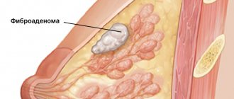
Breast lipoma is a fatty formation, which is why it is often called a wen. This is a benign tumor, but surgeons recommend removing it.
COST OF SOME GYNECOLOGIST SERVICES IN OUR CLINIC IN ST. PETERSBURG
| Price for a gynecologist's appointment | 1000 rub. |
| Extended colposcopy | 1300 rub. |
| Ultrasound of the pelvis (all organs) | 1300 rub. |
| Call free: 8-800-707-1560 *The clinic is licensed to provide these services | |
Reasons for the development of lipoma
The content of the article
There are many hypotheses about the causes of lipomas, the most common:
- Disruption of the metabolic process in the body - various disruptions in the functioning of endocrine organs (thyroid gland, pituitary gland, adrenal glands) can lead to the formation of fat cells.
- Obstruction of the sebaceous glands in the chest area. There is no clinical evidence for this reason yet, but the hypothesis has a right to exist, since it has not yet been refuted.
- Hereditary predisposition is one of the most common theories.
- Hormonal disbalance. The most likely reason, since the formation of lipomas in the breast mainly occurs in women during menopause, when the functioning of the ovaries ceases.
Reasons for the formation of lipomas
Fatty neoplasias have a polyetiological nature of development. It is often difficult to identify a single mechanism. For convenience, mammologists systematize the reasons into several groups:
- Hormonal. During the premenopausal stage, a woman undergoes hormonal changes. Her body performed the function of conception, birth and feeding of offspring. Now the ovaries are shrinking, the endometrium of the uterus is thinning, and the glandular structure of the breast is changing to fatty. If the restructuring is disrupted, a chaotic accumulation of fat cells is possible, from which a lipoma is formed.
Neoplasms in women of reproductive age can occur against the background of prolonged, uncontrolled use of oral contraception or HRT (hormone replacement therapy), or menstrual irregularities.
- Genetic, hereditary. There are systemic diseases - lipomatosis, in which the formation of pathological foci occurs in various organs, including the breast. The development of pathology is associated with a defect in the HMG IC gene.
- Metabolic disorders, in particular fat metabolism. Some of the fat fractions, low-density lipoproteins, accumulate in tissues and are eventually covered with a connective tissue capsule. Defects in lipid metabolism are provoked by an incorrect diet with an excess of animal products, a sedentary lifestyle, and concomitant endocrine diseases.
- External reasons. This category includes factors of mechanical action on the chest: injuries, operations, inconveniently selected underwear. An unhealthy lifestyle (smoking, frequent stress, alcohol abuse), radioactive radiation also adversely affects the general condition of the body.
The combination of several of these causes stimulates excessive division of lipid cells in limited areas. The conglomerate becomes encapsulated over time and separates from healthy tissue. Despite the limited nature of the process, many are concerned about the dangers of breast lipoma. Mammologists do not exclude the possibility of transformation of a benign tumor into liposarcoma. Particular oncological alertness concerns patients with risk factors:
- cases of breast cancer (BC) in the family;
- oncology of other systems in this patient.
Types of lipomas
Lipomas are distinguished according to several characteristics.
By the number of neoplasms:
- Single wen (formed in the form of one compaction);
- Multiple lipomas (formed throughout the body, most often this disease is inherited).
According to tumor consistency:
- Nodular formation (formed from adipose tissue, has a soft elastic consistency);
- A diffuse tumor is divided into: fibrolipomas (in the formation of a tumor, in addition to adipose tissue, connective tissue cells also participate, and this formation has a fairly hard consistency);
- lipofibroma (the wen is formed in exactly the same way as with fibrolipoma, but in this type adipose tissue predominates, so upon palpation the formation is very soft);
- Angiolipoma (the tumor itself has a network of blood vessels).
- Myxolipoma (in addition to blood vessels during the formation of a tumor, there is also mucus tissue).
- Myolipoma (a rare type of tumor that consists of adipose tissue and muscle fibers).
After removal of a breast lipoma
The doctor will give recommendations for treating the postoperative wound. It can be done at home on your own or come to the clinic. Sunbathing, taking a hot bath, or steaming in a bathhouse or sauna in the first weeks after surgery is prohibited.
Sutures from the postoperative wound are removed after approximately 7 days. If swelling, pain or redness appears in the wound area before this time, you should consult a doctor. It is possible that the wound has become infected.
In our Surgery Center, operations are performed by experienced mammologists who follow antiseptic rules and treat patients with care. Therefore, the risks of complications are minimized. You can make an appointment by calling the phone number listed on the website or using the feedback form.
Table: research methods and analyzes - norm and deviations
| Survey | Norm | Deviations |
| Mammography | The structure of the mammary gland is uniform, there are no compactions or darkening. | Visible oval or round formation. In the case of a cancerous tumor, the contours are unclear and uneven. |
| Ultrasound examination of the mammary glands | The thickness of the glandular tissue is no more than 14 mm. The condition of the milk ducts without expansions and deformations in the areolar segment and along the periphery. | Excessive decrease or increase in glandular tissue by more than 20 mm. Formations with a heterogeneous structure are visible. |
| Biopsy | No atypical inclusions or bodies were found. | An abnormal collection of cells, or unusual connections. Obvious changes in cells. |
| Blood for biochemistry | Protein 63-87 g/l. Protein fractions: albumins – 35-45 g/l, globulins – 21.2 – 34.9 g/l. | A deviation is considered to be an increase or decrease in protein levels. |
Treatment
In case of uncomplicated course of the disease, the patient is recommended to undergo dynamic observation by a mammologist. You should visit a doctor and undergo an ultrasound scan at least 2 times a year. If the examination reveals concomitant gynecological or endocrine problems, then a course of specialized therapy is prescribed.
There is no proven effect from conservative treatments. Today, an experimental method of treatment with steroids is being developed. A solution of glucocorticoids is injected into the capsule cavity under ultrasound guidance. So far this direction has not found wide application. So the only radical method of therapy is surgical.
Indications for surgical intervention:
- intensive growth of education;
- persistent pain syndrome;
- obvious cosmetic defect, gigantic size;
- risk of transformation into oncology, family history of breast cancer;
- necrosis.
Surgical treatment options:
- Enucleation - desquamation of the wen along with the capsule. Access is possible using the classical or endoscopic method. The second option involves penetrating the tumor through several punctures of the skin with a flexible hose with a video camera at the end. Lipoma removal is carried out in the classical way, using radio waves or lasers.
- Removal (aspiration) of the capsule contents while preserving the shell. This procedure is minimally invasive, requires a small puncture, and does not leave a scar. But the presence of a capsule does not exclude recurrence of neoplasia.
- Partial breast resection. If the neoplasm is large and there is a risk of malignancy, then they resort to removing the pathological area within healthy tissue. In parallel, a histological rapid analysis is carried out. If cancer is suspected, regional lymph nodes and muscles are immediately removed.
After the operation, the woman is prescribed preventive and supportive therapy. It includes antibiotics, vitamin complexes, immunomodulators. The method of treating breast lipoma depends on the type of wen, location and dynamics of the disease.
Treatment methods for breast lipoma
It is important to know that a tumor forms and grows quite slowly. Formations that reach no more than 2 cm and do not cause any concern do not need to be removed, but regular monitoring is necessary. Lipomas that exceed 2 cm in radius require a special treatment method, which is selected by a mammologist.
The following treatment methods are available:
- Medicinal - a special drug is administered that has a resolving effect.
- Radio wave - the wen is burned out using high purity waves, the method is painless and does not require hospitalization.
- Laser – the formation is removed with a laser beam.
- Puncture-aspiration - the contents of the lipoma itself are sucked out using an electric pump through a special needle.
- Surgical - the wen is removed by cutting the skin.
Symptoms
The main sign of a lipoma is the presence of a tumor-like formation. It is often discovered accidentally during an ultrasound examination of the breast. A woman feels a nodule when its diameter is at least 1.5-2 cm. The favorite localization of the tumor is the upper outer quadrant of the breast. Usually the formations are single, but cases of paired symmetrical development also occur.
Main palpation characteristics of the breast wen:
- round, oval shape;
- the surface is smooth, the contours are clearly defined;
- the consistency of a classic wen is soft-elastic, fibrous lipomas are dense, mixolipomas are doughy;
- pressure is painless.
The disease has no general symptoms. The occurrence of pain syndrome is possible, but extremely rare:
- when squeezing muscle membranes or vascular bundles;
- “old” lipomas with calcification;
- local disturbance of blood flow, which leads to ischemia and necrosis.
Prognosis for cure
Such a formation in the mammary gland occurs in 10% of cases, and this disease is diagnosed more often in women over 40 years of age. In most cases, lipoma is detected in the early stages and can be treated. Without treatment, lipoma can progress to a malignant stage.
To avoid such problems, if symptoms such as cosmetic changes in the breasts occur, or if you feel pain in the mammary glands, you should immediately contact the clinic. The mammologist will conduct all the necessary studies and prescribe treatment. You can seek help from a mammologist at the Diana Clinic in St. Petersburg.
COST OF SOME GYNECOLOGIST-ENDOCRINOLOGIST SERVICES IN OUR CLINIC IN ST. PETERSBURG
| Price for a gynecologist appointment with chairside examination | 1000 rub. |
| Medical abortion (including examination, ultrasound, pills) | 4500 |
| Comprehensive pelvic ultrasound (all organs) | 1300 |
| Call free: 8-800-707-1560 *The clinic is licensed to provide these services | |
Diagnosis of lipoma
There are no typical signs of breast lipoma. It is important to differentiate the neoplasm from other benign tumors and breast cancer.
Patient examination algorithm:
- Consultation with a mammologist, and, if necessary, related specialists (gynecologist, endocrinologist, oncologist).
- Donation of blood tumor marker CA 15-3 to exclude breast cancer.
- Biochemical blood test. Panel of hormones of the reproductive system, thyroid gland.
- Ultrasound of the breast.
- Mammography is an x-ray of the breast. The nodal form is visualized well. The method is not informative if the growth pattern is diffuse; it is technically impossible to carry out with a small size of the gland.
- CT, MRI according to indications. Tomography determines the exact location and blood flow characteristics of the lipoma.
- Biopsy. The procedure for collecting a tissue sample is carried out using a long needle puncture. The cellular structure of the biopsy specimen is studied. A biopsy is required to confirm the benign nature of neoplasia and determine its type.
Indications and contraindications for surgical removal of lipoma
Important!
Although a lipoma is not cancerous and does not cause pain, we recommend seeking medical attention at the first sign of its appearance.
Removal of the wen is mandatory in the following cases:
- The lipoma begins to quickly increase in size.
- If, after an injury, the skin over the wen begins to redden and itch.
- If the neoplasm is a serious visual defect.
- If discharge occurs from the wen.
- If the lipoma reaches a size of more than 4 cm.
- If the neoplasm compresses nearby vessels and nerves.
- If there is a suspicion of malignancy.
- When the wen sag (the appearance of the so-called “pedunculated lipoma”).
Contraindications to surgery to eliminate lipoma are:
- The presence of acute infectious diseases.
- Decompensation of diabetes mellitus.
- Pathologies of the cardiovascular system.
- Exacerbation of chronic diseases.
- Presence of allergic reactions.
- Heat.
The operation must also be rescheduled during pregnancy and breastfeeding. Women are also advised to schedule surgery after the end of menstruation.
Important!
Most contraindications to removing a wen are relative and require only postponing the operation, and not canceling it.
Preparing for the intervention
Before the intervention, the patient is prescribed a series of tests that must be completed. This is necessary so that the doctors of our center can choose the optimal treatment method, determine the presence of contraindications and be able to prevent complications. This is especially important in cases where the removal is performed on a child.
The standard research package includes
- General laboratory blood test.
- Coagulogram to determine blood clotting and estimate bleeding time.
- Checking blood glucose levels.
- Test for infectious diseases (HIV, syphilis, etc.).
In order to accurately determine the location of the lipoma, its size, as well as the characteristics of the blood supply, an ultrasound examination is prescribed. If the pathology is old and located in a critically dangerous area (for example, in the temporal region), MRI and CT are also prescribed.
Important!
You can get all the necessary types of diagnostics at a medical center at affordable prices.
Another important stage of preparation is consultation with an anesthesiologist. The doctor will help you choose the optimal type of pain relief and dosage of the substance. To obtain accurate data, the results of a general urine test and ECG are necessary.
Important!
After surgery, it is recommended to conduct a histological analysis of the removed tissue. Based on its results, the doctor can detect the presence of certain diseases, including cancer.
Diagnostics
A preliminary diagnosis of “breast lipoma” is usually made during an initial examination by a mammologist and palpation of the breast. To accurately establish the nature of the formation, differentiated diagnosis is required, which involves excluding other diseases with similar symptoms. The list of required studies includes:
- Consultation with an oncologist, gynecologist and mammologist.
- Taking a blood test for tumor markers.
- Ultrasound of the breast.
- Blood chemistry.
- Mammography.
- Computed tomography or magnetic resonance imaging.
- Biopsy of tissue obtained by puncture using a long needle.
The results of the conducted studies make it possible not only to identify the nature of the formation, but also, with a high degree of probability, to name the reasons for its occurrence.