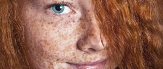
Melanoma is a dark brown malignant formation that appears as a result of certain changes and an increase in melanin synthesis. A dangerous tumor can be located on the legs and cause pain and other discomfort. The anatomical and functional features of the feet require urgent closure of the defects after mandatory excision of the formation in the development of this disease. The complexity of plastic surgery and other reconstructive procedures in this area is associated with age-related pathologies.
Melanoma of the foot is the most aggressive of oncogenic neoplasms. The tumor can metastasize, after which it is considered incurable. In order to prevent this disease, it is necessary to strictly control existing moles and age spots on the legs. This cancer, even if small in size, is very dangerous for human life. After a few months, the oncogenic process spreads to the internal organs.
If melanoma is diagnosed at an early stage, then removing the formation on the feet is not difficult. If the lesion is larger than one centimeter and is characterized by uneven staining with asymmetrical edges, a treatment plan is developed, including chemotherapy in addition to surgery.
Symptoms of foot melanoma
The disease has basic and additional symptoms. The first includes the following:
- the rapid growth of new, recently emerged formations,
- change in the size and structure of the nevus on the leg,
- darkening of the tumor and the appearance of black fragments in it.
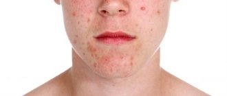
Additional symptoms include the development of an inflammatory process along the edge of the pigment spot, as well as a feeling of itching and bleeding of the skin formation. In frequent cases, pathology occurs on the feet of women. The earlier the diagnosis is made, the greater the chances of a favorable outcome. Treatment of superficial melanomas on the feet in the absence of raised areas usually has a positive result.
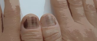
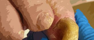
Nodular formations that are raised above the surface of the epidermis are characterized by an aggressive course. They affect approximately 15 percent of patients. Subungual melanoma on the leg is common. It mainly develops on the big toes.
Folk remedies
Along with medications, for red spots on the legs that itch and cause discomfort, it is useful to use traditional medicine recipes. They will be effective for skin irritation, allergies, insect vinegar, frostbite.
Popular folk remedies:
- Herbal decoctions and infusions. In particular, medications based on chamomile, marigold, eucalyptus, and St. John's wort are good at eliminating itching and redness. You can make lotions and compresses, or simply wipe problem areas on your feet. But if the itching is unbearable, then it is recommended to freeze the broth and then wipe the spots with it. This approach will help eliminate discomfort.
- In case of fungal infection, it is recommended to use an infusion of celandine. At 8 tbsp. l. herbs you need to take 3 liters of boiling water and steam. Keep covered for 45 minutes, then strain and add to a bath of water or basin. Immerse your feet in the solution and keep it until it cools completely. The infusion can be used for compresses, but to do this you need to pour 1 spoon of the raw material into a glass of boiling water.
- Viburnum juice. It has antiseptic, anti-inflammatory, analgesic properties. Used to wipe away red spots resulting from allergies, fungus, and skin irritation.
If the red spots on the legs are flaky and occur due to excessive dryness of the skin, then rubbing coconut or olive oil into the epidermis in the morning and evening will help get rid of them. Carry out the procedure after taking a shower or bath.
Preventive measures in this case are simple. It is necessary to carefully monitor body hygiene, use only high-quality and suitable cosmetics, refuse to take medications on your own, and eat a healthy and balanced diet. It is also recommended to avoid contact with stray animals, but if this fails, after coming home you should wash your hands well and treat them with an antiseptic: this will prevent the infection from spreading to other parts of the body.
Causes of foot melanoma
Many people who are faced with this disease are looking for the reasons for its development. These include the following:
1. Exposure of the skin to ultraviolet rays (vacation in the south up to 1-2 times a year, visiting a solarium, getting sunburn), especially for those with fair skin and hair.
2. Congenital or acquired dysplastic pigmented nevi (moles that increase in size).
3. Chronic injury to the mole - this happens if you constantly wear uncomfortable shoes, expose the nevus to friction with items of clothing, or touch the mole while shaving or during hair removal.
4. Genetic predisposition (multiple dysplastic skin nevi syndrome, a history of melanoma of the foot in a close relative)
5. During pregnancy and menopause, hormonal changes occur, which leads to the degeneration of ordinary moles into malignant ones.
Although in reality, the listed factors only trigger a mechanism that was originally inherent at the genetic level in the human body.
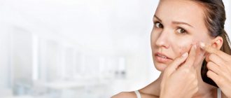
Diagnosis of the disease
Timely diagnosis is the key to the final cure of foot melanoma. It is important to periodically examine the skin yourself, especially if there are moles on the skin. Changes in their shape, shade, edges and growth dynamics should alert you. In generally accepted practice, the international ABCDE rule helps to identify the disease, which is translated from English as: – asymmetry of the neoplasm:
- B – indicates that the edges of the spot are jagged;
- C – change in shade or multicolor (the presence of black, brown, pink and blue colors in one spot);
- D – mole diameter more than 6 mm;
- E - dynamics of changes over time (growth, convexity).
If the above manifestations occur, it is necessary to visit an oncologist, a specialist in the diagnosis and treatment of cancer, as soon as possible.
The oncologist examines the patient, his skin, and the melanoma itself on the leg, examines and palpates regional lymph nodes, and collects anamnesis. After which he prescribes an ultrasound of peripheral lymph nodes, the abdominal cavity and retroperitoneal space, the pelvis, and the chest at the time of the absence or development of metastases.
If necessary, computed tomography (CT) of organs and systems with contrast, magnetic resonance imaging (MRI) of the brain with intravenous contrast, or positron emission tomography of the “whole body” is performed. If enlarged peripheral lymph nodes are detected (if metastases are suspected), it is necessary to prescribe the patient a fine-needle aspiration puncture biopsy using cytological examination.
An important diagnostic criterion is called Breslow thickness; it reflects the rate of division of oncogenic cells. The oncologist examines the extent of the lesion and develops a treatment plan. The thinner the Breslow thickness, the greater the chance of recovery. The indicator is detected and calculated after performing a histological examination of tissue samples. If the indicator is less than 1 mm, then the tumor is considered benign and does not require surgical removal of pigmentation. If the indicator is from 1 mm, then immediate removal of the formation is recommended.
Stages of development of melanoma of the foot
The disease proceeds and develops gradually, the following stages are distinguished:
- Stage 1 - damage to an area no larger than 2 mm in size, the occurrence is localized on the surface of the epidermis, there are no depressions or metastases. The risk of secondary occurrence is rare.
- Stage 2 - the thickness of melanoma on the leg is from 2 mm, ingrowth into soft tissues is observed, without the formation of metastases.
- Stage 3 occurs inflammation and an increase in the size of the lymph nodes. Extension beyond the primary lesion is confirmed by a biopsy of the closest lymph node to the affected area.
- Stage 4: the appearance of metastases in tissues, bones and internal organs are affected. The prognosis for cure is unfavorable and varies within 10%.
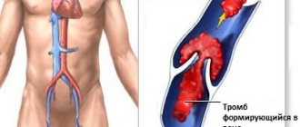
How to remove spider veins on the legs?
What to do if spider veins appear on your legs? Do not panic and consult a phlebologist. An experienced doctor will examine the affected area, ask clarifying questions and prescribe additional research methods to assess the condition of blood vessels (USD) and determine the cause of the disease. Taking into account the characteristics of the patient’s health condition, the doctor will prescribe invasive or conservative treatment to remove spider veins on the legs. Conservative treatment methods include:
- wearing compression stockings;
- drug therapy;
- lymphatic drainage massage;
- physiotherapy.
Treatment methods
Melanoma of the foot develops quickly, metastases form in just a few months. It is difficult to cure the anomaly; it is a multi-step process, including:
- Stage 1. Removal of the formation by surgery, which may affect healthy tissue.
- Stage 2. Carrying out a biopsy. Removal of lymph nodes if they are affected. Alpha interferon drugs are routinely prescribed to reduce the risk of relapse.
- Stage 3. Removal of the tumor and lymph nodes that are located in close proximity. In this case, immunotherapy, radiation, and chemotherapy are used. Interferon and similar medications are prescribed.
- Stage 4. Difficult to heal. The melanoma on the foot is removed; the operation is often accompanied by amputation of the toes; severe pain is relieved by taking strong analgesics. Chemotherapy and potent medications are prescribed. In order to reduce the tumor and improve the patient’s condition, the doctor prescribes the combined use of immuno- and chemotherapy drugs.
Recurrent melanoma on the leg is treated with carefully selected chemotherapy. Malignant tumors that form from melanocytic cells on the lower extremities account for half of the cases. They arise from benign tumors. There are cases when the disease developed from a single melanocyte.
Invasive methods will help more effectively and quickly remove spider veins on the legs:
- sclerotherapy;
- microsclerotherapy;
- microfoam Foam Form sclerotherapy;
- ECHO sclerotherapy;
- percutaneous laser coagulation.
The Medicine and Beauty clinic uses the most modern methods of treating telangiectasia. Among the above, our doctors obtain the fastest visual effect when using percutaneous laser coagulation. This method allows you to eliminate spider veins of any location and size directly during the procedure.
If there are contraindications to the use of a laser, the clinic’s phlebologists will select another suitable method for treating telangiectasia for the patient. All treatment methods are carried out on an outpatient basis and are painless. The clinic’s vascular surgeons have been helping patients with vascular disorders for many years and know how to remove spider veins on the legs forever. Doctors also pay great attention to the prevention of relapses and explain in detail to their patients how to behave after removal of the defect.
The clinic's doctors work only with certified modern equipment and use proven medications.
Forecast and prevention of melanoma development
The further prognosis is determined by the stage of the disease. At the initial stage, melanoma on the leg can be cured. In a complex stage, when the formation has grown into the tissue and the pathology has affected the lymphatic system, the risk of relapse increases markedly, so it is much easier to prevent the disease than to fight it later.
By taking your health seriously, you will avoid most problems, including those whose treatment is not always associated with a favorable prognosis.
To prevent the development of melanomas, it is recommended to sunbathe with caution, especially for those with white sensitive skin. Sunscreen and wearing closed clothing made from breathable natural fabrics on sunny days will help. Regular visits to an oncologist are recommended to promptly identify the development of the pathological process.
It is important to monitor the condition of moles and age spots, monitor the changes that occur and not injure them. Special creams will help to lighten them. A successful preventive remedy that will not allow pigment formation to degenerate into a malignant tumor is Lakshma Maxxi whitening cream. It is one of the most sought after products in the United States and is recommended by the American Dermatological Association. It allows you to eliminate signs of hyperpigmentation on the skin easily, reliably and safely. According to the results of clinical studies, the drug showed high stable results.
Add to cart
Underarm skin brightening treatment
RUB 1,400.00
Add to cart
Whole body skin lightening milk
RUR 3,800.00
Add to cart
Cream Lakshma Maxxi (Lakshma Maxxi)
RUB 2,100.00
Add to cart
Advantages
Today, there are several methods for removing spider veins, but not all of them are equally safe. Often, after the intervention, scars and age spots remain on the skin. The removal of spider veins using laser coagulation is considered the most harmless and effective. This procedure is performed absolutely painlessly and has virtually no contraindications. When spider veins are removed in this way, laser radiation acts on the dilated vessel, heats it and “seals” it. As a result, the surrounding tissues are not affected, and the defects that bother the patient completely disappear. The Medicine and Beauty Clinic offers guaranteed removal of vascular spots and stars on the skin without complications.