First aid for eye injuries
In the event of a serious penetrating injury to the eye, the first step should be to immediately stop the bleeding by applying a sterile bandage to the eye. After this, it is necessary to urgently take the injured person to an ophthalmologist.
Movement of the unaffected eye can aggravate the injury, since the eyes are paired organs and move synchronously, so it is better to apply the bandage to both eyes. Do this if you can wait for an ambulance or if you have the ability to transport a blind person to a medical facility.
If there is a foreign object in the eye, it is strictly forbidden to try to remove it yourself, as this can lead to severe bleeding and even greater injury to the eye tissue. In this case, you need to apply a cotton swab and a sausage-shaped bandage around the foreign object, and then secure it with a bandage.
Possible eye damage
Speaking about injuries to the organs of vision, first of all we mean a violation of the integrity of the cornea of the eye, as well as the upper transparent membrane of the eyeball as a result of exposure to certain external factors. In particular, this could be:
- getting a foreign object (metal shavings, small pebbles, etc.) into the eye;
- a blow to the eye (with a fist, a champagne cork, etc.);
- eye injury;
- chemical or sunburn of the cornea;
- fire damage to the eye;
- eye damage due to a firecracker explosion and much more.
According to statistics, in 90% of cases, patients go to the hospital with very minor injuries and injuries to the eye as a result of being hit with blunt objects, in 8% of cases with burns and in 2% of cases with serious penetrating injuries.
First aid for foreign particles entering the eye
Often small foreign bodies get into the eye, for example: plant thorns, glass fragments, pieces of plastic, metal, etc.
You can only remove small particles that do not have sharp edges yourself.
Trying to remove sharp particles yourself is strictly prohibited, because this can cause even greater trauma to the eye tissue and lead to severe consequences. The longer a foreign object remains in the eye, the more severe visual impairment will occur later, so it is very important to see a doctor as soon as possible.
To remove the particle yourself, you can use 1% boric acid solution (dissolve 1/2 teaspoon of powder in a glass of water) or a weak solution of potassium permanganate (no more than 2 crystals per glass of water). With a moistened cotton swab, carefully rinse the eye, running along the eyelid from the outer corner of the eye to the bridge of the nose.
Prevention of eye injuries
To protect your eyes from possible damage, you should adhere to the following rules:
- When doing construction and welding work, be sure to wear safety glasses;
- do not be in the sun without sunglasses;
- Follow labor safety rules when working in manufacturing plants.
In fact, labor protection is the most important system for preserving the life and health of workers in the process of work.
Managers of any enterprises must provide employees with training on industrial safety and labor protection in order to minimize the occurrence of emergency situations. Clean View Eye Clinic diagnoses and successfully treats any eye and eyelid injuries. You can find out more details and make an appointment by phone.
First aid for a chemical burn to the eye
If you accidentally get any caustic liquid (stain remover, bleach, paint, etc.) into your eye, you should rinse your eye as soon as possible under the tap or with a water bottle. To do this, you need to carefully open your eyelids and direct a stream of water in the direction from the bridge of your nose to the outer corner of your eye.
After this, you need to drip eye antiseptic from the first aid kit and cover the eye with a clean cloth. If there is a chemical burn around the eye, that too needs to be treated.
Do not rub the affected eye with your hands or apply ice to it (this may cause injury to the cornea).
You must urgently consult a doctor. It’s good if you can show the specialist a sample of the liquid that has entered the eye so that he can more accurately prescribe treatment.
Remember that the sooner you contact a specialist for any eye damage, the higher the chances of a successful outcome and preservation of vision.
How is eye injury treated?
Treatment is selected based on the type and extent of eye damage. In mild cases, we manage with drug therapy, which includes antibacterial drugs, non-steroidal anti-inflammatory drugs, painkillers and hormonal eye drops. For moderate and severe burns, the patient is hospitalized; for serious wounds, surgery is performed. Treatment after an eye injury involves using drops recommended by your doctor.
Consequences of eye injury
The consequences of damage depend on its extent and the quality of assistance provided. Doctors treat wounds and carry out all necessary manipulations, so if you quickly go to the emergency room, there will be fewer complications. Undesirable consequences are associated with infection entering the blood or even greater injury as a result of rash actions.
If a person does not receive timely medical care, scars remain, the soft tissues around the eye are deformed, and visual acuity decreases until it is completely lost.
If the infection enters the blood, sepsis develops - inflammation that is dangerous to the entire body. Accumulating pus affects the internal structures of the eye, sometimes reaching the brain, causing inflammatory processes in the body.
Sympathetic ophthalmia
Dependence of CO on the characteristics of injuries:
— Penetrating wounds, — Repeated operations or operations on the injured eye, — Localization in the anterior section, mainly in the corneoscleral zone, — Prolapse, entrapment of the uveal membranes, — Aseptic autoimmune post-traumatic uveitis, traumatic cataract, — Retinal detachment.
Dependence of SB on the timing of PSO:
- For patients, this is late PSO - from 2 days to 5 weeks;
— Insufficiently qualified wound treatment; — Frequent development of complications (traumatic cataracts, secondary glaucoma, retinal detachment and eye subatrophy with loss of all visual functions). Factors contributing to the development of sympathetic ophthalmia: 1. Violation of the hemato-ophthalmic barrier during penetrating trauma of the eyeball, complicated by prolapse and damage to the uveal membranes, migration of “beyond-barrier” uvearetinal antigens along the lymphatic pathways to the lymphoid organs and the development of autoimmune post-traumatic uveitis. 2. Violation of the immunoregulatory mechanisms of the immune system, the formation of cellular and humoral autoimmune reactions to uvearetinal antigens, retinal S-ag. 3. Various factors of the external and internal environment, including immunogenetic ones, influencing the state of the body’s immunity. When examining patients with OM (L.T. Arkhipova et al., 1976-1997), disorders of the immune status were revealed, characterized by: - A decrease in the number of T-lyphs, mainly against the background of a sluggish or recurrent process, after long-term treatment with steroids; — A decrease in the number of T-helpers and T-suppressors (CD4 and CD8) with an imbalance in the Tx/Tc immunoregulation index; — A decrease in the functional activity of T-lf (in the RBT test for the nonspecific mitogen PHA) more often in a chronic course; — Decrease in the concentration of IgG, A, M and dissymmunoglobulinemia. The immunosuppressive treatment system includes 3 components: 1. Enucleation of the blind sympathetic eye as the primary source of antigenic autosensitization of the body; 2. Immunosuppressive treatment with steroids and cytostatics that suppress the body’s immunopathological reactions; 3. Anti-relapse immunocorrective treatment for chronic forms of sympathetic inflammation. On the issue of enucleation of the sympathetic eye
Most literature sources and analysis of the Helmholtz Institute show that the prognosis of sympathetic ophthalmia improves when enucleation of the blind injured eye is performed in the early stages after the onset of the disease (from 1-2 days to 3-4 weeks). However, after studies conducted at the Helmholtz Institute in patients with a preserved sympathetic eye (with visual acuity in it from objective to 1.0), it turned out that they had a more favorable course and high visual acuity than in the group of patients whose eye was removed. This is believed to be due to the fact that in this group the injured eye had less severe inflammation. Marak GE (1979) showed that the prognosis of the disease is worse if the process in the injured eye spreads to the posterior part of the uvea and the retina. At the Helmholtz Research Institute, L.T. Arkhipova and others developed a treatment regimen for CO with steroids. They are prescribed locally from the first days of the disease (in instillations, under con-vu and parabulbar), given that in more than 80% of patients the inflammatory process spreads to the posterior part of the uvea and the retina. Principle of systemic treatment: 1. Average therapeutic doses - 25-60 mg (in terms of prednisolone) depending on age, clinical form and severity of the process. 2. Gradual reduction in dosage: first by 5 mg (1 tablet) of prednisolone every 5 days, and then starting from 30 mg to 2.5 mg (1/2 tablet), and from 15 mg to 1.25 mg (1/2 tablet). 4 tablets) every 5 days. 3. Steroid withdrawal occurs under the guise of non-steroidal anti-inflammatory drugs such as indomethacin. Those. when prednisolone is reduced to 10 mg, indomethacin 50 mg per day is additionally prescribed (2 tons x 2 times a day). In general, the course of treatment is 4-6 months, of which 1.5-2 months are steroids, and 2.5-3.5 months are indomethacin. In severe forms, treatment begins with intravenous injections of steroids at a rate of 1.0 in 10.0 saline solution No. 3-4, followed by a transition to tablet forms. For posterior forms of sympathetic ophthalmia and exudative chorioretinitis, pulse therapy is performed (IV drip in rheopolyglucin 16 mg every other day No. 3, then 8 mg No. 3 every other day, followed by switching to tablets with 60 mg.). Treatment with steroids is carried out against the background of giving potassium and calcium salts and an antidiabetic diet. The duration of local treatment is on average 8-10 months, total 5-6 months. Treatment is carried out under clinical and immunological control until reactions to uvearetinal tissue antigens are persistently suppressed.
Non-perforating wounds of the cornea and sclera
Corneal erosion is accompanied by significant pain, photophobia, lacrimation, and blepharospasm. A fluorescein test facilitates 100% diagnosis of this condition. The main task is to relieve pain and prevent infection of the defect. First aid, in addition to instillation of dicaine, consists of instilling drops containing antibiotics or sulfonamides and placing eye ointment behind the eyelids. Urgent surgical procedures are required in 2 cases: 1. A scalped wound of the cornea, when a layer of surface tissue has separated from it or the flap tends to wrap its edges and shift. 2. Foreign body in the layers of the cornea. It is removed with a spear or a needle, followed by scraping out the rust rim, otherwise the healing of the defect will take a long time. After removing foreign bodies, it is necessary to perform a Seidel color test, instill drops with an antibiotic or sulfonamides, apply an ointment and apply a bandage.
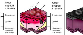
Radiation burns (sun, welding)
Ultraviolet radiation (powerful light with an ultra-short wavelength invisible to the eye) primarily affects the skin of the eyelids, the conjunctiva and the cornea; the latter is the most dangerous. When damaged by light radiation in the infrared (thermal) range, the destructive flow reaches the retina and blood vessels.
These types of burns are usually associated with visiting solariums, unprotected presence in the area of electric welding or the action of a quartz lamp, as well as prolonged exposure to intense sun in blinding snow (especially in the polar regions). Symptoms usually develop several hours after the actual burn.
Radiation damage to the retina is a typical result of careless handling of laser equipment or unprotected contemplation of solar eclipses. In such cases, it may take several days for specific symptoms and/or decreased vision to develop.
Symptoms
Clinically, a corneal burn always manifests itself clearly and unambiguously: even without special knowledge, it is difficult to mistake it for another injury. The most striking symptoms of chemical and thermal burns are:
- acute, almost unbearable pain localized in the damaged eye and eyelid;
- reflex closure of the eyelids, photophobia;
- intense lacrimation (if the tear ducts are damaged, the sign may be absent);
- hyperemia of the skin, mucous membranes, conjunctiva;
- clouding of the cornea (noticeable with a severe burn).
Radiation damage may not be accompanied by unpleasant symptoms immediately after receiving it. Burning sensation, lacrimation, photophobia, hyperemia and photophobia appear after a few hours.