The article was checked by an ophthalmologist, Ph.D. Ismailova D.M. , is for general informational purposes only and does not replace specialist advice. For recommendations on diagnosis and treatment, consultation with a doctor is necessary.
The most common inflammatory diseases of the eyelids include blepharitis and stye.
Blepharitis is inflammation of the edges of the eyelids. There are scaly, or simple, and ulcerative blepharitis.
Barley is an acute purulent inflammation of the sebaceous or sweat gland of the eyelid. There are external and internal barley.
What is a chalazion?
In ophthalmology, a chalazion of the eyelid is a benign tumor-like growth
. It is a capsule closed in the thickness of the mobile eyelid, filled with sebaceous secretion, sometimes pus and dead particles of the epithelium. Upon closer examination, it turns out that a chalazion is an inflamed meibomian gland, connected to the surface by thin ducts that open onto the mucous membranes of the eyelids in contact with the eye. These structures are located in the cartilage of the lower and upper eyelids and are responsible for protecting the cornea. They secrete a secretion that forms a protective film that prevents the evaporation of tears and reduces friction between the eyelid and the surface of the eye.
Under certain conditions, these ducts become blocked and a chalazion begins to develop. First, the secretion it produces accumulates in the gland inside the eyelid, then it stretches its walls, which provokes low-grade inflammation. At the initial stage, the process looks like barley, but is located not along the edge of the eyelid, but at some distance from it. Unlike stye, a chalazion of the eye is not accompanied by pain and looks like a normal flesh-colored lump on the outside and grayish on the side of the mucous membrane in contact with the eye.
Despite the absence of an acute inflammatory process, the chalazion cavity may contain purulent exudate.
Children and adults over 35 years of age are at greater risk of finding out what a chalazion is on the eye. This pathology occurs extremely rarely in adolescents and young adults. When making a diagnosis, the doctor will have to differentiate chalazion from barley, uveitis, keratitis and other inflammatory diseases on the eyelids and outer membranes of the eye.
Causes
The main cause of a chalazion is a blockage of the meibomian gland duct.
followed by an inflammatory process. In most cases, the source of the problem is primary inflammation of the eyelid margin or the gland itself. Chalazion also has secondary causes, that is, not related to eye diseases. Among them, doctors call somatic pathologies:
- the digestive system, in which lipid metabolism is disrupted, and the synthesized fat secretion becomes excessively thick, viscous, and prone to accumulation in the ducts;
- chronic infectious skin diseases - seborrhea and demodicosis, in which parasites and inflammatory processes lead to scarring of the ducts, their gradual narrowing and disrupt the outflow of secretions;
- endocrine diseases, in particular diabetes mellitus, in which immune defense is disrupted and the activity of microorganisms on the surface of the eye and mucous membranes of the eyelids increases;
- deficiency of certain vitamins;
- congenital features associated with increased activity of the sebaceous glands - according to statistics, those with oily skin are more likely to suffer from eye chalazion.
Another common cause of chalazion
is a violation of eye hygiene:
- improper storage and installation of contact lenses;
- contact of dirty hands with eyes;
- using low-quality or expired eye drops;
- using non-sterile tools to create makeup, etc.
Chalazion can also develop against the background of oncology of other organs or the eyelid itself. In the first case, the neoplasm occurs after chemotherapy due to a general decrease in immunity, and in the second it can be a malignant neoplasm, which is mistaken for a chalazion.
Stages of development
Regardless of the observed causes of chalazion, it always develops according to the standard pattern. Ophthalmologists distinguish 3 main stages in the formation of a chalazion on the eye:
- Zero stage
, blockage of the duct outlet. There are no external manifestations, since the size of the neoplasm does not exceed 2 mm in diameter. At this stage, sebaceous secretion accumulates inside the gland, inflammation is absent or mild, and there is no pain. Pathology at this stage is discovered accidentally during cleansing of the eyelid, applying makeup and other manipulations. - Stretching of the meibomian gland
with the formation of a noticeable hailstone on the eyelid. The sebaceous secretion thickens, the chalazion increases in size and becomes harder. Due to the pressure created by the neoplasm on surrounding tissues, minor pain and discomfort in the eye may occur when trying to palpate the pea. When an infection occurs, swelling and slight hyperemia of the eyelid over the neoplasm may be observed. The size of the chalazion at this stage does not exceed 3-4 mm. - Encapsulation of the chalazion
, further growth of the hailstone or its spontaneous resorption.
Sometimes, in the absence of therapy and the persistence of the factors that influenced the formation of a tumor in the eye, the growth process of the chalazion is complicated by intense inflammation. At the same time, in addition to fatty secretion, purulent exudate can accumulate in the capsule. It can melt the shell of the chalazion and lead to spontaneous leakage of its contents onto the outer surface of the eyelid or, more often, onto the cornea of the eye. In the first case, a lesion remains at the site of the neoplasm, a fistula is formed - a duct, which periodically opens, and the contents of the gland emerge from it. In the second case, in addition to the formation of a fistula, there is a possibility of infection spreading to the membranes of the eye.
Fungal diseases
Actinomycosis. The causative agent is radiata fungi (actinomycetes). A dense, painless nodule appears in the outer or inner corner of the eyelid, which then softens, forming an intradermal infiltrate.
A breakthrough of the infiltrate leaves a long-term non-healing fistula, secreting pus with tangles of fungal threads. Treatment includes the use of antibiotics and actinolysate. Conducting radiotherapy, opening the abscess and curettage it.
Candidiasis (candidiasis) is a disease caused by yeast-like fungi of the genus Candida. When affected, swelling of the skin of the eyelids, hyperemia, and rashes of small pustules are observed.
Treatment includes lubricating the lesions with a solution of brilliant green prepared in water, using nystatin ointment and taking nystatin orally.
Trichophytosis (ringworm). The disease occurs as folliculitis and is caused by fungi of the genus Trichophyton. It is customary to distinguish between superficial and deep trichophytosis. At the same time, the edges of the eyelids are hyperemic and swollen, contain pustules, in yellowish crusts. In some areas of the eyelids there are no eyelashes, in others they are broken off. There is a coating of spores on the eyelashes.
The process may be accompanied by conjunctivitis or keratoconjunctivitis. As healing occurs, scars appear.
During treatment, the affected areas of the skin are wiped with an alcohol solution of iodine or methylene blue. Griseofulvin is prescribed orally.
Symptoms
With chalazion, the symptoms are not similar to inflammatory diseases of the eyelids, although the appearance of the neoplasm at different stages of development may resemble barley, blepharitis, conjunctivitis and other pathologies. It is distinguished from them by its slow development and the absence of a clinical picture characteristic of acute inflammation.
Chalazion can be distinguished from the mentioned pathologies by the following signs:
- absence of acute inflammation in the tissues surrounding the hard capsule, expressed by redness and swelling;
- no pain when blinking;
- absence of purulent discharge in the lumen of the duct;
- mobility of the hard capsule when palpated.
The main symptoms of this eye disease are
ophthalmologists call the gradually increasing sensation of a foreign object under the eyelid, in the place where the altered meibomian gland is located. Visually, the eye may appear smaller than the other due to the thickening of the eyelid. This feeling appears when the pea reaches a diameter of about 3-5 mm. At the same time, the patient may be concerned about:
- itching of the eye in the place where the pea is located;
- peeling and thinning of the skin over the lesion;
- mild hyperemia, which appears only during periods of increased inflammation of the gland;
- moderate lacrimation.
When the diameter of the neoplasm exceeds 5 mm, the chalazion of the eyelid not only irritates the membranes of the eye, but also puts constant pressure on them. In addition to the already mentioned itching, watery eyes and flaking of the skin, such a large neoplasm can affect visual acuity. It puts pressure on the eyeball, which leads to increased eye pressure and blurred vision. Chalazion can also cause damage to the cornea with subsequent thickening or clouding. All this provokes a significant deterioration in vision.
The most pronounced discomfort is caused by a chalazion of the upper eyelid.
Larger capsules form on it. The degree of discomfort and the participation of the eyelid in moistening the eye are affected - the upper mobile skin fold makes movements with a wider amplitude than the lower one, so any swelling on it is felt better. This type of chalazion is also more noticeable visually - when the upper part of the palpebral fissure is affected, the organs of vision look asymmetrical, and the blinking mechanism is disrupted.
During periods of exacerbation, the chalazion increases in size, tightens the skin and lifts the inner lining of the eyelid, which causes the patient to worry about:
- burning, feeling of sand in the eye due to insufficient hydration of the cornea;
- inflammation, damage to the conjunctiva, which is expressed by redness of the eye, lacrimation, discharge of pus and pain;
- inversion of eyelashes into the eye, resulting in irritation of its membranes, formation of a vascular pattern on the sclera or pinpoint hemorrhages.
Most often, the described symptoms appear in winter, after hypothermia. Chalazion also worsens in the summer after the opening of the beach season, among vacationers in other climatic regions. The statistics are explained by a temporary decrease in immunity and poor eye hygiene when relaxing in open water. In children, chalazions in the eyes often become inflamed in the summer, when children play in sandboxes, dry pools, etc.
Classification
The disease has different localization and is divided into the following forms according to location:
- Marginal anterior (the process involves the ciliary edge);
- Posterior marginal (with damage to the lacrimal glands and lacrimal sacs);
- Angular (inflammation is localized in the corners of the eyes).
The following classification of blepharitis depending on clinical manifestations is generally accepted:
- Simple form;
- Demodectic mange (caused by a tick-borne agent);
- Osteofolliculitis (ulcerative-necrotic manifestations);
- Seborrheic (caused by dermatic lesions of the eyelid epithelium);
- Allergological;
- Rosacea-blepharitis;
- Mixed form.
Diagnostics
The use of instrumental methods to diagnose chalazion is not required, since even an inexperienced ophthalmologist can distinguish this pathology from other eye diseases. When interviewing the patient, the doctor will find out:
- is there pain when blinking - its absence is interpreted in favor of blockage of the meibomian gland duct;
- is there any sticking of the eyelid with yellow, white or greenish pus in the morning - with a chalazion this phenomenon should not occur;
- how long ago the tumor was first noticed - a period of more than 2 weeks is interpreted in favor of the described pathology.
Next, the doctor examines the eye: palpates the affected eyelid, turns it inside out to examine the inner surface. To determine the causes of chalazion, the ophthalmologist may prescribe additional tests:
- laboratory blood tests (general clinical, biochemistry) to identify endocrine, metabolic, infectious and other disorders;
- bacteriological blood culture for pathogens of systemic infections;
- culture of a smear from the eyelid for staphylococcal infection;
- inoculating a smear from the eyelid for pathogens of gonococci, trichomonas and other pathogens of STIs that can affect the mucous membranes of the eyes;
- stool analysis for eggs of worms, lamblia and other parasites;
- microscopic analysis of epithelial scrapings for demodex and other skin parasites;
- immunological screening.
The doctor may not necessarily prescribe all of the tests listed. It is enough to limit ourselves to the most probable causes of the pathology identified during the collection of individual and family analysis. In most cases, the doctor limits himself to a general clinical blood test and referring the patient to a consultation with a dermatologist and therapist.
Treatment
According to statistics, treatment of chalazion takes from 2 weeks when diagnosed in the early stages to several weeks if the disease is diagnosed in an advanced form. If you contact an ophthalmologist before the formation of a dense capsule, treatment is carried out using simple means and the tumor is eliminated in the same period as with barley, that is, 2-5 days. However, in practice this happens extremely rarely, because before the gland is encapsulated, the chalazion is practically invisible and does not cause discomfort.
At the initial stages, chalazion treatment is carried out using antiseptics, antibiotics and anti-inflammatory drugs in the form of drops and ointments. They can be single-component or contain additional substances that enhance the effect of the main components.
The most effective are:
- ointments and drops with antibiotics - “Vigamox” (active substance moxifloxacin), “Floxal” (ofloxacin), “Oftaquix” (levofloxacin), “Tsiprolet” (ciprofloxacin);
- combination drugs with antibiotics and hormonal anti-inflammatory substances - “Tobradex” (tobramycin with dexamethasone), “Floxadex” (ciprofloxacin with dexamethasone), “Garazon” (betamethasone with gentamicin).
In most cases, doctors tend to use combination drugs, especially if the chalazion has become chronic or recurrent. Such agents actively suppress pathogenic microflora, reduce inflammation and prevent purulent complications. When prescribed in a timely manner, medications from this group relieve swelling from the walls of the meibomian gland ducts and restore the outflow of secretions.
If the neoplasm is not bothered by unpleasant symptoms and there are no signs of inflammation, ophthalmologists recommend physiotherapy, combining gentle dry heating of a pea with subsequent massage of the eyelid. Warming can be performed in a clinic or private clinic with special equipment. At home, it can be replaced in the following ways:
- place a fabric bag with salt or sand heated in a frying pan on the eyelid for 5-10 minutes;
- iron the cotton fabric with a heated iron and apply it to the eye until it cools, repeat 5-7 times.
Gentle heating helps to soften the thickened secretion, which comes out of the duct during subsequent massage.
Important to remember
that this method cannot be used to treat a chalazion that has arisen against the background of an infection of the eye, as well as one that occurs with obvious inflammation of the meibomian gland. The methods are suitable for eliminating pathology caused by metabolic disorders, chronic gastrointestinal diseases, etc.
After the procedure, you need to rinse the eyelid with warm water using a cotton pad or sterile napkin. After washing, you can treat the eye with any antiseptic. You can also use:
- “Blepharogel” with hyaluronic acid to restore the structure and moisture of the eyelids;
- “Blephaclean” eye hygiene wipes with antiseptics, hyaluronic acid, antioxidants and substances that regulate the activity of the sebaceous glands.
These products can be used not only after a massage, but also during it. The active substances in their composition help soften the sebaceous secretion of the glands and remove it from the ducts of the glands. Despite the enormous benefits for eye health, it is recommended to use them no more than twice a day.
If the chalazion is opened on your own, it is recommended to continue therapy according to the regimen prescribed by the doctor. To prevent infection of the resulting wound, quickly and efficiently cleanse the capsule and tissue regeneration, it is recommended to use:
- drops with antibiotics - “Signitsef”, “Tobrimed”, “Uniflox”;
- ointments with antibiotics - tetracycline, Floxal, erythromycin;
- corticosteroids in the form of ointment - “Hydrocortisone” 0.5%, “Combinil Duo”, “Floxadex”;
- homeopathic ointment "Traumel".
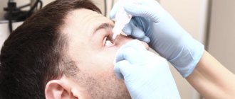
If conservative methods do not lead to improvement or there is a tendency for the tumor to grow, surgical methods are used to treat chalazion. For children, operations are performed under general anesthesia, and for adults, under local anesthesia. There are several methods for removing tumors:
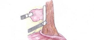
- Classical desquamation of the capsule and its contents.
To carry it out, the doctor makes an incision in the skin of the eyelid and through it, using a curette (a curved sharp instrument), scrapes out the chalazion, then cleans the cavity, treats it with antiseptics and applies sutures. The intervention lasts no more than 15 minutes. The sutures are removed after 5-7 days. - Enscaping the capsule through an incision on the inside of the eyelid
. The doctor everts the eyelid, makes an incision and performs the procedure as in the previous case. There are no stitches with this method. For better healing and prevention of infection, apply antibiotic ointment behind the eyelid and cover the eye with a sterile bandage. - Laser removal of chalazion
. The doctor makes an incision on the outer part of the eyelid and evaporates the capsule with its contents layer by layer. Treatment with antiseptics and sutures are not necessary for this type of intervention - they will be replaced by a scab formed by the action of the laser on the tissue.
After any intervention, slight swelling remains in the eye for a week, and the patient may experience moderate pain.
Rules for the use of drugs
In order for the prescribed drops and ointments to act purposefully to achieve a faster effect, they must be administered correctly. So, it is more convenient to do this with a special spatula - it can be purchased at any pharmacy. You should squeeze out about 5-6 mm of ointment from the tube onto your shoulder blade, carefully pull back the lower eyelid and place the ointment there, being careful not to touch the eye tissue with the tip of the tube. Then you need to close your eyelids for a while so that the product is evenly distributed inside.
When instilling drops, make sure that they fall exactly into the conjunctival sac. Do not exceed the recommended dosage of medications, acting strictly as prescribed in the prescription. Excessive amounts of medication may harm your eyesight. It is better to instill in a horizontal position so that the liquid does not leak out.
The duration of use of antibacterial drugs is determined by the attending physician. However, a visible effect should occur within 3-4 days from the start of their use. If this does not happen, then you need to seek advice from a specialist - you may need to change your medication. It should be remembered that if antibiotics are used for too long, there is a risk of developing superinfection - the growth of pathogenic microorganisms that are immune to them, including fungi.
It contains mild, soothing herbal ingredients: green tea, chamomile, witch hazel extract, and effectively cleanses the eyelid skin of impurities. Apply a little lotion to a cotton swab and lubricate their edges at the base of the eyelash row, trying to remove all traces of discharge. After this, medications can be administered.
In the allergic form of blepharitis, moisturizing preparations that imitate natural human tears will help relieve dryness, itching, and irritation: Hilo-chest of drawers, Systane, Artelak Balance and others.
You should not make a diagnosis yourself and self-medicate by seeking advice from friends or using information from the Internet. Only a specialist, after a competent diagnosis, will select effective medications to eliminate inflammation of the eyelids. An inappropriate remedy for a particular type of blepharitis can only aggravate the course of the disease and thereby delay recovery.
Prevention
To prevent chalazion, it is enough to maintain visual hygiene:
- thoroughly clean your eyelids after sleep;
- remove makeup before going to bed;
- do not apply cosmetics and creams along the edges of the eyelids and in the eyelash line;
- store contact lenses in sealed, sterile containers with fresh solution;
- pick up and install lenses with clean hands or a sterile instrument;
- do not touch your eyes with dirty hands;
- if you are prone to inflammation of the eyelids, use “Blepharogel” and “Blephaclean” wipes for daily eye care;
- promptly eliminate problems with digestion, metabolism and hormonal imbalances;
- avoid hypothermia.
If you find a small hailstone on your eyelid, you should immediately go to an ophthalmologist with the problem in order to have time to deal with it using conservative methods.
Hygiene procedures for blepharitis of the upper eyelid
Blepharitis is difficult to cure if hygiene procedures are not followed. Their implementation plays an important role in the treatment and prevention of this ophthalmological disease. The main hygienic procedure is daily eye washing. It is best to use sterile dressings and exclusively boiled water for this. Medicinal infusions of chamomile, marshmallow, rose hips, thyme or calendula, which have an anti-inflammatory effect, will have a good effect.
The prepared solution should be at room temperature. This will avoid damaging the damaged skin of the eyelids. It is necessary to rinse both eyes at once, even if only the eyelid is inflamed. Individual cotton swabs or bandages should be used for each. Reuse of hygiene products is prohibited. This can lead to infection of the visual organs and cause serious complications.
Consequences and complications
With timely diagnosis and treatment, chalazion has no consequences. If you ignore a hailstone for a long time and allow it to grow to 5 millimeters or more, there is a risk of complications:
- transfer of infection to the conjunctiva, cornea;
- chronic compression of the eyeball and irreversible decrease in visual acuity;
- formation of ulcers on the cornea;
- development of phlegmon of the century;
- fistula formation.
Treating these pathologies is tens of times more difficult and expensive than eliminating a small chalazion. But even with a successful outcome, scars will remain and the shape of the palpebral fissure will change. Therefore, you should not hope that the tumor will resolve on its own. It is better to enlist the help of an ophthalmologist and undergo a course of treatment.
List of sources
- Maychuk Yu. F. Blepharitis. Modern drug therapy: a manual for doctors, 2nd edition. - M., 2013. - 25 p.
- Egorov E.A. Blepharitis // Ophthalmology. National leadership. Ed. S.E. Avetisova, E.A. Egorova, L.K. Moshetova, V.V. Neroeva, Kh.P. Takhchidi. – M. – 2008. – Section 5.4. – pp. 342-347.
- Therapeutic eyelid hygiene is the basis for the prevention and treatment of blepharitis / Polunin G.S., Polunina E.G., Zabegailo A.O., Pimenidi M.K. // Collection of scientific papers of the scientific and practical conference of the Russian National Ophthalmological Forum. — M2008. — P.334-348.
- Koroev O.A. // Blepharitis. Vladikavkaz. – 2005. – 83 p.
- Dolzhich G.I., Dolzhich R.R. Ophthalmology 2008, “Phoenix” 286 p.