To the reference book The inner ear, also called the labyrinth, is an auditory and vestibular analyzer.
Author:
- Sokolova Alla Vasilievna
otorhinolaryngologist, otosurgeon, doctor of the highest category
(Voted by: )
- Outer ear
- Middle ear
- Inner ear
, the inner ear is otherwise called a labyrinth.
Located in the thickness of the pyramid of the temporal bone, it consists of 3 parts:
- vestibule;
- snails;
- semicircular canals.
The vestibule is considered the center of the labyrinth and has two pockets. The first, spherical in shape, is located closer to the cochlea. The second, elliptical, is adjacent to the semicircular canals, which are located in three mutually perpendicular planes. Blood supply is carried out through the labyrinthine artery.
The main functions are auditory and vestibular. The auditory analyzer allows you to perceive sound vibrations and ensures the transmission of nerve impulses to the auditory nerve centers, where recognition of the received information occurs. The vestibular analyzer implements sensory, somatic and other reactions.
Causes of the disease
Patients of any age are susceptible to the disease, but children suffer from it more often than adults, since they have the anatomical features of the Eustachian tube: it is smaller and located more horizontally. So why does fluid accumulate behind the eardrum in adults and children:
- any types of allergies;
- viral and infectious diseases, sinusitis suffered the day before;
- polyps in the nasal cavity, enlarged tonsils or adenoids, as well as other processes that may cause blockage of the tube;
- exposure to external factors (for example, tobacco smoke);
- trauma to the ear cavity that is caused by changes in pressure (barotrauma, which occurs under water pressure when diving or on an airplane).
1.General information
The appearance of any discharge where normally there should not be one is always a symptom, i.e. a sign of pathology, and in all cases this symptom is quite serious. With regard to the hearing organs, vigilance should be maximum, taking into account their location, anatomical complexity, intensity of innervation and blood supply. Any, even mildly symptomatic or latent pathology in this zone is dangerous. There are many cases where the primary problem could be eliminated relatively quickly and simply, provided timely contact with a doctor, but in the absence of medical care, complications turned out to be extremely severe, disabling, and sometimes fatal.
The discharge of liquid or mucopurulent discharge from the ear canal in medicine is called otorrhea. The reasons for this condition are very diverse, but it is certainly necessary to have at least a general idea of them.
A must read! Help with treatment and hospitalization!
Fluid in the middle ear: symptoms
Patients often complain that there is a feeling of cotton wool in the ear. This is one of the signs of the disease. If we talk about young children, then in them this disease can be asymptomatic. Adults usually experience minor discomfort, but some patients complain of severe pain and general malaise. So, the accumulation of fluid in the ears can manifest itself:
- pain and a feeling that the ears are “plugged”;
- unpleasant ringing;
- muffled sounds or deafness;
- feeling of fullness in the ears;
- less often the disease is accompanied by dizziness and loss of balance.
Diseases
Regardless of the nature of the disease, the above analyzers are necessarily involved, therefore the patient’s condition is characterized by vestibular or cochlear symptoms. With such disorders, there is usually a violation of two functions of the ear labyrinth at once.
Diseases can also be divided into inflammatory and non-inflammatory:
- labyrinthitis - inflammation of the inner ear, in which cochleovestibular dysfunction is observed (often occurs as a complication of chronic otitis media; the prevalence is limited and diffuse; occurs in acute and chronic forms; taking into account the clinical manifestations, serous, purulent and necrotic types of lesions are distinguished);
- sensorineural hearing loss (taking into account the degree of damage to the auditory analyzer, there are cochlear, retrocochlear, central and mixed; according to the speed of development, sudden, acute and chronic forms are distinguished);
- Meniere's disease (characterized by periodic attacks of systemic dizziness, deterioration of hearing, first of the conductive type, then of the mixed type, the appearance of tinnitus; the etiology of the disease remains unclear);
- benign paroxysmal positional vertigo (BPPV);
- otosclerosis (a limited process of dystrophic damage to the bone tissue of the labyrinth, develops against the background of a genetic predisposition, with metabolic disorders and hormonal disorders).
Symptoms of inner ear diseases
General symptoms are damage to the facial nerve, dizziness, dyspeptic disorders (nausea, vomiting), imbalance, tinnitus, hearing loss.
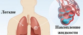
Fluid has accumulated in the ear: what to do?
First of all, the patient needs qualified diagnostics, which can be performed in a specialized clinic. This is especially important for young children because, as already mentioned, they may have no symptoms. To diagnose this disease, an otoscope is used - a special device for examining the middle ear, which allows you to make the most accurate diagnosis. The doctor visualizes the eardrum and can assess the level of fluid or the bubble in which it is accumulating.
Tympanometry is also often prescribed as an additional examination - a special “mirror” is placed in the ear, which measures the pressure inside the ear. Based on these studies, the doctor chooses a way to remove fluid from the ear.
Inner ear
, membranous labyrinth, the main part of the hearing organ and the organ of static sense in vertebrates and humans.
V. u. filled with fluid - endolymph and immersed in a cartilaginous or bone skeletal labyrinth. A slit-like cavity between the V. at. and the skeletal labyrinth is filled with perilymph; in terrestrial vertebrates, this cavity communicates with the lymphatic cavities of the head through the perilymphatic duct. In the skeletal labyrinth of terrestrial vertebrates, two openings, or windows, are formed. The oval window includes the base of the auditory ossicle (stapes) from the middle ear
. Below is a round window, covered with an elastic membrane that allows liquid to enter the V. move when the stirrup moves.
V. u. appears as a depression in the ectoderm at the back of the head. During the development of the embryo, the rudiment of V. takes the form of a vesicle, communicating with the external environment through a thin endolymphatic duct, and then completely separating from the ectoderm. Subsequently, the rudiment of V. differentiates into upper and lower sections, connected to each other. In the upper part of all vertebrates there are three semicircular canals.
(in cyclostomes - 1-2 canals);
At one end of each channel a swelling is formed - an ampoule. The rest of the upper section, connecting the semicircular canals with each other, is called the oval sac (utriculus). In the lower section of the V. a round sac (sacculus) is formed with a special swelling - lagena, or cochlea
.
Sensitive (receptor) epithelium of V. u. distributed unevenly and forms in both the oval and round sacs the so-called auditory spots (macula), the sensory cells of which are equipped with short hairs, and auditory (ampullary) ridges, protruding in the form of plates into the internal cavity of the ampullae of the semicircular canals; the sensory cells of the ridges are equipped with long hairs. In most vertebrates, the cochlea has a receptor apparatus in the form of a primary auditory papilla, which arises by separating a round sac from the auditory spot. In fish, amphibians and some other vertebrates, there is one small auditory spot near the junction of the oval and round sacs. In amphibians, the main auditory papilla is separated from the primary auditory papilla of the cochlea and the corresponding part of its wall forms the so-called main (basal) membrane. In reptiles, the projection of the sac is more developed; in crocodiles it turns into a long and somewhat curved canal of the cochlea; the development of their main membrane and the sensitive hair cells located on it leads to the division of the cochlear canal into the upper (scalena vestibule) and lower (scalena tympani) sections. As the receptive auditory apparatus becomes more complex, a covering plate develops above the main membrane and hair cells. Birds and monotreme mammals have a curved cochlear canal, separated from the round sac by a narrow canal. The hearing organs reach their highest degree of development in viviparous mammals and humans. The cochlear canal becomes even more elongated and bends in a spiral, forming 1.5-5 turns. The primary auditory papilla disappears, and the main one is transformed into the organ of Corti
.
The bases of receptor cells of all structures of V. at. contact with short processes (dendrites) of nerve cells, the bodies of which are grouped in the so-called cochlear ganglion, and long processes ( axons
) form the auditory nerve, which transmits excitation to the vestibular and auditory centers of the brain.
In the endolymph of V. u. There are calcareous deposits characteristic of the static sense organs - otoliths
(statoliths) of various sizes and often replaced by a mass of small grains - otoconia.
In cyclostomes, calcareous deposits V. u. appear in the protoplasmic reticulum in the form of otoconia, which can merge into the otolith. In most fish and all terrestrial vertebrates, large otoliths are placed in sacs, and small calcareous inclusions are often found in other parts of the ventricle. (for example, in the endolymphatic duct). Calcareous inclusions, cupules in the ampoules of the semicircular canals, together with accumulations of ciliated cells and endolymph that perceive their effects, form the structural and functional basis of the vestibular apparatus
.
Lit.:
Shimkevich V., Course of comparative anatomy of vertebrate animals, 3rd ed., M. - P., 1922; Shmalgauzen I.I., Fundamentals of comparative anatomy of vertebrate animals, 4th ed., M., 1947; Prosser L. and Brown F. Comparative physiology of animals, trans. from English, M., 1967; Kislyakov V. A. and Orlov I. V., Physiology of the vestibular system (current state of the problem), in: Issues of physiology of sensory systems, [in 1], M. - L., 1966.
G. N. Simkin.
Table of contents
3. Symptoms and diagnosis
The amount, color, clarity, smell, consistency of ear discharge, as well as accompanying symptoms, vary widely. So,
- the discharge of pus is inherent mainly in infectious and inflammatory processes of bacterial or fungal etiology;
- an admixture of blood in one concentration or another is characteristic of otorrhea due to trauma or cancer;
- clear flowing discharge is typical for allergic external otitis, exudative otitis media, traumatic liquorrhea (see above).
In most cases, otorrhea is accompanied by the following symptoms (single or in various combinations):
- decreased hearing acuity, feeling of congestion or a foreign body, perception of external sound as if through cotton wool or from under water;
- otalgia (pain syndrome localized in the external or internal structures of the ear);
- headache;
- vestibular disorders (dizziness, incoordination, loss of balance, etc.);
- various types of noise in the ear;
- rhinitis (runny nose), often with symptoms of sinusitis (inflammation of the paranasal sinuses);
- low-grade fever, less often high body temperature.
Diagnosis requires, first of all, the collection of complaints and anamnesis. Then an otoscopy is required, and in some cases a microotoscopy. Various laboratory tests are prescribed (to identify the pathogen in infectious and inflammatory processes, assess the intensity of the immune response, identify tumor markers, etc.). When examining for a head injury or ear injury, as well as for diagnosing bone abnormalities and tumor processes, CT, MRI, and radiography are used.
About our clinic Chistye Prudy metro station Medintercom page!
2. Reasons
The least dangerous cause can be considered abnormally liquid sulfur, as the secretion of the ceruminous glands of the external auditory canal is colloquially called. Due to individual characteristics and/or under the influence of certain irritating factors, the sulfur glands can produce protective “lubricant” in excess quantities and with atypical rheological properties. This situation, however, also requires mandatory consultation with an otolaryngologist.
Relatively rare is cholesteatoma, a complex encapsulated formation of dead cells, connective tissue and cholesterol; this is a pseudotumor, which, in the case of an associated infection, can lead to otorrhea (discharge from cholesteatoma, as a rule, has a specific putrefactive odor).
However, in the vast majority of cases, when examined for otorrhea, the causes are identified as inflammatory processes in various parts of the hearing organs, which are infectious (bacterial, fungal, viral, combined), allergic or mixed infectious-allergic in nature:
- otitis externa;
- acute otitis media;
- chronic purulent otitis media;
- exudative otitis media (accumulation of volumes of non-purulent fluid requiring release);
- mastoiditis (inflammation of the mastoid process of the temporal bone);
- furunculosis of the external auditory canal (a bacterial, usually staphylococcal infection of the hair follicles and/or sebaceous glands in the ear canal, which is often one of the first symptoms of diabetes).
The immediate cause of otorrhea in purulent and exudative “internal” otitis is perforation of the eardrum, which provides an outlet for the accumulated masses.
Relatively less common:
- traumatic otoliquorrhea (leakage of cerebrospinal fluid from the ear canal, usually due to a fracture of the base of the skull);
- otorrhea caused by a progressive tumor process.
Visit our Otolaryngology (ENT) page
Treatment
Treatment for Meniere's disease is as follows:
- Intravenous drips with 5-7% sodium bicarbonate, 100-150 ml. The speed should be quite high - about 120 drops per minute (the norm is 1 drop per 1 second.) The number of such droppers can vary from 1 to 15, depending on the severity of the condition and the stage of the disease.
- Taking B vitamins, as well as Aevita (vitamins A and E), Belloid (ergot alkaloids) and Bellataminal, which is a sedative.
- A 20 ml Glucose solution can be injected intravenously in a slow stream. The number of intravenous infusions will depend on the severity of the patient’s condition and the stage of the disease at which he presented.
- Since attacks of Meniere's disease are associated with dizziness and an unstable, shaky gait, during them it is necessary to maintain complete rest and inject a subcutaneous solution of atropine sulfate in the dosage prescribed by the ENT doctor.
- Reflexotherapy is also used to treat Meniere's disease: acupuncture massage, electro-apuncture, magnetic apuncture and other types of acupressure on certain areas of the human body.
- If drug treatment and reflexology do not bring the desired results (which happens only in rare cases), then treatment of Meniere's disease is carried out by surgical intervention in the labyrinth located in the inner ear of a person. It must be carried out in an ENT hospital, followed by a mandatory rehabilitation period.