Do you quickly feel short of breath? This is not normal for a healthy person. Maybe lung disease is behind this.
In this article you will learn about what types of lung diseases exist and how to deal with them.
Take a very deep breath, then exhale quickly. It was easy? Congratulations, your lungs are likely healthy.
Do you hear wheezing in your lungs or have you had a coughing fit? Perhaps your breathing was heavier than usual?
Then you should read this article carefully because lung disease is usually a gradual process that you often only notice when it's too late.
What are the symptoms of lung disease?
Scientists say the signs are the same for most lung diseases: cough, shortness of breath and expectoration. Don't worry, a cough or cold is not always a reason to panic.
However, if symptoms persist for a long time, become unusually severe, develop a fever, or even cough up blood, you should definitely see a doctor.
In most cases, you can get a lung test, which will tell you if you have lung problems. The pulmonologist can then make a more extensive diagnosis by checking your lung capacity.
Lung diseases: symptoms and signs
If you experience even minor symptoms, you should visit your doctor, as early detection can improve treatment outcomes and potentially save lives. Here are some warning signs of lung disease to watch out for:
- Coughing. As a rule, a normal cough due to a cold gradually disappears within a maximum of 2 weeks. But if the cough symptoms are persistent and there are no signs of their severity decreasing, you should consult a pulmonologist. In more serious cases, the cough may be accompanied by bloody sputum. This indicates damage to the airways, which can be caused by the patient's geographical location, medications, the cough itself, or lung cancer. Consideration should be given to whether such problems arose after the person encountered a potential trigger in everyday life. For example, if he has been spending more time with smokers lately. Informing your doctor about any lifestyle changes will be helpful in identifying the source of the problem;
- Labored breathing. If sudden shortness of breath is not due to obesity or heart problems, it may indicate asthma or COPD. According to experts, this can also be an early sign of lung collapse, airway obstruction, pneumonia, the presence of a blood clot in the pulmonary artery, etc. If your breathing sounds unusual, resembling a wheezing noise (a high-pitched whistling sound), there may be inflammation, a tumor, or even a foreign object blocking the airway;
- Pain in different parts of the body. Are you constantly feeling tired? Close attention should be paid if it does not go away after taking some measures and is accompanied by pain. For example, pulmonary fibrosis can affect body tissue, leading to muscle and joint pain. Lung cancer is associated with back pain, which worsens with deep inspiration and occurs even at rest. This pain often spreads to other areas of the body and affects the chest and shoulder areas. When lung cancer is the cause of chest pain, discomfort may result from enlarged lymph nodes or metastases in the chest wall, pleura, or lungs.
Smoking:
“In our practice, the most common cause of lung disease is smoking,” the researchers say. Whether it's a cigarette, hookah or e-cigarette, many toxins from smoke enter the lungs and cause irreparable damage.
Burnt tobacco contains at least 100 substances that have been shown to be carcinogenic. Especially if you already have an affected lung, such as from asthma or just a cold, smoking can significantly complicate your symptoms or make your condition worse. Smoking a hookah is also very dangerous.
Infections:
If minor infections are not properly treated in time, they can become more serious illnesses. “If you do not treat a cold promptly, your weakened immune system becomes more vulnerable, which can lead to a chronic form of pneumonia,” warns a pulmonologist.
Although it is relatively rare, you should take every infection seriously and treat it completely. An annual flu shot and pneumococcal vaccine can help prevent the disease. This way you can quickly get rid of a cold.
What are the most common lung diseases?
Basically, the lungs consist of blood vessels and small sacs (alveoli), which allow gas exchange between carbon dioxide and oxygen, as well as air channels (bronchi). All these diseases can be acute or chronic.
At the beginning of the disease, the symptoms seem harmless, and you are unlikely to pay attention to it. But the longer you remain inactive, the more severe the disease becomes and can even be fatal. Therefore, you should know these 6 lung diseases:
Lung pain
Since the lungs do not have pain-sensing nerves, there is no pain in the lungs. Symptoms of pulmonary diseases are characterized by coughing and difficulty breathing. The only part of the lungs that can cause pain is the pleura, trachea and large bronchi. In the differential diagnostic plan, assessment of pain intensity , its localization, connection with breathing and cough, the appearance of shortness of breath, and the effectiveness of pain-relieving drug therapy . Intense pain in the lungs indicates an acute disease and, as a rule, it is combined with shortness of breath, intensifies with breathing, this is pleural pain. In acute tracheitis, the pain is localized behind the sternum and is intense, aggravated by coughing.
Chest pain accompanied by shortness of breath is a serious sign, especially if it is difficult to treat. You should always clarify the dependence of pain on the position of the patient’s body and the effect of movement on the intensity of pain. This pain occurs with the following diseases:
- intercostal neuralgia;
- pathologies of the thoracic spine;
- radiculitis;
- muscle diseases;
- pleurisy.
You should also remember about projected and radiated pain. Chest pain occurs with the following diseases:
- pericarditis;
- aortic diseases;
- pulmonary embolism, gastric and duodenal ulcers;
- pancreatitis;
- subphrenic abscess.
Pain behind the sternum, in the heart area, in the left half of the chest, between the shoulder blades, often radiating to the left arm, is characteristic of coronary heart disease.
Causes of pain in the lungs
Dry pleurisy develops during a tuberculous process in the lungs. Pleurisy also occurs with the following diseases:
- pneumonia;
- lung abscess;
- pulmonary infarction;
- tumors affecting the pleura;
- echinococcosis, with chest injuries;
- rheumatism;
- diffuse connective tissue diseases (collagenosis);
- uremia, blood diseases.
Chest pain is usually one-sided, stabbing, worsens with deep breathing, coughing, or changing body position. The most typical location is the lower and lateral parts of the chest. In the position on the affected side, the pain subsides, which is associated with a decrease in the movements of the pleura, so the patient instinctively takes this position.
Exudative pleurisy
In most cases, the cause of exudative pleurisy is tuberculosis, but it can also be a consequence of other diseases. At the beginning of pleural exudation, there is pain in the side, limited respiratory mobility of the affected side of the chest, pleural friction noise, and a dry, painful cough occurs. As the effusion accumulates, the pain in the side disappears, a feeling of heaviness appears, some bulging and smoothing of the intercostal spaces on the painful side appears. Lobar pneumonia is an acute infectious disease of the lungs, affecting one or more lobes. The most common pathogen is pneumococcus. The disease begins with the appearance of stabbing pain in the chest, accompanied by the following symptoms:
- a sharp rise in temperature;
- chills;
- increasing symptoms of intoxication;
- cheek hyperemia;
- herpes on the lips;
- shortness of breath, cyanosis.
A dry, painful cough is also characteristic, followed by a cough with the release of rusty sputum, a viscous consistency mixed with blood.
With spontaneous pneumothorax, air enters the pleural cavity until the pressure in it reaches atmospheric pressure or the lung collapses. Spontaneous pneumothorax can develop in the following diseases:
- chest injury;
- pulmonary tuberculosis;
- suppurative lung diseases;
- lung cancer;
- bullous emphysema;
- breakthrough of echinococcal bladder and air cyst.
There are open, closed and valve pneumothorax. With an open pneumothorax, the pleural cavity communicates with atmospheric air constantly, both during inhalation and exhalation. Closing the perforation leads to the establishment of negative pressure in the pleural cavity . The most dangerous is valvular pneumothorax, in which air enters the pleural cavity during inhalation, and during exhalation the hole closes and the air is retained. As air accumulates, the pressure in the pleural cavity increases, the mediastinum shifts to the healthy side, the lung on the affected side collapses, and hemodynamic disturbances occur.
Spontaneous pneumothorax occurs mainly in young men and is manifested by sudden sharp pain in the chest, aggravated by breathing, talking, and physical stress. The pain is usually long-lasting and is accompanied by the following symptoms:
- severe pallor of the skin;
- weakness;
- cold sweat with low rapid pulse;
- drop in blood pressure.
The patient is bothered by severe shortness of breath and dry cough. Tachycardia and cyanosis appear. Patients prefer to be in a sitting position. Respiratory movements of the chest are superficial. There is a lag in breathing and often an expansion of the corresponding half of the chest. There is no vocal tremor on the affected side.
With lung cancer, chest pain may occur late in the disease. Chest pain may occur late in the disease. Other clinical symptoms depend on the location of the tumor, its proximity to the bronchial tree, the rate of tumor growth and the involvement of nearby organs in the process. If the inflammatory perifocal process in lung cancer reaches the pleura, then hemorrhagic exudative pleurisy develops.
The nature of the pain is different: acute, stabbing, girdling, aggravated by coughing and breathing. The pain can cover a certain area or half of the chest, and it can radiate to the arms, neck, and abdomen. The pain becomes especially intense and excruciating when the tumor grows into the ribs or spine with compression of the nerve roots. There is a special form of lung cancer (Penkosta) with painful plexitis.
Pain may be in the spine and limbs, which is associated with tumor metastasis. In the late stage, the following symptoms characteristic of the development of the disease appear:
- dyspnea;
- cough;
- hemoptysis;
- fever;
- increasing weakness;
- weight loss.
The patient's condition is extremely serious. Decreased breathing is heard with a large number of moist fine rales.
For pain of bone origin, the following pathological processes may be the cause:
- injury;
- inflammatory diseases:
- osteomyelitis of the ribs and sternum;
- swelling;
- hyperemia;
- fluctuation.
Palpation of the ribs is sharply painful. Fistulas may form. As a rule, the process is accompanied by an increase in temperature and signs of intoxication.
Tuberculosis of the ribs (the result of hematogenous dissemination), which is characterized by a more sluggish course, typically the formation of a cold abscess, and subsequently a fistula.
Actinomycosis of the ribs , developing as a complication of actinomycosis of the lungs; characterized by a hard deep infiltrate, fistulas, pus on the skin surface.
Pain appears only when the tumor is significant. Chondrosarcoma is usually localized in the posterior rib. Rarer forms include myeloma, Ewing endothelioma, fibrosarcoma, and neurosarcoma.
Pain in the lungs is also affected by metastatic bone damage and metastases of the lungs , breast, and prostate gland. In addition, pain in the lungs is caused by:
- tumor-like processes;
- dystrophic processes;
- osteoporosis;
- osteomalacia of bones.
The above diseases are most often observed in endocrine diseases and as a complication of corticosteroid therapy.
arthritis of a rheumatic and infectious-allergic nature can be causes of chest pain More common are arthritis of infectious-metastatic origin, developing with osteomyelitis, tuberculosis, actinomycosis and syphilis. The following symptoms are characteristic:
- hyperemia;
- swelling of the joints;
- severe pain on movement and palpation of the joints;
- arthrosis;
- joint tumors.
The cause of pain of muscle origin is often myositis and the following diseases:
- acute infections (influenza, gonorrhea, typhoid fever);
- chronic infections (tuberculosis, syphilis);
- metabolic diseases (diabetes mellitus, gout);
- muscle fatigue, injury.
Characteristic symptoms are swelling, induration, and soreness of the affected muscle during movement and palpation. Pain in the left side of the chest may occur with excessive hypertrophy of the anterior scalene muscle.
Gas pain
The large intestine reaches the level of the chest. Passing through the upper part of the abdominal cavity behind the stomach, it reaches the upper point behind the lower left part of the chest. This turn is located behind the spleen, which is why it is called the splenic flexure of the large intestine. Gases tend to rise upward, so they accumulate at this top point.
Compressed by the powerful muscles of the colon, the trapped gases cause severe pain; this happens so often that the phenomenon has received its own name - air (gas) accumulation syndrome in the splenic flexure. It happened that people with this syndrome were brought to hospitals with suspected heart attacks or kidney stones.
After finding out the true cause of the pain, patients experienced great moral relief, while doctors who made an incorrect diagnosis felt just as much embarrassment and shame. Pain from gas accumulation syndrome in the splenic flexure of the large intestine manifests itself to the left of the sternum or in the left upper part of the abdominal cavity, and an experienced doctor in this case will immediately make the correct diagnosis. But do not self-diagnose if the pain is severe and acute.
Asthma
During an asthma attack, your bronchi suddenly constrict and you can't breathe. This happens most often at night, or when you are under stress, also during physical activity.
In case of an acute attack of asthma, an inhaler helps, thanks to the spray of which the bronchi expand again and you get air.
If you suffer from asthma, you need to always breathe clean air. Experts recommend strengthening your lungs with moderate physical activity, not smoking, and not living in big cities.
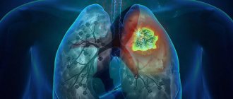
Lungs' cancer
Lung cancer is the third most common cancer in the world, and it is almost always malignant. Today, smoking (both active and passive) is the most common cause of lung cancer.
Toxic substances can even end up on clothing or hair and then end up in the air we breathe. You should also be especially vigilant if you have a history of lung cancer in your family, because then your own body is at greater risk.
When you find out you have cancer, it is often too late. Chemotherapy or radiation therapy can only extend your lifespan. Therefore, the most effective method of combating lung cancer is prevention, and most importantly, quit smoking.
Bronchitis
Acute bronchitis is usually caused by infectious viruses or, less commonly, bacteria, which excite the bronchi and cause the typical cough. This may continue for several weeks until the tissue is completely restored.
If your cough doesn't go away, you may have chronic bronchitis. This can happen if you do not treat an acute infection properly. According to experts, the risk of the disease becoming chronic also increases if the bronchi are exposed to multiple pollutants. Thus, hand washing actually protects against infection.
What can help? You must definitely cure acute bronchitis, get plenty of rest, eat plenty of vitamins and drink as much fluid as possible. You usually don't need antibiotics unless you have bacteria in your lungs in addition to a viral infection.
For chronic bronchitis, your doctor may prescribe a cough suppressant or short-term, low-dose inhaled cortisone therapy to help the inflammation go away. However, it is important that you protect your lungs from environmental toxins and allow them to heal fully.
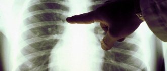
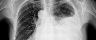
Otherwise, there is a risk that the disease will become acute chronic. Only an x-ray can provide a reliable diagnosis of lung diseases. Pneumonia can definitely be seen on an x-ray.
Symptoms of a tumor in the lungs - stages and treatment
Cough
A cough stimulates the sensitive nerve endings in the lining of the respiratory tract anywhere from the vocal cords to the bronchi.
Coughing is a reflex action, although it can be initiated voluntarily. When coughing, the pressure in the chest increases significantly and the sudden release of air during the cough reflex serves to clear the airways and to free the lining of the airways from mucous secretions. An irritant that affects the mucous membrane of the airways affects the nerve endings, which leads to coughing. However, the cough cannot remove the primary source of irritation - cancer, which leads to persistent coughing phenomena. Any bleeding intensely irritates the lining of the bronchi and causes hemoptysis.
Coughing attacks occur when irritation intensifies and does not go away. These attacks can be so severe that they can lead to broken ribs and also damage the small blood vessels of the lungs. Cough caused by lung cancer is not specific, but most often it occurs at night or immediately after waking up. Over time, the cough becomes periodic and paroxysmal throughout the day. Physical activity or increased breathing often leads to coughing attacks.
Symptoms of a tumor in the lungs in the early stages of the disease are characterized by a dry cough, which is subsequently joined by whitish sputum. As infection develops, the sputum becomes purulent in nature with a greenish or yellowish tint and the possible presence of hemoptysis. Hemoptysis is usually accompanied by the presence of blood streaks in the sputum. However, when a small vein is damaged, fresh blood is coughed up. In these rare cases, severe bleeding may occur. Hemoptysis is a very important symptom of a lung tumor and should not be ignored. In about a quarter of people over 40 years of age, hemoptysis is a sign of lung cancer.
Discomfort and pain
Discomfort and pain are the second most common among the initial manifestations of tumor symptoms in the lungs. They occur in approximately 20% of patients. The nature of pain and discomfort in the chest can vary greatly. The most common descriptions of symptoms include a feeling of fullness and tightness, which intensifies in certain positions, as well as with deep breathing or coughing.
Sometimes there is sharp, localized chest pain when breathing or coughing. This is called pleural pain, which is caused by inflammation in the lining of the lungs (pleura). Tumors located high in the upper lungs can affect the large nerve roots (brachial nerve cluster), which can lead to increasing pain in the upper chest and shoulder.
Infections and airway deterioration
The growth of a tumor in larger parts of the airways of the lungs can gradually lead to disruption of their patency. This may prevent the normal passage of mucus from the lungs. As a result, unresolved sputum can become a source of infection. In such cases, symptoms of a tumor in the lungs include general malaise, chills, fever, night sweats, and loss of appetite and weight. There may be a poor response to treatment with standard antibiotics.
If complete airway obstruction is present, this may result in shortness of breath, cough and fever. The nature of symptoms and their degree depend on the extent of lung damage. With incomplete obstruction, wheezing develops caused by the passage of air through the blockage. They increase with deep breathing, for example, after exercise.
Dyspnea
Symptoms of a malignant lung tumor are rarely characterized by shortness of breath as the first manifestation of this disease. However, this condition often develops over the course of the disease. In lung cancer, the causes of shortness of breath may be:
- asthma-like attacks caused by spasm of the airways;
- lung collapse caused by obstruction of a large airway;
- pulmonary infection;
- pleural exudate (fluid accumulation);
- superior vena cava syndrome (compression of the main veins surrounding the heart);
- pericardial effusion (a buildup of fluid around the heart that limits the organ's ability to function normally).
Hoarseness
One of the nerves leading to the voice box curves down to the base of the left lung. A tumor growing in this part of the chest can damage this nerve, which can negatively affect the functioning of the vocal cords. Sometimes this leads to a weak and hoarse voice, as well as the development of a characteristic cough with a metallic tint.
Chronic obstructive pulmonary disease (COPD)
Chronic obstructive pulmonary disease is characterized by constant and irreversible destruction of lung tissue. COPD often occurs due to bronchitis.
Smoking is by far the most common cause of this disease, with about 20 percent of smokers developing COPD. Exhalation is especially difficult for patients, because the cells of the bronchi are destroyed after exhalation.
What can help? The effects of COPD can only be reversed to a small extent. The sooner you learn about the disease, the sooner you can limit its further development. In addition, medications can relieve symptoms such as shortness of breath or coughing spells. You must avoid environmental toxins, smoking and infections, otherwise your quality of life and longevity will decline rapidly!
Chronic nonspecific lung diseases (CNLD)
Chronical bronchitis
Like other chronic nonspecific lung diseases, it is often a consequence of a protracted course of acute bronchitis of viral etiology (developed against the background of influenza, measles, adenovirus or RS infection) or bacterial origin (caused by long-term persistence in the bronchi of Haemophilus influenzae, pneumococcus, etc.).
It can develop as a result of prolonged exposure to the airways of chemical and physical factors (smoking, dusty air, pollution from industrial waste). The prevalence can be local or diffuse; by type of inflammation - catarrhal or mucopurulent; by the presence/absence of bronchial obstruction - obstructive and non-obstructive; according to the nature of morphological changes in the bronchi - atrophic, polypous, deforming. Clinical criteria for chronic bronchitis are 2-3 exacerbations of the inflammatory process per year for 2 years with an annual duration of at least 3 months. Patients are bothered by a constant cough with phlegm. During exacerbations, the cough intensifies, the sputum becomes purulent, low-grade fever and sweating occur. The outcomes and complications of chronic bronchitis can include chronic pneumonia, pulmonary atelectasis, emphysema, and pneumofibrosis.
Bronchial asthma
It is the second most common form of chronic nonspecific lung diseases. It is characterized by hyperreactivity of the bronchial tree, leading to hypersecretion of bronchial mucus, edema and paroxysmal spasm of the airways. The main clinical types include nonatopic, atopic, mixed, aspirin-induced, and occupational BA.
Clinically, asthma of any origin is manifested by repeated attacks of expiratory dyspnea. There are 3 periods in their development: precursors, suffocation and reverse development. Harbingers signaling an approaching attack of asthma can include coughing, mucous discharge from the nose, conjunctivitis, and restlessness. During an attack of suffocation, wheezing, sudden shortness of breath with prolonged exhalation, diffuse cyanosis, and nonproductive cough appear. Patients take a forced vertical position with a raised shoulder girdle. In a severe attack, the patient's death may occur from respiratory failure. During the period of reverse development of the attack, sputum begins to separate when coughing, the amount of wheezing decreases, breathing becomes free, and shortness of breath disappears.
Between attacks, the condition of patients with asthma is quite satisfactory. With a long history of chronic nonspecific lung disease, obstructive emphysema, cor pulmonale, and pulmonary heart failure develop.
Chronic obstructive pulmonary emphysema
It is a chronic nonspecific lung disease, the morphological basis of which is a persistent expansion of the lumen of the respiratory bronchioles and alveoli as a result of chronic obstruction of the airways against the background of chronic bronchitis and bronchiolitis obliterans. The lungs acquire increased airiness, become overinflated, and increased in size.
Clinical manifestations of emphysema are caused by a sharp reduction in the area of gas exchange and impaired pulmonary ventilation. Symptoms increase gradually as pathological changes spread over a large area of lung tissue. Worrying about progressive shortness of breath, cough with scanty mucous sputum, weight loss. Note the barrel-shaped expansion of the chest, bluishness of the skin, thickening of the nail phalanges of the fingers like drumsticks. With emphysema, infectious complications, pulmonary hemorrhages, and pneumothorax are common. The cause of death is severe respiratory failure.
Bronchiectasis
The morphological substrate of this form of chronic nonspecific lung diseases is saccular, cylindrical or spindle-shaped expansion of the bronchi. Bronchiectasis can be local or diffuse, congenital or acquired in origin. Congenital bronchiectasis is caused by disturbances in the development of the bronchial tree in the prenatal and postnatal periods (as a result of intrauterine infections, Sievert-Kartagener syndrome, cystic fibrosis, etc.). Acquired bronchiectasis can develop against the background of recurrent bronchopneumonia, chronic bronchitis, or prolonged presence of a foreign body in the bronchi.
Main respiratory symptoms include a persistent cough, yellow-green, odor-bearing sputum, and sometimes hemoptysis. Exacerbations occur like exacerbations of chronic purulent bronchitis. The extrapulmonary symptom complex in bronchiectasis is represented by deformation of fingers in the form of drumsticks and nails in the shape of watch glasses, and “warm” cyanosis. Complications of chronic nonspecific lung disease can include pulmonary hemorrhage, lung abscess, cardiopulmonary failure, amyloidosis, purulent meningitis, and sepsis. Each of these conditions poses a potential threat to the life of a patient with COPD.
Chronic pneumonia
Pathomorphological changes in chronic pneumonia combine an inflammatory component, carnification, chronic bronchitis, bronchiectasis, chronic abscesses, pulmonary fibrosis, therefore, at present, this chronic nonspecific lung disease as an independent nosology is not recognized by all authors. Each exacerbation of chronic pneumonia leads to the appearance of new foci of inflammation in the lung tissue and an increase in the area of sclerotic changes.
Constant symptoms accompanying the course of chronic pneumonia include cough with sputum (mucopurulent in the remission phase and purulent in the acute phase) and persistent wheezing in the lungs. In the acute period, body temperature rises, chest pain occurs in the projection of the infiltrate, and respiratory failure occurs. The disease can be complicated by pulmonary heart failure, abscess formation, pleural empyema, pulmonary gangrene, etc.
Pneumosclerosis
A chronic nonspecific lung disease that occurs with the replacement of functioning parenchyma by connective tissue is called pneumosclerosis. It is a consequence of inflammatory-dystrophic processes, leading to wrinkling, airlessness and compaction of the lung tissue. Often develops as a result of chronic bronchitis, EBD, chronic pneumonia, COPD, pneumoconicosis, pleurisy, fibrosing alveolitis, tuberculosis and many others. etc. Based on the prevalence of changes, local (focal) and diffuse pneumosclerosis are distinguished. According to the severity of connective tissue proliferation, three stages of the pathological process are distinguished - pneumofibrosis, pneumosclerosis, pneumocirrhosis.
It manifests itself both as signs of a causally significant disease and as signs of respiratory failure (shortness of breath, cough, bluish discoloration of the skin, “Hippocratic fingers”). In the stage of lung cirrhosis, the deformation of the chest is sharply expressed, and atrophy of the pectoral muscles is noted. The patient is weakened, gets tired quickly, and loses weight. The course of the underlying disease leads to the progression of pneumosclerosis, and pneumosclerosis aggravates the underlying pathology.
Pneumonia
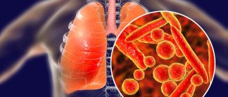
In the case of pneumonia, it is not the airways or bronchi that are affected, but the lung tissue itself. Small bubbles in the lungs become inflamed and stick together, preventing oxygen from reaching the blood. Like bronchitis, pneumonia is caused by contagious germs in the air we breathe. When a doctor listens to a patient's lungs, he hears them cracking when he breathes because the nodes are coming off.
In this case, you need to act immediately because pneumonia is number 1 among deadly infectious diseases. How to fight pneumonia? If you act immediately, your chances of recovery are high. Most germs that cause pneumonia are easy to treat. It's even better if there is no infection at all. Therefore, strengthen your immune system through exercise and a balanced diet.
You can also protect yourself with vaccinations, since pneumococcus and influenza are the leading causes of pneumonia. Modern medicine has vaccines against both.
Pulmonologist – who is he and what diseases does he treat?
Table of contents
- What diseases does it treat?
- In what cases should you contact this specialist?
- How to make a diagnosis
- Treatment methods
- Prevention
- Advantages of MEDSI
Pulmonology is a field of medicine that studies diseases of the human respiratory system, their diagnosis and treatment.
It was first identified as an independent industry only in the second half of the last century. Until this time, science had been accumulating and expanding knowledge about the human respiratory organs, the patterns and characteristics of diseases affecting this area. A pulmonologist is a specialist who has all the knowledge and achievements obtained by medical science today. Call now (495) 7 800 500
Leave a request
You will receive an automatic call back, wait for the operator to respond.
* By clicking the “Order a call” button, you agree to the processing of your personal data by JSC Group
Policy regarding the processing of personal data Consent to the processing of personal data
Make an appointment
The human respiratory system means:
- Respiratory tract (upper (nose, pharynx) and lower (larynx, trachea, bronchi, right and left lungs))
- Pleura (visceral membrane lining the inside of the chest cavity and the outer surface of the lungs)
- Muscles involved in the act of breathing
- The respiratory center in the medulla oblongata, which is responsible for timely exhalation and inhalation
A pulmonologist is a doctor who studies and treats diseases of all parts of the respiratory system, considering them in close connection with the entire body. Some respiratory diseases are treated by a pulmonologist together with doctors of other specialties:
A pulmonologist is a doctor who has all the knowledge and achievements obtained by medical science today.
- Surgical interventions on the chest organs are performed by a thoracic surgeon
- Threatening conditions associated with acute respiratory failure are treated by a resuscitator
- Pulmonary tuberculosis is treated by a TB doctor in a specialized clinic
What diseases does it treat?
The doctor’s competence extends to a fairly wide range of diseases, from acute respiratory infections to cancer and congenital diseases, however, in some cases, assistance will be limited only to consultation with a pulmonologist and diagnosis of the disease; for further treatment, the patient is transferred to specialized specialists.
A pulmonologist treats:
- Chronic and acute (simple and obstructive) bronchitis
- Pneumonia (lung inflammation)
- Alveolitis (inflammation of the alveoli - the smallest structural units of the lung)
- Pleurisy (inflammation of the membranes of the lung)
- COPD (chronic obstructive pulmonary disease)
- Bronchial asthma
- Allergic and occupational diseases (including pneumoconiosis, silicosis)
- Sarcoidosis, cystic fibrosis and other fibrosis (connective tissue growths)
Can diagnose, but does not cure:
- Tuberculosis (it is treated by a phthisiatrician)
- Diseases of the upper respiratory tract (they are treated by otolaryngologists)
- Lung cancer (treated comprehensively with the participation of a pulmonologist, oncologist, surgeon, chemotherapy)
- Emphysema
The doctor’s competence extends to a fairly wide range of diseases
: from chronic rhinitis (runny nose) to lung cancer.
In what cases should you contact this specialist?
Usually, a therapist (or general practitioner) refers to a pulmonologist, since it is quite difficult for a patient without a medical education to independently determine the disease due to the similarity of symptoms: a cough can be accompanied by influenza, pharyngitis and laryngitis (inflammation of the larynx), which do not require consultation with a pulmonologist.
You are referred to a pulmonologist if you have the following symptoms, one way or another related to the cough reflex:
Cough may be accompanied by influenza, pharyngitis and laryngitis (inflammation of the larynx), which do not require consultation with a pulmonologist
.
- A hacking, nonproductive cough accompanied by symptoms of respiratory failure
- Persistent cough with or without sputum production
- Changes in the nature of sputum: the appearance of pus, blood, drops of blood when coughing, discharge of pink foam
- Acute chest pain, aggravated by breathing and accompanied by increasing respiratory failure
- An increase in temperature accompanied by a cough, the appearance of shortness of breath, weakness, sweating and other signs of intoxication
- Choking, worsening cough in the evening/night
- The appearance of spasms, difficulty breathing under certain conditions (odors, dust pollution, changes in air temperature)
- Constant irritation of the upper respiratory tract, prolonged profuse runny nose with clear discharge, laryngospasms (breathlessness)
How to make a diagnosis
A pulmonologist has in his arsenal a wide range of diagnostic methods, which include analysis of biological fluids (blood, sputum) for the presence of bacteria and markers of inflammation and visual hardware studies. Among them:
- Radiography and fluoroscopy are two methods of radiological diagnostics. As a result of the first, a static picture (photograph) of the lungs is obtained, in the second case, the organ is illuminated for some time, and the result is broadcast on the screen in real time, making it possible to evaluate the respiratory function and the distribution of contrast in the vessels
- Bronchoscopy is an endoscopic visualization that allows you to examine the bronchial mucosa from the inside, take material for research, and administer a drug.
- Computed tomography is a cross-sectional image of organ tissue in order to search for neoplasms (abscesses, fibrous cysts, cavities, foci of necrosis or purulent melting, tumors, etc.)
- Functional diagnostics: peak flowmetry (assessment of expiratory speed), spirometry (assessment of breathing volume and speed), spirography and others
Call now
(495) 7 800 500
Leave a request
You will receive an automatic call back, wait for the operator to respond.
* By clicking the “Order a call” button, you agree to the processing of your personal data by JSC Group
Policy regarding the processing of personal data Consent to the processing of personal data
Make an appointment
Biomaterial research:
- A clinical blood test shows the presence of inflammation (with an increase in the erythrocyte sedimentation rate - ESR), the acute or chronic phase of the process (according to the leukocyte formula), the presence of allergic reactions (with an increase in the number of eosinophils), general weakness of the body (decreased hemoglobin or red blood cells)
- Sputum culture will help isolate pathogenic microflora and identify the causative agent of the disease in order to select the most effective antibiotic
- Allergy tests will establish the range of allergens that provoke deterioration
- Genetic diagnostics will help identify mutations characteristic, for example, of cystic fibrosis
a wide range of diagnostic methods in his arsenal
.
Treatment methods
Based on the results of the examination, the pulmonologist can prescribe radical (surgical) or conservative (drug, hardware) treatment, adherence to a certain regimen and diet, and can refer for further examination to other specialists if the problem is beyond his competence (for example, if a tumor is detected or suspected tuberculosis).
Treatment with medications can also be very varied and, depending on the results of studies and clinical symptoms, include:
- Antibacterial therapy (for bronchitis and pneumonia of bacterial etiology)
- Anti-inflammatory therapy using NSAIDs and steroids (for severe swelling)
- Antitussives (suppressing the cough reflex) or, conversely, expectorants (increasing bronchial secretion)
- Drugs that eliminate bronchospasm and dilate the bronchi: adrenomimetics, anticholinergics, antispasmodics
- Antiseptics can be administered endoscopically or using pleural puncture (for pleurisy, pneumothorax)
Treatment with medications
can be very diverse.
Hardware and instrumental treatment may include:
- Electrophoresis with various medicinal solutions
- Warming up
- Bronchoscopy with the introduction of antiseptics
- Pleural puncture (performed by a thoracic surgeon) with pumping out fluid or pus, washing the cavity with antiseptic solutions, intracavitary administration of antibiotics
Any prescription is made by a doctor after carefully studying each case of the disease. Many procedures are performed only in a hospital, and some diseases require long-term hospitalization.
Prevention
Consultation with a pulmonologist can also be preventive. This is how chronic patients regularly go to see a pulmonologist. The purpose of these visits is to assess the current state of the body, prolong the period of remission (absence of symptoms), and mitigate the symptoms of future relapses (exacerbations).
To avoid bronchopulmonary diseases, your doctor may advise you to harden yourself and lead an active lifestyle.
To avoid bronchopulmonary diseases, the doctor may advise all patients without exception:
- No smoking
- Protect yourself from infections (vaccination and personal hygiene will help)
- Avoid potential allergens
- Eliminate the influence of damaging factors (when working in production with high levels of dust or toxic substances, carefully follow safety precautions and use protective equipment)
- Eat well
- Train the respiratory system (through breathing exercises, singing, playing wind instruments)
- Temper yourself and lead an active lifestyle
Advantages of MEDSI:
Leading pulmonologists in Moscow are world-class doctors
with vast experience and experience.
- The ability to make an appointment with a pulmonologist at any time convenient for you or to see a specialist without an appointment for acute complaints
- Availability of a clinic in your area (wide network of branches)
- Availability of all hardware tests and therapeutic procedures
- Own clinical and bacteriological laboratories
- Feedback from the patient – adjustment of prescriptions and assessment of their effectiveness during treatment
- Possibility of staying in the hospital of our clinic during the entire course of treatment
- Leading pulmonologists in Moscow are world-class doctors with extensive experience and experience
It’s easy to sign up for a consultation – just call 8 (495) 7-800-500 (calls are accepted 24 hours a day).