Interstitial lung diseases (ILD) are various lesions of the pulmonary system, the pathomorphological basis of which is chronic inflammation of the alveoli, small bronchi, and capillaries of the lungs, leading to fibrosis. ILD includes more than a hundred diseases with clear and unclear etiology.
In patients who are hospitalized in the pulmonology department of the Yusupov Hospital, certain interstitial lung diseases are diagnosed in 20% of cases. The disease is more common in men under the age of 70 who have been smoking cigarettes for many years. Some diseases in this group have a reversible course and a relatively favorable prognosis, while others lead to early disability and even death.
Pulmonologists at the Yusupov Hospital have extensive experience in diagnosing and treating interstitial lung diseases. Patients are examined using modern devices from leading global manufacturers. The hospital cooperates with leading medical universities in Moscow and specialized pulmonology centers. For this reason, patients have the opportunity to undergo complex examination and treatment. The Yusupov Hospital has all the conditions for a comfortable stay for patients: cozy rooms, polite medical staff, competent doctors.
Classification of IBL
Currently, the classification developed in 2002 by the American Thoracic Society (ATS) and the European Respiratory Society (ERS) is adopted as the basis. According to this classification, there are interstitial lung diseases. With established etiology:
- Radiation, medicinal, toxic;
- Pneumomycoses associated with HIV infections
- Interstitial lung diseases against the background of collagenosis and pneumoconiosis;
- Interstitial lung diseases due to infections;
- Interstitial lung diseases against the background of allergic alveolitis.
Idiopathic interstitial pneumonia:
- Nonspecific, lymphoid, acute, desquamative, cryptogenic organizing;
- Idiopathic pulmonary fibrosis.
- Granulomatous: interstitial diseases against the background of sarcoidosis, exogenous allergic alveolitis, hemosiderosis.
Associated with other diseases:
- Pathology of the liver, intestines, kidneys;
- Hereditary diseases;
- Malignant neoplasms.
Other: associated with primary pulmonary amyloidosis, pulmonary proteinosis.
Pathologists distinguish the following types of fibrosis in interstitial lung diseases:
- Simple;
- Desquamative;
- Lymphocytic;
- Bronchiolitis obliterans with pneumonia;
- Giant cell.
Make an appointment
Idiopathic interstitial pneumonia
The group of interstitial lung diseases (ILDs) includes several dozen separate nosological forms that differ in etiology, features of pathogenesis and morphological picture, having different clinics and prognosis. Terminological and classification approaches to these diseases have changed repeatedly, further complicating the already difficult diagnostic work with this category of patients. Even today, despite the existence of a generally accepted classification of ILD, the term “interstitial pneumonia” is associated by the doctor with a viral infection rather than with a disease that requires mandatory morphological verification and has a rather serious prognosis [1].
Let us recall the basic definitions.
Pneumonia is a group of acute infectious (mainly bacterial) diseases characterized by focal damage to the respiratory parts of the lungs with the obligatory presence of intra-alveolar exudation. The category “pneumonia” excludes diseases caused by physical (radiation pneumonitis) or chemical (“gasoline” pneumonia) factors, as well as those having an allergic (“eosinophilic pneumonia”) or vascular (pulmonary infarction due to pulmonary embolism) origin. Inflammatory processes in the lungs with measles, rubella, influenza, etc. are considered not under the heading “pneumonia”, but within the framework of the corresponding nosological forms [2].
Pneumonitis (alveolitis) is an inflammatory process, often of an immune, non-infectious nature, affecting predominantly the parenchymal interstitium (alveolar walls) and extra-alveolar connective tissue of the lungs without obligatory intra-alveolar exudation. A number of authors distinguish between the concepts of “pneumonitis” and “alveolitis”, suggesting that in alveolitis the inflammatory process is localized mainly in the alveoli, and in pneumonitis the inflammation also affects other structures of the lung parenchyma, however, such a division has no practical significance, and the terms are often used as synonyms [ 3].
The term “pneumonitis” does not reflect a specific nosological form, but rather the characteristics of the pathological process. Damage to the lungs like pneumonitis (alveolitis) can develop in a variety of diseases: idiopathic interstitial pneumonia, drug-induced lung lesions, systemic connective tissue diseases, hypersensitivity pneumonitis, sarcoidosis, etc. It should be noted that in each of these diseases pneumonitis is mandatory, but far from not the only manifestation of lung damage. The causes, clinical manifestations, treatment directions and prognosis for these diseases are different, therefore, when identifying signs of pneumonitis, morphological verification and further nosological diagnosis are of such great importance.
From a modern point of view, ILDs represent a heterogeneous group of diseases, the common features of which are damage to the interstitial tissue of the lungs such as productive pneumonitis with the subsequent formation of fibrosis, progressive dyspnea on exertion, nonproductive cough, crepitus, diffuse changes in radiography and computed tomography of the lungs, restrictive ventilation disorders, decreased diffusion capacity of the lungs and increasing respiratory failure [4].
Currently, in most countries, including Russia, the classification of ILD adopted by the Consensus Commission of the American Thoracic Society and the European Respiratory Society (ATS/ERS, 2002) is used [5]. According to this classification, four groups of ILD are distinguished: ILD of known etiology, granulomatosis, idiopathic interstitial pneumonia, and other ILD (Fig.).
Idiopathic interstitial pneumonia (IIP) is a typical representative of the group of ILD of unknown etiology, having many similar clinical, radiological and functional signs, but a fundamentally different morphological picture, which determines the clinical features, response to therapy and prognosis. The principle of constructing the clinicopathological ATS/ERS classification is that each clinical form of IIP corresponds to a certain histological variant of IIP (Table 1).
Clinical features of patients with various types of IIP are shown in Table. 2.
Idiopathic pulmonary fibrosis
Idiopathic pulmonary fibrosis (IPF) is one of the most common diseases from the IIP group. A synonym for IPF is “idiopathic fibrosing alveolitis,” a term traditionally used in our country.
The disease most often occurs in patients over 50 years of age. The main complaints of patients are increasing shortness of breath and nonproductive cough. The onset of the disease is usually imperceptible, the disease progresses quite slowly, patients manage to adapt to their shortness of breath and at the time of presentation have a history of the disease lasting up to 1–3 years. Fever and hemoptysis are not typical for patients with IPF. Other symptoms may include general weakness, arthralgia, myalgia, and changes in the nail phalanges in the form of “drumsticks.” A typical auscultatory phenomenon in IPF is inspiratory crepitus, which has been compared to the “crackling of cellophane.” As the disease progresses, signs of respiratory failure and cor pulmonale appear, as well as weight loss and cachexia. Laboratory findings are nonspecific. IPF is a restrictive pulmonary disease; therefore, the characteristic functional features of the disease are a decrease in static lung volumes, detected by body plethysmography. One of the early signs of the disease is a decrease in DLCO. Spirometric FEV1/FVC is within normal limits or elevated.
The most common radiographic signs of IPF are bilateral reticular changes, more pronounced in the lower parts of the lungs. In the early stages of the development of the disease, only a slight decrease in the volume of the lung fields and a decrease in the transparency of the lungs like “ground glass” may be observed. As the disease progresses, the reticular pattern becomes coarser and heavier, and round cystic clearings appear, reflecting the formation of a “honeycomb lung.” To clarify the X-ray picture, it is advisable to conduct a multispiral computed tomography of the chest organs.
Since the diagnostic capabilities of IIP are limited, and examination data are not always specific, the “gold” diagnostic standard for all IIP is a lung biopsy: open or thoracoscopic. A special need for a biopsy arises in cases where there is a not quite typical clinical and/or radiological picture, the patient’s age is less than 50 years, the presence of systemic signs of the disease, or rapid progression of the disease. A necessary condition is the predominance of the benefits of making a correct diagnosis over the risk of surgical manipulation.
There is a diagnostic approach that makes it possible to establish a diagnosis of IPF with a high probability in cases where a biopsy is not possible. To do this, the patient must have four of the four major criteria and at least three of the four minor criteria.
Large criteria
- Exclusion of other ILDs due to known causes, such as drug injury, CTD, etc.
- Changes in respiratory function, including restrictive changes and impaired gas exchange.
- Bilateral reticular changes in the basal regions of the lungs with minimal ground-glass changes on high-resolution computed tomography.
- There are no findings on transbronchial biopsy or bronchoalveolar lavage that suggest an alternative diagnosis.
Small criteria
- Age over 50 years.
- Invisible, gradual onset of dyspnea during physical activity.
- The duration of the disease is more than 3 months.
- Inspiratory crepitus in the basal regions of the lungs.
Modern therapy for IPF is based mainly on anti-inflammatory therapy (corticosteroids and cytostatics (CS)), i.e., drugs that can affect the inflammatory and immunological components of the development of the disease. The basis for this approach is that chronic inflammation precedes and inevitably leads to fibrosis and that aggressive suppression of inflammation can block the subsequent formation of fibrotic changes.
Three modes of anti-inflammatory therapy are widely used: monotherapy with glucocorticosteroids (GCS), a combination of GCS with azathioprine, and a combination of GCS with Cyclophosphamide. ATS/ERS recommends combination regimens as preferable [6]. Therapy is carried out for at least 6 months. Careful monitoring of side effects of therapy is mandatory. When prescribing cytostatics, patient monitoring should include a complete blood count weekly for the first month, then once every 2–4 weeks; During treatment with Cyclophosphamide, a weekly urine test for hematuria is required.
If GCS monotherapy is chosen, the initial daily dose of prednisolone is 1 mg/kg of ideal weight per day (maximum up to 80 mg/day). After 4 weeks, the tolerability of such therapy is assessed. If there is an improvement or stabilization of functional indicators, then over the next 3 months the daily dose of prednisolone is reduced. If there is no response to steroids, azathioprine is added [7].
An alternative approach that focuses on reducing excess matrix deposition in the lungs or accelerating collagen breakdown is antifibrotic therapy. Antifibrotic drugs include D-penicillamine, colchicine, interferon gamma-1 b, and pirfenidone.
It has been proven that the effectiveness of therapy is increased when N-acetylcysteine is added to anti-inflammatory drugs at a dose of 600 mg 3 times a day. Currently, leading experts in the treatment of IPF prefer a regimen that includes prednisolone, azathioprine and N-acetylcysteine [8].
In addition to drug therapy, as with other lung diseases, oxygen therapy is used when hypoxemia develops. With the development of pulmonary hypertension, in addition to oxygen therapy, the use of vasodilators is possible. The development of infections of the tracheobronchial tree requires the use of antibacterial and antifungal drugs. Regular vaccination with influenza and pneumococcal vaccines is recommended for all patients with IPF.
Other idiopathic interstitial pneumonias (non-IPF)
Nonspecific interstitial pneumonia (NSIP), along with IPF, is one of the most common forms of IIP. NIP can be idiopathic; this form is included in the IIP group. However, a morphological picture corresponding to the NIP pattern also occurs in cases of lung damage in patients with CTD, hypersensitivity pneumonitis, radiation pneumonitis, etc.
Clinical, laboratory and functional indicators for NIP are nonspecific. Chest radiography most often reveals bilateral ground-glass changes and reticular changes in the lower lungs.
The prognosis of patients with NIP is more favorable than with IPF. The clinical course and survival of patients depend on the severity of pulmonary fibrosis. The ten-year survival rate for NIP is about 35%. Spontaneous cases of recovery without treatment in NIP are unknown; GCS therapy without or with the addition of cytostatics leads to improvement or stabilization in approximately 75% of patients [9].
Cryptogenic organizing pneumonia
Synonyms for cryptogenic organizing pneumonia (COP) are the terms “bronchiolitis obliterans with organizing pneumonia” and “proliferative bronchiolitis.” COP has clear clinical and morphological differences from “isolated” bronchiolitis obliterans: along with damage to the bronchioles, there is involvement of the alveoli in the inflammatory process with the presence of organized exudate in their lumen. COP in most cases is idiopathic, i.e. the cause remains unknown. Among the established causes, the most important are CTD (rheumatoid arthritis, etc.), complications of drug therapy (amiodarone, gold drugs, etc.).
The disease most often develops in people aged 50–60 years; men and women get sick equally often. COP is characterized by an acute or subacute course, the clinical picture often resembles bacterial pneumonia. The average duration of symptoms until diagnosis is 2–6 months. Routine laboratory tests reveal peripheral blood leukocytosis (50%), increased ESR and C-reactive protein (70–80%).
A typical radiological sign of COP is the presence of spotty, bilateral (less often unilateral) dense foci of consolidation of subpleural localization. In COP, migration of pulmonary infiltrates has been described, most often from the lower to the upper sections. The differential diagnosis of COP, in addition to bacterial pneumonia, is carried out with chronic eosinophilic pneumonia, bronchoalveolar cancer and pulmonary lymphoma.
Spontaneous improvement in COP has been described but is rare. The treatment of choice for COP is oral corticosteroids. Clinical improvement occurs within 1–3 days from the start of the first dose, radiological changes usually disappear after a few weeks, the total duration of GCS therapy ranges from 6 to 12 months. When the dose of GCS is reduced, relapses of the disease occur quite often; in this situation, the dose of steroids is increased again. The prognosis for COP is usually favorable; most patients are completely cured when taking GCS. However, in rare cases, there is a poor response to steroids and a steadily progressive course of COP. In such patients, the use of cytostatics is recommended [10].
Desquamative interstitial pneumonia
Desquamative interstitial pneumonia (DIP) is a fairly rare disease from the IIP group. Among all patients with DIP, more than 90% were smokers. In addition, rare cases of DIP associated with other conditions - CTD, reactions to drugs, exposure to environmental factors - have been described.
The clinical picture of the disease is typical for IIP. Laboratory, functional and radiological indicators for DIP do not provide additional information.
If there is a questionable picture, a lung biopsy is recommended to exclude more aggressive forms of ILD.
Quitting smoking is the first step in the treatment of DIP, as it has been shown that this measure often leads to reversal of the disease. For most patients with DIP, the main treatment is prednisolone therapy at a dose of 40–60 mg/day. The initial dose of prednisolone is usually prescribed for a period of 1-2 months, and then the dose of the drug is gradually reduced over 6-9 months. During GCS therapy, clinical improvement or stabilization of the disease is observed in approximately two thirds of patients with DIP. The significance of cytostatics in this form of IIP is not yet clear. The 5- and 10-year survival rates for DIP are 95.2 and 69.6%, respectively [9].
Respiratory bronchiolitis associated with interstitial lung disease
Respiratory bronchiolitis associated with interstitial lung disease (RB-ILD) is a disease from the IIP group in which respiratory bronchiolitis is combined with damage to the alveoli and pulmonary interstitium.
This disease occurs in smokers with a smoking history of more than 30 packs/years. The average age of patients ranges from 30 to 40 years. The clinical picture and laboratory and instrumental examination data are typical for IIL.
Quitting smoking often leads to complete resolution of the disease; in some cases, small doses of corticosteroids may be required. The prognosis for RB-ILD is more favorable than for IPF, but still, this disease in some cases can have a steadily progressive course and cause death in patients [11].
Lymphocytic interstitial pneumonia
Lymphocytic interstitial pneumonia (LIP) is one of the most rare diseases from the IIP group. As the name suggests, the disease is based on widespread homogeneous lymphocytic infiltration of the pulmonary interstitium. The morphological diagnosis of LIP is very complicated, since some diseases associated with massive lymphocytic infiltration of lung tissue have a similar histological picture: pseudolymphoma, primary lymphoma, lymphomatous granulomatosis, etc.
LIP occurs most often in women, usually between the ages of 40 and 60 years. Most patients with LIP are non-smokers. The onset of the disease is most often unnoticed and gradual. The X-ray appearance of LIP is nonspecific.
To make a diagnosis of LIP, an open lung biopsy is required in all cases. The basis of LIP therapy is GCS. Doses and duration of therapy are approximately the same as for other cellular forms of IIP, such as DIP. Against the background of anti-inflammatory therapy, improvement or stabilization of the disease is observed in the majority of patients (about 80%), although in a small group of them there is a slow but steady progression of the disease. In addition to GCS, therapy with azathioprine, cyclophosphamide, methotrexate and cyclosporine was attempted in patients with LIP [12].
Acute interstitial pneumonia
The first mention of AIP dates back to 1935, when Hamman and Rich described four patients with rapidly progressive respiratory failure, which led to the death of the patients within 6 months of the onset of the disease. At autopsy, severe widespread pulmonary fibrosis was found [13]. For a long time, diseases with a chronic course (primarily IPF) were also called Hamman-Rich syndrome, but currently only AIP can be classified as Hamman-Rich syndrome [14].
In modern guidelines, AIP is considered as a disease characterized by progressive respiratory failure, leading in most cases to death. The clinical picture resembles acute respiratory distress syndrome (ARDS), but with AIP the cause of the disease is unknown and there is no involvement of other body systems in the process (multiple organ failure). Currently, about 150 cases of AIP have been described in the world literature, which is associated not so much with the rarity of the disease, but with the complexity of its diagnosis [15].
AIP is characterized by a very rapid increase in symptoms of the disease. The period from the appearance of the first symptoms to seeking medical help in most patients is no more than 3 weeks and very rarely exceeds 2 months. The disease can develop at any age and occurs equally often in men and women. The most common symptoms of AIP are nonproductive cough and dyspnea, fever, myalgia, headache, and weakness. Upon examination, attention is drawn to tachypnea, tachycardia, and cyanosis. During auscultation, crepitus is heard, and less often, dry wheezing.
Functional tests are nonspecific and reveal a picture typical of other IIPs, but a full functional study is not always possible. A characteristic feature of AIP is severe hypoxemia, often refractory to oxygen therapy; therefore, the majority of patients described in the literature required mechanical ventilation.
The X-ray picture of AIP reveals bilateral spotted reticulonodular shadows, spreading to almost all pulmonary fields, with the exception of the costophrenic sinuses, and dense infiltrates (consolidation). Typical findings of computed tomography of the lungs are areas of reduced transparency of the parenchyma like “ground glass”, dilatation of the bronchi and disruption of pulmonary architecture. Ground glass changes most often have a patchy distribution (“geographic map”).
For morphological verification of the diagnosis, it is possible to perform an open or thoracoscopic lung biopsy. However, unfortunately, due to the extreme severity of patients with AIP, this diagnostic procedure is most often impossible. All morphological changes in UIP described in the literature are based on data from autopsy or open lung biopsy performed while the patient was undergoing mechanical ventilation.
The disease is characterized by a fulminant course, the prognosis is poor, and the mortality rate of patients with AIP is extremely high and averages 70% [16]. The differential diagnosis of AIP is most often made with bilateral bacterial pneumonia or ARDS. With ARDS, the cause is usually known (sepsis, trauma, shock, etc.); in addition, ARDS is most often one of the components of multiple organ failure.
There is currently no effective therapy for AIP. Mandatory components of AIP therapy are oxygen therapy and respiratory support.
Literature
- Shmelev E.I. Differential diagnosis of interstitial lung diseases // Consilium medicum. 2003. T. 5. No. 4. pp. 15–19.
- Community-acquired pneumonia in adults: practical recommendations for diagnosis, treatment and prevention. A manual for doctors. Smolensk: MAKMAKH, 2010. 80 p.
- Ilkovich M. M. Interstitial lung diseases. In the book: Diseases of the respiratory system. St. Petersburg, 1998. pp. 109–318.
- Avdeev S. N. Interstitial idiopathic pneumonia. In the book. Respiratory medicine. Ed. A. G. Chuchalina. M.: “Geotar”, 2007. T. 2. P. 217–250.
- American Thoracic Society/European Respiratory Society International Multidisciplinary Consensus Classification of the Idiopathic Interstitial Pneumonias // Am J Respir Crit Care Med. 2002. V. 165. P. 277–304.
- Johnson MA, Kwan S. et al. Randomized controlled trial comparing prednisolone alone with cyclophosphamide and low dose prednisolone in combination in cryptogenic fibrosing alveolitis // Thorax. 1989. V. 44. P. 280–288.
- American Thoracic Society. Idiopathic Pulmonary Fibrosis: Diagnosis and Treatment. International Consensus Statement // Am J Respir Crit Care Med. 2000. V. 161. P. 646–664.
- Behr J., Maier K. et al. Antioxidative and clinical effects of high-dose N-acetylcysteine in fibrosing alveolitis. Adjunctive therapy to maintenance immunosuppression // Am J Respir Crit Care Med. 1997. V. 156. P. 1897–1901.
- Carrington CB Gaensler EA et al. Usual and desquamative interstitial pneumonia // Chest. 1976. V. 69. P. 261–263.
- Cordier JF Cryptogenic organising pneumonia // Eur Respir J. 2006. V. 28. P. 422–446.
- Epler GR, Colby TV et al. Bronchiolitis obliterans organizing pneumonia // N Engl J Med. 1985. V. 312. P. 152–158.
- Cha SI, Fesler MB et al. Lymphoid interstitial pneumonia: clinical features, associations and prognosis // Eur Respir J. 2006. V. 28. P. 364–369.
- Hamman L., Rich AR Fulminating diffuse interstitial fibrosis of the lungs // Trans Am Clin Climat Assoc. 1935. V. 51. P. 154–163.
- Olson J., Colby TV, Elliott CG Hamman-Rich syndrome revisited // Mayo Clin Proc. 1990. V. 65. P. 1538–1548.
- Bonaccorsi A., Cancellieri A., Chilosi M., Trisolini R., Boaron M., Crimi N., Poletti V. Acute interstitial pneumonia: report of a series // Eur Respir J. 2003. V. 21. P. 187 –191.
- Quefatieh A., Stone CH et al. Low hospital mortality in patients with acute interstitial pneumonia // Chest. 2003. V. 124. P. 554–559.
M. V. Vershinina, Candidate of Medical Sciences, Associate Professor
State Budgetary Educational Institution of Higher Professional Education Omsk State Medical Academy of the Ministry of Health and Social Development of Russia, Omsk
Contact information about the author for correspondence
Causes
To date, the etiological mechanisms are not fully understood. We can only speak reliably about causes with a known etiology:
- Inhalation of inorganic substances;
- Organic dust;
- Mercury vapor;
- Taking toxic medications;
- Radiation therapy.
The development of such diseases may be based on recurrent bacterial, fungal and viral pneumonia, tuberculosis, respiratory distress syndrome, and cancer.
In addition, interstitial lung diseases can be accompanied by blood diseases, hereditary diseases, and others. The most important risk factor is smoking. The pathogenesis of interstitial lung diseases is divided into acute, chronic and terminal stages. In the acute stage, the pulmonary capillaries and epithelium are affected, and edema develops. During this period, reversibility of changes or their progression is possible. In the chronic course, there is extensive damage to the lungs. In the terminal stage, the alveoli and capillary network are replaced by fibrous tissue with the formation of dilated cavities.
Symptoms
Despite the diversity of etiological forms of interstitial lung diseases, their symptoms are largely similar and are characterized by general and respiratory symptoms. Often the disease begins gradually, the manifestations are not typical. General symptoms:
- Fever;
- Malaise;
- Fast fatiguability;
- Loss of body weight;
- Shortness of breath, which is accompanied by wheezing, which is why it is often mistaken for bronchial asthma;
- Unproductive cough - dry or with scanty mucous sputum;
- Cyanosis;
- Changing fingers to the “drumstick” type and nails to the “watch glass” type;
- Chest deformity;
- Pulmonary heart failure (in severe forms).
Diagnostics
When examining a patient, the pulmonologist pays attention to tachypnea (rapid breathing), a discrepancy between the severity of shortness of breath and physical changes in the lungs.
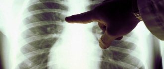
During auscultation, crepitating rales are heard during inspiration. A general blood test revealed moderate leukocytosis and increased ESR. The gas composition of the blood changes. Arterial hypoxemia (lack of oxygen) is determined, which can be replaced by hypercapnia (excessive carbon dioxide in the blood) in the terminal phase. CT diagnostics, as well as lung radiography, are of great information value for interstitial lung diseases. The Yusupov Hospital has the most modern equipment - a multislice computed tomograph, thanks to which the research is safe, fast and of high quality. In the early stages of the disease, images can show deformation and intensification of the pulmonary pattern, decreased transparency of the lung fields, and finely focal shadows. Next, a picture of interstitial fibrosis and “honeycomb lung” develops.
According to spirometry, disturbances in pulmonary ventilation and a decrease in volumes are detected. The ECG shows myocardial hypertrophy in the right heart. The most important diagnostic method is histological confirmation of the diagnosis. To obtain material at the Yusupov Hospital, the following methods are used:
- Cytological examination of pleural fluid;
- Percutaneous fine needle biopsy of the lung;
- Study of bronchial and bronchoalveolar lavages;
- Brush biopsy and pinch biopsy of the bronchi;
- Biopsy of lung tissue, pleura and mediastinal lymph nodes during videothoracoscopy, thoracoscopy or mediastinoscopy.
The collection of material for research is carried out by highly qualified specialists who are fluent in the methodology of performing the procedure. To examine patients, they use modern equipment from leading companies in the world.
2. Causes and symptoms of diseases
Causes of interstitial lung diseases.
The causes of damage to lung tissue can be different. So, interstitial pneumonia
may be caused by bacteria, viruses or fungus.
Other interstitial diseases may be associated with regular inhalation of irritating substances
- asbestos, quartz dust, talc, coal and metal dust, grain dust.
In rare cases, lung diseases in this group may develop due to exposure to certain drugs
.
The peculiarity of interstitial lung diseases is that the above factors, in fact, cause only some of the diseases. In most cases, the exact cause of lung disease remains unknown.
.
Symptoms of interstitial lung diseases.
The most common symptom of all forms of the disease is shortness of breath, which may worsen over time. In most diseases, shortness of breath develops quite slowly, over about a month. In the case of interstitial pneumonia or acute interstitial pneumonia, symptoms can develop very quickly, in just a few days or even hours.
Other symptoms of the disease may be
- The cough is usually dry and unproductive;
- Weight loss;
- Labored breathing.
Visit our Pulmonology page
Treatment and prognosis
The first step in the treatment of interstitial lung diseases should be giving up bad habits - smoking, interaction with toxic production factors, dangerous drugs.
All subsequent treatment is carried out in parallel with the treatment of the underlying disease. The first-line drugs for the treatment of IBL are corticosteroids (prednisolone), which are prescribed in large dosages for three months with a gradual transition to a maintenance dose. If positive dynamics are not observed throughout the year, cytostatics (cyclophosphamide, azathioprine, chlorambucil) are prescribed. Other pharmacological drugs include bronchodilators (orally and inhaled). They are effective only at the stage of reversible bronchial obstruction. For arterial hypoxemia, oxygen therapy is used. In severe cases of interstitial lung disease, the only effective treatment may be a lung transplant.
The outcomes of interstitial lung disease can be positive dynamics, stabilization of the condition, progression in the form of complications, death, and less often - spontaneous regression of changes. The average life expectancy of patients ranges from 1 year with Hamman-Rich disease to 10 years or more with respiratory bronchiolitis. Prevention of interstitial lung diseases is possible only if the cause of their occurrence is established.
The best specialists in the country work at the Yusupov Hospital. Doctors select therapy and monitor patients’ condition in accordance with international and Russian standards. The Yusupov Hospital uses innovative diagnostic equipment that detects changes in lung tissue at the earliest stages. You can make an appointment and consultation with a specialist at the Yusupov Hospital online on the website or by calling.
Make an appointment
What is dissemination in the lungs?
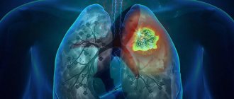
Dissemination consists of multiple pathological foci (compressions) with a diameter of 1-10 mm. On CT scans they appear as light spots, while normally the lung tissue is visualized as an almost uniform dark color. The lesions can be completely different in size, shape (ellipsoid, with uneven edges) and morphology. Perifocal inflammation is often found around the lesions. They can merge and in this case resemble infiltrative processes in pneumonia. Dissemination also manifests itself in the form of focal micro lesions with blood and swelling.
During disseminating processes in the lungs, the respiratory organ partially (depending on the volume of damage) ceases to perform its main function - breathing and transporting oxygen to other organs, in particular to the heart and brain. With total disseminated lung damage, the patient may die.