To the reference book The middle ear is an intermediate link between the outer and inner ear and is involved in the processes of converting sound vibrations.
Author:
- Sokolova Alla Vasilievna
otorhinolaryngologist, otosurgeon, doctor of the highest category
(Voted by: )
- Outer ear
- Middle ear
- Inner ear
The middle ear is a part of the human auditory system, which is located between the outer and inner ear and is formed by the tympanic cavity with the auditory ossicles (hammer, incus, stapes). It is entrusted with the function of converting air vibrations into liquid vibrations, which are perceived by the hearing aid in the inner ear.
Middle ear device
Just behind the eardrum is the tympanic cavity, which contains three small auditory ossicles - the malleus, the incus and the stapes. With the help of a system of these bones, sound vibrations are removed from the eardrum and transmitted to the inner ear. The tympanic cavity is connected to the nasopharynx by the auditory (or Eustachian) tube. This connection with the nasopharynx makes the ear sensitive to diseases of the nose and throat.
Dizziness and tinnitus
S.Ya.Kosyakov, G.Z.Piskunov Dizziness
Introduction
Dizziness is a general term that characterizes a large number of symptoms.
In general, it means a pathological sensation of movement, it can also mean imbalance, lightheadedness, darkening of the eyes, disorientation, weakness and other sensations. Symptoms can vary in intensity from mild and short in duration to severe attacks of rotation accompanied by nausea and vomiting. To more accurately define symptoms, the following definitions are used: Dizziness:
a general concept describing symptoms of imbalance and stability.
Balance problems:
Difficulty maintaining balance, especially while standing and walking.
Presyncope:
A feeling of being “switched off”, similar to the sensation that occurs when holding your breath for a long time.
Systemic dizziness:
sensation of rotation, spinning, twisting of surrounding objects.
The ability to maintain balance is the result of a complex interaction of various organs and systems. The brain is the main center for processing all information about balance coming from the senses to the muscles that maintain balance. Information in the form of nerve impulses comes from the main systems: visual, vestibular, proprioceptive and tactile (joints and feet). Visual information is the most important for the brain and signals movement in relation to surrounding objects. Anatomy
There are two components of hearing: mechanical and electrical (neural).
The mechanical component ensures the delivery of sound waves through the external auditory canal, the movement of the eardrum and the three auditory ossicles in the middle ear. The inner ear is represented by a cochlea, consisting of two halves connected to each other and filled with liquid. The cochlea is responsible for the electrical component of hearing and converts a mechanical signal into an electrical one, which in turn goes to the brain. The other part of the inner ear is responsible for balance and the vestibular system. The three semicircular canals are located in mutually perpendicular planes. Depending on the direction of movement of the head, the fluid moves in the channels, the resulting electrical impulse is transmitted to the brain through the vestibular nerve, transmitting information about the direction of movement. The fluid in the inner ear is renewed daily. Its source of origin is cerebrospinal fluid, absorption occurs in the endolymphatic sac. In Meniere's disease, the absorption capacity of the endolymphatic sac deteriorates. Increased pressure in the inner ear leads to dizziness and decreased hearing. The facial nerve exists in close relationship with the ear. The facial nerve carries out movements of the facial muscles and allows the tip of the tongue to distinguish taste. When it is affected, the eye closes poorly, fluid pours out of the corner of the mouth, and facial movements on the affected side are impossible. Balance function
The balance function is ensured by the interaction in the brain of nerve impulses coming from the inner ear, neck muscles, muscles and joints of the lower extremities.
Disturbances in any of these systems can lead to a subjective feeling of dizziness and instability. General problems with body functions (such as low or high blood pressure, nearsightedness, and many others) can lead to dizziness by affecting the coordination of impulses in the brain. The brain's response to distorted or inconsistent impulses can lead to false sensations of movement (dizziness), which in turn leads to unsteady gait and falls. Dizziness is often accompanied by cold sweats, nausea and vomiting. Visual and muscle and joint signals (tactile and proprioceptive) to the brain warn us that we are moving on the right path or that our head is tilted. The brain interprets this information along with information from the vestibular system and gives the appropriate command to the muscles to maintain balance. Dizziness occurs when sensory information is distorted. Some people experience dizziness when they are in a high place, for example. This is partly due to the inability to focus on nearby objects. While standing on the ground, a person may sway slightly. A person maintains balance, identifies his body position relative to something. When standing in a high place, it is difficult for a person to correlate the position of his body relative to objects in the distance and, accordingly, it is more difficult to maintain balance. As a result, anxiety, fear, and dizziness may occur, which sometimes forces the person to sit down. There is an opinion that motion sickness, a disorder that occurs during sea motion, in a car, or in flight, occurs when the brain receives conflicting sensory information about the movement and position of the body. For example, when reading while driving in a car, the inner ear perceives the movement of the vehicle, but the gaze is fixed on a stationary book that does not move. As a result, sensory conflict can lead to typical symptoms of motion sickness, dizziness, nausea, and vomiting. Another form of dizziness occurs when you spin repeatedly and suddenly stop. Rotation causes movement of the endolymph. The movement of the endolymph causes impulses, which in turn tells the brain that we are moving, but other sensory systems report that we have stopped, so the patient feels dizzy. Causes of Dizziness
Dizziness can be classified into types depending on the part of the vestibular system that is not working properly.
Disturbances can occur at the level of the inner ear, brain, eyes and limbs (muscles of the back, neck, legs and joints that react to maintain our position). Dizziness due to the inner ear
Part of the inner ear (cochlea) is used for hearing, the other part is used for balance (labyrinth).
If there are disturbances in the labyrinth or in the nerve connecting it to the brain, this leads to dizziness. Various types of disorders in the inner ear can lead to vertigo, including Meniere's disease, labyrinthitis, positional vertigo, vestibular neuronitis, and nerve tumors. These disorders typically cause problems with balance, a sensation of spinning objects, and nausea. Also, these phenomena may be accompanied by tinnitus and hearing loss on the corresponding side. Dizziness of a central nature
The cause of dizziness of a central nature is usually a disturbance in the area of the brain responsible for balance.
Symptoms may include lightheadedness, confusion, unsteadiness, and sometimes loss of consciousness. Causes of central vertigo include low blood sugar, low blood pressure, stroke, multiple sclerosis, migraines, head injuries, tumors and age-related changes. Treatment of this type of dizziness is usually associated with the elimination of problems leading to disruption of brain function. Muscular-articular dizziness
This type of dizziness is rare.
If there are diseases of the muscles, joints, or the sensitivity of the lower extremities is impaired, then difficulties arise in the body’s reaction to movement and in maintaining an upright position. Musculo-articular dizziness can be caused by: atrophic changes in muscles (muscular dystrophy), severe forms of diabetes, arthritis, joint implantation and trauma. Symptoms: Typically unsteadiness and poor balance. Visual dizziness
Unsteadiness of the eye muscles and poor vision can impair balance function.
The brain relies on visual information to maintain balance. Motion sickness in a car or at sea are examples of visual vertigo because the eyes are constantly fixated on a moving object and “confuse” the vestibular part of the brain. This leads to dizziness, nausea and vomiting. Dizziness is not a fatal condition and may improve with treatment, but balance problems may remain. Diagnosis of dizziness
Dizziness can be caused by various disorders in the body.
Based on the medical history and examination data, the doctor selects the required scope of examination to obtain a more complete picture of the disease. The usual set of examinations includes testing of hearing and vestibular function, computed tomography and nuclear magnetic resonance, blood tests, and ultrasound examination. The most commonly used test for dizziness is electronystagmography (ENG). This test measures inner ear endurance and eye coordination. The method involves observing eye movements while cold and warm air is blown into the external auditory canal. This usually causes a brief feeling of dizziness. It is important not to take medications before the test that could affect the test results (for example, Valium, alcohol, etc.). When ordering such an examination, it is necessary to find out from the doctor the effect of the medications taken on the test results. Transcranial Doppler sonography is another test specific for the examination of dizziness of vascular origin. It is a safe, quick way to see disturbances in blood flow in the parts of the brain responsible for balance. In addition, a computed tomography (CT) scan of the temporal bones and, in some cases, magnetic resonance imaging (MRI) may be performed. The purpose of these examinations is to achieve confidence in the absence of a life-threatening pathology and to determine the exact location of the disorder. This is the basis for effective treatment. The scope of the examination is determined by the doctor in each specific case. Several tests are necessary to diagnose the cause. Perseverance and understanding are necessary for both the doctor and the patient, which is also the basis for effective treatment. The most common types of vertigo Benign paroxysmal positional vertigo (BPPV)
BPPV is the most common type of vertigo.
With this disease, dizziness occurs only when the head position changes (usually when turning in bed, tilting the head backwards or forwards). This type of dizziness is caused by microcrystals that float in the fluid of the inner ear and cause a spinning sensation. The most common cause of BPPV is head trauma or viral infections, but sometimes it begins without any obvious cause. Treatment for BPPV consists of certain exercises to return the crystals to a place where they will not cause dizziness. When left at rest in a certain position for 48 hours, they often lock into place. Exercise may reduce symptoms. If these actions are ineffective, then surgical treatment (for example, occlusion of the posterior semicircular canal) may be necessary. Vestibular neuronitis
Neuronitis (nerve inflammation) usually occurs due to a viral infection and can affect the balance centers or the vestibular nerve.
When this happens, the balance centers in the brain are overstimulated, resulting in significant imbalance and systemic dizziness. Fortunately, vestibular neuronitis usually subsides over time and does not recur. Drugs such as betaserc help in the initial stage and reduce the severity of the main symptoms; later, vestibular rehabilitation exercises can speed up the recovery process. In some cases of persistent disease, surgical treatment is recommended. Meniere's disease (Endolymphatic hydrops)
Meniere's disease is a consequence of disorders in the inner ear due to increased pressure in the endolymphatic space.
This is usually due to increased sodium concentration in the fluids of the inner ear. In addition to imbalance that lasts for hours, patients may experience hearing fluctuations, tinnitus, and a feeling of fullness in the affected ear. Sometimes the lesion affects both ears. The full cause of this disorder is not fully known. Sometimes attacks can be caused by excessive salt intake, anxiety, changes in weather, and other reasons. Treatment usually includes limiting salt intake and using diuretics, fluid restriction, sedatives and some other vestibular suppressants. Betaserc is the only drug created for long-term treatment of dizziness. Treatment helps reduce the severity of attacks, but complete cure of the disease cannot be achieved. Vestibular rehabilitation exercises can speed up the recovery process and increase the patient's resilience to vestibular disorders. All prescriptions of drugs should be carried out only by a doctor. For severe cases of Meniere's disease, surgical treatments are available. The list of these methods is long and more often they are destructive in nature for the structures responsible for balance. Vascular vertigo
The correct functioning of the balance system requires not only the flow of information into the inner ear, but also the appropriate transmission of impulses along the nerves to the brain.
If there is not enough blood flow to the areas of the brain responsible for balance, even for a short time, dizziness can occur. The causes of vascular dizziness are different. The phenomena of osteochondrosis in the cervical spine can lead to compression of the arteries leading to the brain; atherosclerotic plaques can narrow the arteries, also causing a decrease in blood flow. Often, blood pressure in the vessels leading to the brain may temporarily decrease when standing up suddenly, especially in older patients receiving blood pressure-lowering medications. Special examinations such as MRI or Doppler sonography help in diagnosing such diseases. Another rather rare cause of dizziness is Perilymphatic Fistula
. The inner ear is a space filled with fluid located in the temporal bone.
If fluid leaks from the structures of the inner ear, hearing loss may occur, which may be greater or less, and dizziness. Most often, fluid leaks through the membranes of the windows of the inner ear, which can occur after physical activity or injury. In some cases, there are congenital disorders that characterize an enlarged connection between the inner ear and the brain ("enlarged vestibular aqueduct"). Sometimes this can be seen with a special x-ray examination - computed tomography. Sometimes the site of the membrane rupture heals on its own, sometimes minor surgery is required. A perilymphatic fistula, or as it is also called, a labyrinthine fistula, can occur as a result of chronic inflammation of the middle ear, especially with cholesteatoma. Cholesteatoma is compacted skin scales. If there is a hole in the eardrum, the skin grows into the cavity of the middle ear, and its metabolic products, like the formation of a pearl, form a lump of cholesteatoma, which presses on the walls of the middle ear cavities and destroys the bone, in particular, the semicircular canal. Therefore, the treatment of chronic otitis media is very important, and when the hole is localized in the upper part of the eardrum (epitympanitis), it must be surgical, because Most often, cholesteatoma is found in these cases. Tumors
Rarely, tumors may be the cause of dizziness.
Most tumors are benign. Acoustic neuroma is a benign tumor of the vestibular nerve. The presence of a neuroma can lead to instability, hearing loss and noise. The most effective treatment method is surgery. Treatment of dizziness All questions regarding the treatment of dizziness and, in particular, taking medications should be discussed with your doctor.
Treatment in each specific case is selected individually and depends on age, severity of dizziness, concomitant diseases and many other factors.
Ear noise
Ear noise is a very common symptom.
The noise can be constant or periodic, of varying severity and frequency. The noise can be subjective (audible only to the patient) or objective (audible to others), and may or may not be associated with hearing loss. Noise is a symptom and not a disease and can occur in various diseases, such as pain in the arm or leg are symptoms of various diseases. Noise appears when the auditory nerve is irritated for various reasons. The noise may or may not be accompanied by hearing loss. Hearing is measured in decibels (dB). A hearing level of 0 to 25 dB is considered normal for the perception of spoken language. Hearing mechanisms
to understand the possible causes of noise in the ear, it is necessary to have some idea of hearing mechanisms.
The mechanism of auditory perception is provided by the five main components: the outer ear, the middle ear, the inner ear, the paths and the brain. The outer ear
of the outer ear consists of an auricle and an external auditory pass.
These structures collect sound waves and transmit them to the eardrum. middle ear
is located between the eardrum and the inner ear.
This space contains three auditory bones: a hammer, an anvil and a stirrup. The fluctuations of the eardrum are transmitted through the auditory bones on the liquid of the inner ear. The middle ear lined with the mucous membrane is identical to the nose and contains mucous glands and blood vessels. The drum cavity is connected to the rear parts of the nose using the Eustachian pipe. Eustakhiev’s pipe is served to maintain equal pressure between the middle ear and the outer atmosphere. The feeling of clicking or congestion when height changes is a demonstration of the ventilation function of the Eustachian pipe. The inner ear
in the inner ear is in a dense bone capsule and contains liquids and auditory cells.
The cells are covered with delicate membrane with microscopic blood vessels. In the inner ear, fluctuations in the fluid as a result of the movements of the stirrup, are converted into electrical impulses in the nerve. Electric impulses arising in the inner ear are transmitted to the brain along the auditory nerve. The auditory nerve going into the brain is located in a small bone channel along with the vestibular and facial nerve. The brain
of the auditory nerve by reaching the brain is divided into many internal connections.
In the brain, nerve impulses are recognized as recognizable sounds. Ear noise,
most ear noise are heard only by patients - this is a subjective noise.
The noise that the patient himself hears, and someone is also called objective. Objective noise can be as a result of muscle cramps in the middle ear or auditory pipe, or due to anomalies of blood vessels of the surrounding the ear. Ear noise of muscle nature,
noise can be the result of muscle spasm attached to one of the auditory bones or the result of a spasm of muscles attached to the auditory pipe.
In the middle ear, there are two muscles: a stirmer that is attached to the stirrup and muscle pulling the drum overlain attached to the hammer. Usually these muscles quickly decline in response to loud noise or at fear. Sometimes one or two of these muscles begin to rhythmically contract for no apparent reason. These abbreviations can cause repeated noise in the ear. An annoying clicking usually passes on its own. The ear noise of muscle nature as a result of a spasm of various muscles of the pharynx is quite rare, but sometimes it can be if muscle spasm is long, then drug treatment (muscle relaxants) or surgical treatment (intersection of spasmodic muscles) is used. The ear noise of vascular nature
has two large blood vessels closely related to the middle and outer ear: jugular vein and sleepy artery.
These are large blood vessels supplying the brain with blood and carrying out its outflow. To hear your own heartbeat or noise of blood passing through these large vessels is not a normal phenomenon. Sometimes this phenomenon can occur at high temperature, infection of the middle ear, after intense physical activity. The noise of circulation in these situations is temporary and is not heard by others. Sometimes the noise of blood circulation becomes heard by another. This can happen due to the thickening of the wall of the blood vessel, the presence of bending or narrowing in the vessel. Further examination is necessary to identify the cause and choose the treatment of this pathology. Ear noise due to the outer ear
is the closure of the external auditory passage with a gray, foreign body, edema is leading to a decrease in hearing and pressure on the eardrum.
Often this leads to a pulsating type. Ear noise due to the middle ear,
impaired function of the middle ear may be the result of an allergic reaction, infection, injury, cicatricial changes and limitations of the mobility of the auditory bones.
These disorders often lead to hearing impairment and ear noise. However, there is no direct dependence between the degree of hearing loss and noise intensity. Ear noise due to the inner ear
any condition that violates the balance of fluid pressure in the inner ear can lead to an ear noise.
This may be the result of an allergic reaction, infection, circulatory disorders, which lead not only to changes in the liquids of the labyrinth, but also in the membrane structures of the inner ear. Ear noise due to damage to the tracking paths,
the most delicate structures of the mechanism responsible for the hearing.
Hair cells convert fluctuations in the fluid into nerve impulses. The most insignificant edema and violation of interference in the hair cells, regardless of the cause, lead to function and irritation. This can happen for various reasons: an allergic reaction, infection, edema, systemic diseases, both acute and chronic, toxic effects, sudden loud sounds and sensitive subjects, injuries, the effects of medications, minute changes in blood supply and diet changes. Pressure changes can cause swelling both outside and inside the nerve when passing in the bone tunnel to the brain. In these cases, ear noise occurs on one side. Because The bone tunnel cannot stretch, then due to compression, not only the auditory and vestibular functions suffer, but also the facial nerve. The gap or spasm of a small vessel that occurred somewhere in the auditory path causes compression and violation of circulation. Accordingly, under such conditions, a sudden noise may occur with complete or partial loss of auditory function. If the blood clot, then it can resolve with minimal consequences. Auro noise of brain nature
as a result of edema, pressure or circulation disorders for hypertension, atherosclerosis, as a result of the consequences of injuries, one or more complexes of the passing paths at the entrance and end of their brain can be involved.
In such situations, symptoms are usually localized on the one hand, in addition, the development of symptoms and signs can tell the doctor the place and prevalence of the lesion. The ear noise accompanying the decrease in
ear noise can be associated or not with hearing impairment.
With the coexistence of ear noise and hearing loss, the intensity of ear noise is not an indicator of the further development of hearing loss. Many patients with the advent of ear noise are afraid of the progression of hearing loss. However, these are often not interconnected things. All issues of ear noise treatment should be discussed with your doctor.
Thus, the treatment of dizziness and ear noise is a difficult task, which is possible only by the joint efforts of the doctor and the patient. High -quality hearing diagnosis plays a very important role in such a treatment. The sequence in identifying the causes of these conditions, in treatment and rehabilitation is an integral condition for achieving success.
Causes of otitis media
Otitis media can develop as a complication of otitis externa, but more often the infection enters the middle ear from the nasopharynx through the Eustachian tube. In this case, otitis media is a complication of colds. It is enough to blow your nose incorrectly when you have a runny nose (without pinching one nostril), and discharge from the nose with a current of air can enter through the auditory tube into the tympanic cavity. Normally, the middle ear is sterile, so the penetration of bacteria from the nasopharynx can easily lead to the development of inflammation. If this happens against the background of a weakened immune system, the development of otitis media is very likely. acute otitis media usually occurs
which in a significant number of cases quickly turns into a purulent form.
The development of inflammation can be facilitated by the accumulation of fluid (exudate) in the middle ear. This fluid is constantly produced. Normally, it should leave the middle ear through the auditory (Eustachian) tube. This evacuation can be difficult due to diseases and pathologies of the nose (rhinitis, adenoids, deviated nasal septum), as well as diseases of the auditory tube itself (eustachitis, tubootitis). In this case, the fluid fills the space of the middle ear, becomes viscous, and interferes with the functioning of the auditory ossicles. This condition is defined as exudative otitis media.
.
Infection of the accumulated fluid leads to the transition of exudative otitis media to chronic purulent otitis media
.
What is the organ of hearing?
Air and bone sound conduction
Sound energy travels to the structures of the inner ear through air conduction and bone conduction.
Airborne sound conduction is the usual way for sound vibrations to enter the ear - through the auricle and external auditory canal, sound comes to the eardrum. Further, vibrations of the eardrum through the chain of auditory ossicles are transmitted to the fluids of the auditory cochlea - peri- and endolymph, causing the main membrane and structures of the organ of Corti to vibrate.
Bone conduction is the conduction of sound vibration from the surface of the head directly into the cochlea of the inner ear, bypassing the middle ear. When sounds enter the ear through bone conduction, sound vibrations travel through the bones and tissues of the head. Under the influence of bone-conducted sounds, the walls of the cochlea of the inner ear vibrate, which is transmitted to the fluids that fill it. This, in turn, causes oscillatory movements of the basilar membrane and the organ of Corti. Then everything happens in the same way as with airborne sound transmission.
We hear our own voice precisely through bone conduction: the sounds of the voice pass to the cochlea of the inner ear through the tissues of the head. This is why we hear our voice differently than in the recording. This is caused by the fact that the bones of the skull conduct low frequencies better than high frequencies. Therefore, during sound pronunciation, people perceive their own voice as lower and deeper than others perceive it.
Since bone sound conduction practically excludes the middle ear from the process of sound transmission, the study of the auditory perception of air- and bone-conducted sounds during audiometry is very important in diagnosing hearing.
In addition, in cases where it is impossible to use air conduction hearing aids, in particular, in case of certain diseases and after certain operations on the middle ear, the doctor considers the possibility of bone conduction hearing aids.
Intermediate (conducting) section of the hearing organ
The intermediate (conducting) section of the hearing organ begins with the auditory nerve and ends in the cerebral cortex. The neuron bodies of the auditory nerve are located spirally along the axis of the cochlea and form the so-called spiral ganglion. And their long processes - axons - form the auditory nerve, which transmits nerve impulses “up” to the brain. The right and left auditory nerves are called the eighth (VIII) pair of cranial nerves.
The axons of the auditory nerve, like other neurons, are covered with a layer of special tissue - the myelin sheath, which contains “intercepts” - bare sections of the axon. This sheath and its “intercepts” play a key role in the transmission of nerve impulses by the neuron.
The neurons of the auditory nerve switch to the neurons of the medulla oblongata - the cochlear nuclei. Moreover, the cochlear nuclei are the last formations of the auditory analyzer, receiving nerve impulses from only one ear.
The pathways and subcortical centers of the auditory analyzer are part of the central nervous system (CNS) and include the ascending (afferent) and descending (efferent) systems. Anatomically, it is located in the brain stem, subcortical structures of the brain. A simplified diagram of the ascending auditory system is shown in the diagram.
As can be seen from the diagram, the number of nerve cells (neurons) increases many times as one rises from the auditory nerve to the cerebral cortex. There are approximately 35 thousand of them in the auditory nerve, and more than 12 million in the auditory cortex. In addition, as one rises to the auditory cortex, the connection of auditory neurons increases both between both sides of the brain and with neurons of other sensory systems, memory areas, speech and many others.
It is noteworthy that above the right auditory nerve and the cochlear nuclei, in which its neurons switch to the next level, the bulk of the ascending auditory neurons move from this ear to the left side of the brain. And vice versa. Thus, a “crossing” of the conductive pathways of the auditory analyzer occurs, which is clearly visible from the diagram of the brain stem.
Central part of the hearing organ
The central (cortical) section of the auditory analyzer is located in the temporal lobes of the cerebral cortex. Nerve impulses from the right ear go mainly to the left hemisphere of the brain, and vice versa, from the left ear to the right. This is of great importance in hearing care, and here's why. The auditory zones of both hemispheres perform, although similar, but different work.
Research in the 1960s and 70s showed that in most right-handed people, the left hemisphere is better at processing high-frequency, rapidly changing sounds, and is better at perceiving individual sounds, syllables and words of speech. That is why the left hemisphere and, accordingly, the right ear were called dominant in speech perception. And that is why, for the majority of right-handed people, if binaural hearing aid is not possible, hearing aid for the right ear is preferable. For left-handed people, it’s usually the other way around. But since there are many individual differences, an audiometric examination must determine which ear responds better to verbal tests. Assessing the perception of whole speech is a rather long and difficult psychoacoustic study and is not used in audiological practice.
Later studies in the 1970-80s showed that not everything is so simple with the speech dominance of the hemispheres.
Experiments by many scientists have shown that the hemisphere opposite to the dominant one when perceiving individual words (in most right-handed people - the right) perceives intonation and speech rhythm much better, which are necessary for understanding whether the speaker is stating something or asking, whether he is speaking seriously or joking . That is, it better understands sentences as a whole. Moreover, it is the opposite dominant hemisphere that connects all sentences into the general meaning of everything said, for example, the entire story, the entire conversation as a whole. Thus, the hemisphere that was considered “dominant” (the left in right-handed people) carries out a sequential analysis of individual sounds, and the hemisphere that was considered “non-dominant” carries out a holistic perception of speech messages.
Otitis media in children
Otitis media is a common childhood disease. Children suffer from otitis media more often than adults, this is caused by a number of reasons:
- The Eustachian tube in children is shorter and wider than in adults, and is located more horizontally. All this facilitates the entry of mucus from the nasopharynx into the middle ear;
- children suffer from colds more often;
- children often cry and sniffle at the same time, as a result of which nasal secretions can get into the auditory tube;
- Children often have adenoids - a pathologically enlarged pharyngeal tonsil that blocks the Eustachian tube. Difficult ventilation of the tympanic cavity contributes to the development of infection.
Breasts are even more vulnerable. In newborns, there is a special (myxoid) connective tissue in the middle ear, loose, gelatinous, with a small number of blood vessels, which is a very favorable environment for the development of harmful bacteria. Breasts spend most of their time lying down, which interferes with the outflow of mucus. When regurgitated, milk can enter the auditory tube and tympanic cavity.
Otitis media in young children appears suddenly. The temperature rises to 39-40° C. The baby begins to cry in pain; He can't sleep and refuses to suckle. He constantly touches the sore ear - but this is only from the age of four months, and before that it is possible to establish what exactly the child is hurting in only by indirect signs. The picture is also complicated by possible gastrointestinal manifestations of otitis media – diarrhea and vomiting.
Therefore, it is very important to show the child to the doctor. If your child has a high temperature, call a doctor at home. If a child complains of ear pain, it is necessary to take him to an ENT doctor.
Types of ear diseases
Hearing diseases are divided according to the following categories:
- Congenital - arise due to the initially incorrect structure of one or another part of the hearing organs. They can be either hereditary or acquired as a result of intrauterine development disorders.
- Traumatic - usually a consequence of mechanical impact on the hearing organs.
- Infectious - are a consequence of the impact on the hearing organs of various infections and are usually characterized by inflammation.
Diseases of the hearing organs are divided according to how they arise and proceed:
- Acute – symptoms occur suddenly;
- Chronic – can accompany a person for several years in a weaker form.
Almost any hearing disease can lead to various types of hearing loss and characteristic hearing impairments.
Diseases of the hearing organs are grouped according to the specific affected area.
Inner ear diseases
The inner ear is a department that, in addition to hearing, is responsible for the vestibular apparatus. Therefore, the consequence of a disease in this department can be not only hearing loss, but also problems with coordination and balance. Treatment of these diseases depends on the severity and characteristics of the pathology.
Middle ear diseases
The middle ear is a section that communicates not only with other parts of the hearing organ, but also with the nasopharynx. Therefore, diseases of this department can be a consequence of inflammatory processes in the nasopharynx. Conversely, the condition of this department can affect the condition of the throat, nose, and head as a whole.
Problems with this particular department are the most common cause of hearing disease in children. Otitis often occurs due to chronic rhinitis, a common disease in the first years of a child’s life. But often adults also suffer from inflammation of the middle ear resulting from a respiratory disease. One of the common symptoms of middle ear diseases is the so-called autophony (a stronger than usual perception of the sound of your voice in one ear).
Diseases of the middle ear are quite dangerous for hearing in general. So, if purulent secretion has accumulated in the ear cavity (one of the consequences of inflammatory processes), the eardrum may become distorted or even rupture. And the consequence of inflammation or tumor of this section can be deformation of the auditory ossicles - and this problem can only be solved through surgery.
Diseases of the external ear
Diseases of the outer ear are not as common as diseases of other parts of the hearing organ, since this part of the ear is best adapted to environmental influences. However, the abuse of antibiotics, unfavorable environmental conditions and violation of ear hygiene rules threaten the appearance of a very unpleasant disease - inflammation of the external auditory canal, or otitis externa. Its main symptom is itching and pain that occurs when something touches the ear.
Hearing diseases are quite unpleasant, and their consequences can be very, very serious. specialist at the Audionica hearing clinic, audiologist Elena Yurievna Kropacheva
Another discomforting disease of this part of the hearing organ is boils that appear in the auricle. They appear when there is a metabolic disorder. If this problem occurs, it is important not to self-medicate and under no circumstances try to squeeze out the boil yourself - this action can aggravate the process and lead to the introduction of new infections into the ear.
Symptoms of otitis media
Acute otitis media is usually manifested by the following symptoms:
Earache
Ear pain with otitis media can be pulsating, “shooting,” or vice versa. nagging, incessant pain can radiate (give) to the temple and back of the head, and teeth may begin to ache.
Hearing impairment
Hearing deterioration in otitis media is typical for the initial period of the disease, but can be observed throughout the entire course of the disease. The ear, as they say, “blocks up.” The patient may hear extraneous noises or his own voice as if from within (autophony effect).
Temperature
Body temperature can rise to 39° C.
More about the symptom
Ear discharge
Serous or purulent discharge from the diseased ear may be observed.
General weakness
The disease is characterized by general malaise; Nausea and sometimes vomiting are possible.
Symptoms of hearing disease
Depending on the specific department and etiology of hearing impairment, the symptoms of the disease can be very different. When diagnosing, the rate of development of a particular symptom is taken into account (whether it appeared suddenly or appeared gradually), the intensity and nature of the pain (strong, dull, stabbing or intermittent), duration, localization and changes in the condition when exposed to various factors.
Depending on the specific part and cause of the ear disease, the symptoms of hearing diseases can be very different. When diagnosing, the rate of development of a particular symptom is taken into account (whether it appeared suddenly or appeared gradually), the intensity and nature of the pain (strong, dull, stabbing or intermittent), duration, localization and changes in the condition when exposed to various factors.
Symptoms of hearing problems may include:
- fever - most often a consequence of an infectious disease;
- vomiting is a symptom of various inflammations in the organ of hearing;
- rhinitis and sore throat are also a consequence of inflammatory processes in the hearing organ, most often in the middle ear;
- sneezing, coughing – a consequence of otitis media, sometimes occurs when there is a foreign body in the ear canal;
- dizziness, noises, ringing in the ears - a sign of primary otalgia;
- congestion in the ear, partial hearing loss - most often due to wax plugs or otitis media;
- purulent secretion released from the ear canal is a consequence of inflammation of one or another part of the hearing organ;
- bloody discharge from the ear canal is a consequence of mechanical impact, injury;
- various changes in the skin of the auricle - fungi, eczema, erysipelas, etc.
Treatment methods for otitis media
Severe, “shooting” pain in the ear, characteristic of acute otitis media, leaves no other option but to immediately begin treatment of the disease. However, it is highly desirable that treatment of otitis occurs under the guidance of a physician.
Left without proper treatment, otitis media can become chronic. In this case, the pain in the ear ceases to be painful. For a long time, the patient may not feel pain at all. This is explained by the fact that in chronic otitis media the eardrum remains perforated, and through this hole pus can flow into the ear canal without creating excess pressure in the eardrum. A constant focus of inflammation and impaired integrity of the eardrum contribute to the development of hearing loss.
Another danger: improper treatment can lead to the formation of adhesions and scars in the tympanic cavity, disrupting the functioning of the auditory ossicles and blocking the auditory canal. This is the so-called adhesive otitis media , which leads to persistent hearing loss and the appearance of tinnitus.
Other complications are also possible. A timely visit to a doctor will help avoid complications and maintain hearing acuity. The usual course of treatment for otitis lasts ten days.
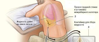
Specialist consultation
If two days have passed and your ear is still bothering you, you should definitely see an ENT specialist. There is a possibility that the disease may progress to the next stage, in which purulent discharge begins to accumulate in the middle ear. Accumulated pus can rupture the eardrum. It is advisable to avoid this. If, nevertheless, pus needs to be removed from the tympanic cavity, then it is better for a doctor to do this by making a careful puncture of the eardrum, the so-called paracentesis.
Make an appointment Do not self-medicate. Contact our specialists who will correctly diagnose and prescribe treatment.
Rate how useful the material was
thank you for rating
Diagnosis of diseases
The most effective diagnostic methods include:
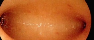
- otoscopy (used to determine the condition of the eardrum, which can be hyperemic, perforated with the release of purulent exudate);
- probing of the tympanic cavity (helps diagnose cholesteatoma, bone caries);
- radiography of the temporal bone (carried out in two projections - according to Schüller and Mayer, in some cases irradiation is also required along the oblique, that is, Stenvers; allows to identify all destructive changes);
- audiometry (consists of examining hearing, establishing hearing loss, type of hearing loss).
In medical practice, traditional methods of palpation and percussion are also used. With their help, it is possible to determine the degree of swelling of the examined areas, which has developed against the background of edema or due to infiltration of tissues in the postauricular area.
If you experience pain, ear congestion, or other subjective symptoms of pathology, you should make an appointment with an otolaryngologist. Our clinic is equipped with modern equipment, which, in the hands of certified personnel, will allow you to get a detailed picture of the condition of the middle ear and associated structures. A correct diagnosis and timely treatment increase the chances of a speedy recovery and reduce the risk of developing serious complications.
Prevention of hearing diseases
Hearing diseases are quite unpleasant, and their consequences can be very, very serious. Specialist at the Audionics hearing clinic, audiologist Elena Yurievna Kropacheva gives the following recommendations:
- Protect your ears from the cold (wear a hat), avoid drafts - so as not to “cold” your ears;
- Do not use hard objects (including cotton swabs) to clean your ears - this will not help clean the ear canal, but will only increase the production of wax and thicken the wax plug; In addition, there is a high risk of injury to the outer part of the ear and infection. If your ears need to be cleaned, it is best to consult an appropriate doctor.
- If your type of activity involves loud sounds and noise, use hearing protection, such as earplugs.
- Take diseases of the nasopharynx and throat more seriously - their inflammation can spread to the ears. Be sure to consult a doctor, do not self-medicate, and do not abuse traditional medicine. Remember that the success of treatment largely depends on how timely assistance was provided.
- Be sure to regularly visit an otolaryngologist - if there are no problems with hearing, then as a preventive measure (once every year or two), but if you are at risk - once every six months.