Causes of dacryoadenitis
Dacryoadenitis is an inflammatory lesion of the lacrimal gland, which comes in two forms:
- Spicy. Develops after infectious diseases caused by bacterial, viral or fungal agents. Thus, acute dacryoadenitis is a consequence of influenza, sore throat, pneumonia, cytomegalovirus, scarlet fever, and infection in the gastrointestinal tract. Most often, the development of the disease occurs against the background of mumps (popular name - mumps), in which it takes on a bilateral character. The inflammatory process simultaneously affects the parotid and submandibular salivary glands.
- Chronic. Develops in chronic infectious diseases in the active stage - tuberculosis, blood pathologies, brucellosis. In addition, chronic dacryoadenitis can occur due to systemic diseases: Wegener's granulomatosis, Sjögren's syndrome, reactive arthritis, etc.
Forecast
Most often, acute dacryoadenitis has a benign course - the inflammatory infiltrate resolves and the disease resolves within 10-15 days. If an abscess of the gland forms, the disease becomes more severe. Treatment is lengthened if the abscess resolves on its own with pus draining into the upper conjunctival fornix or out through the skin of the eyelid, but the prognosis for recovery is favorable. With adequate treatment of canaliculitis, complete recovery occurs. But if the inflammatory process in the lacrimal canaliculi takes a chronic course, then a disturbance in the outflow of tears is observed.
Symptoms of dacryoadenitis
Clinical manifestations vary depending on the form of dacryoadenitis. Acute is accompanied by the following symptoms:
- pain when pressed;
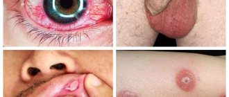
- hyperemia, swelling and drooping of the outer edge of the upper eyelid;
- deformation of the palpebral fissure (becomes similar to the letter S) or complete closure.
The disease begins abruptly, and swelling may spread to the temples and half of the face, causing the palpebral fissure to completely close.
Acute dacryoadenitis causes enlargement of the lymph nodes, after which they begin to hurt. The eyeball becomes deformed, deviating towards the lower and inner side, and also protrudes slightly from the orbit, causing double vision. There is a disturbance in the movement of the visual organs to the outer and upper side. If you pull back the upper eyelid, you can see that the conjunctiva is swollen and red.
If acute dacryoadenitis occurs in a child with weak immunity, then in severe cases an abscess or phlegmon of the gland may develop, spreading to the space behind the organ of vision.
In addition, symptoms of acute dacryoadenitis include a general deterioration in health:
- temperature increase;
- the appearance of headaches;
- sleep and appetite disturbances.
The duration of the disease is usually 7-21 days.
Chronic dacryoadenitis does not manifest symptoms of an acute inflammatory process. The lacrimal gland thickens and enlarges, and pain occurs less frequently when pressed. The color of the skin of the affected area does not change. Due to the enlargement of the lacrimal gland, the palpebral fissure on the outside may narrow. The visual organs move as usual. Clinical manifestations do not develop immediately, so it may take quite a long time before the patient sees an ophthalmologist.
Dacryoadenitis with syphilis
The disease may develop with primary and tertiary syphilis. In the first case, the gland enlarges and thickens, but there is no pain. In the second, a soft formation forms in the affected area. Diagnosis of dacryoadenitis is carried out by collecting anamnesis, during which the doctor identifies signs of syphilis from other body systems.
For Mikulicz's disease
This disease is a chronic lymphostasis of the lacrimal and salivary glands. It develops against the background of leukemia and pseudoleukemia. In this case, the symptoms of dacryoadenitis are that the glands are enlarged on both sides (mainly submandibular, in more rare cases - parotid, sublingual). The magnification is so strong that the organ of vision shifts to the lower and inner side. The eye may bulge forward. The eyelids droop, the lymph nodes enlarge, causing the palpebral fissures to become narrow. The patient experiences dryness in the oral cavity and organs of vision. This is due to the fact that the function of the glands decreases.
Pseudotumorous pathology
The disease belongs to the group of orbital pseudotumors. Such diseases arise due to the inflammatory process, and their course resembles the development of a malignant tumor. Pseudotumors are considered to be autoimmune pathologies, despite the fact that to date it has not been precisely established why they appear.
Inflammation of the lacrimal gland is characterized by the following symptoms:
- iron increases noticeably;
- when pressed, a dense, smooth formation is felt, there is no pain;
- the upper eyelid swells and droops slightly.
With a long course, the inflammatory process spreads to adjacent tissues. Then the connective tissue grows and scars appear.
For sarcoidosis
This systemic disease belongs to granulomatosis. It is not known exactly what exactly causes them. With this disease, a large number of small nodes form in the skin, lymphatic system, and internal organs. Formations in granulomatosis are of the same type and clearly limited from adjacent tissues. Dacryoadenitis, as a rule, develops against the background of general symptoms of the disease, but can appear without involving other organs. In the first stages, the disease does not manifest itself in any way and is characterized by a long course. The lacrimal gland usually enlarges evenly, the sarcoid node is not clearly visible. There is no pain when pressed. Diagnosis is difficult.
For chronic tuberculosis
The lacrimal gland swells, it gradually increases in size, and pain is felt when pressed. In addition, other symptoms of tuberculosis are noticeable - the cervical lymph nodes are enlarged, changes in the lungs are visible on x-rays.
Complications
If treatment is not carried out, the risk of infectious complications increases:
- Abscess of the upper eyelid;
- Orbital phlegmon.
Symptoms of an abscess are as follows:
- Severe bursting pain;
- Headache;
- Severe swelling and redness of the eyelid, which causes it to close (narrow eye);
- Skin tension and redness, but in some cases it may become jaundiced;
- Zone of fluctuation (softening) in the area of the upper eyelid (its appearance is associated with purulent melting of subcutaneous fat);
- Body temperature rises, but visual functions do not decrease.
Orbital phlegmon is a more dangerous complication and is manifested by other signs:
- Rapid development in a couple of hours;
- Intense pain in the eye;
- Red-violet widespread swelling of the eyelids;
- Pronounced narrowing of the eyelids;
- Infringement of the conjunctival membrane in a sharply narrowed palpebral fissure;
- Double vision and severe decreased vision (a person may go blind);
- Almost complete immobility of the eyeball;
- Intense headache;
- Fever and chills.
Diagnosis of dacryoadenitis
If characteristic symptoms occur, you must visit an ophthalmologist who will conduct an examination. Diagnosis of the acute form of dacryoadenitis requires the following procedures:
- visometric and tonometric examination;
- biomicroscopy of the organ of vision.
Symptoms of the acute stage are usually severe, so the diagnosis is made quickly.
To diagnose chronic dacryoadenitis, additional measures are required:
- Ultrasound of the eyeball;
- magnetic resonance or computed tomography (to exclude a tumor of the eyelid or lacrimal gland).
Also, in case of chronic dacryoadenitis, in order to clarify the etiology of the inflammatory process, a chest X-ray is taken (this makes it possible to detect changes in the lung tissue), the Mantoux test and other studies.
Material and methods
A study was conducted in 26 patients (29 orbits) in the period 2007-2012. There were 7 men, 19 women. The age of the patients was 59.08±15.42 years (28-81 years). Pathological changes in the SG on the right occurred in 9 patients, on the left in 14, and in three cases the process was bilateral. The duration of the disease before seeing a doctor was 17.78±26.19 (1.5-120) months. Ultrasound scanning, CT and remote thermography were used as instrumental research methods. 22 people (24 orbits) were operated on (extirpation of the gastrointestinal tract). All surgical material was subjected to pathohistological examination. Tear production was examined in all cases using a single method, including sequential use of the following tests: tear fluid osmolarity test, Schirmer I test, Jones test, corneal staining with 1% fluorescein solution and Norn test. For monolateral lesions, the contralateral side served as control.
Treatment of dacryoadenitis
If an acute form of the disease develops, hospitalization of the patient will be required; therapy in most cases is conservative. Treatment of chronic dacryoadenitis is selected depending on the underlying pathology.
Treatment of acute dacryoadenitis
The patient is prescribed antibacterial drugs. For maximum effectiveness, it is recommended to simultaneously use sulfonamides - antimicrobial medications that temporarily suppress the proliferation of bacterial agents.
In addition, it is necessary to take non-steroidal anti-inflammatory drugs - Indomethacin, Voltaren, Diclofenac.
For two to three weeks, drops are dripped into the conjunctival cavity without fail and local agents are applied:
- solutions of glucocorticosteroid drugs - to relieve inflammation and allergic reactions;
- solutions of non-steroidal anti-inflammatory drugs;
- antiseptic drugs;
- antibiotic ointments.
Treatment of the chronic form
To cure chronic inflammation of the lacrimal gland, it is necessary to correct the underlying disease. Consequently, therapy is selected by several specialists - a venereologist, hematologist or phthisiatrician.
Physiotherapy may be prescribed, for example, UHF is a procedure that has an intense absorbable effect. If therapy does not bring results, the affected area is irradiated with x-rays.
In addition, for the treatment of chronic dacryoadenitis, medications necessary for the treatment of the underlying disease are prescribed. For example, if the disease develops against the background of tuberculosis, then the patient is prescribed medications by a phthisiatrician.
General information
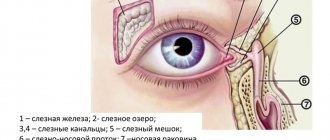
The lacrimal apparatus of the eye includes several links: the lacrimal gland, which is located in the upper part of the orbit from the outside, and the lacrimal ducts. The gland produces tear fluid and enters the upper fornix of the conjunctiva, washing the anterior surface of the eye. Next, the tear enters the lacrimal lake in the inner corner of the eye, where the openings of the lacrimal canaliculi open - there are two of them, upper and lower. The lacrimal canaliculi flow into the lacrimal sac, which passes into the nasolacrimal canal, which opens in the nasal cavity. Thus, the tear travels from the lacrimal gland to the nasal cavity.
Diseases of the lacrimal organs occur in a third of ophthalmic patients. The prevalence of inflammatory diseases out of the total number of pathologies is 12%. At any level, the lacrimal apparatus can be involved in the inflammatory process. Inflammatory diseases are most often of an infectious nature and include:
- Dacryoadenitis is an inflammation of the lacrimal gland. Inflammation of the lacrimal gland most often occurs as a complication of common infections ( scarlet fever , influenza , tonsillitis , typhoid fever , pneumonia , mumps ).
- Canaliculitis is inflammation of the tear ducts. Always occurs secondary to disease of the eyelids, lacrimal sac or conjunctiva. It occurs in acute and chronic forms. In chronic canaliculitis, either the superior or inferior tubule is affected, but unilateral damage to both tubules at once is common. Women aged 50-80 years are most often affected. According to the latest data, the condition of the lacrimal canaliculi is reflected in the production of tears and any inflammation affects its quantity - either in the direction of increase or decrease.
- Dacryocystitis is an inflammation of the lacrimal sac, which occurs when the outflow of tears is disrupted due to narrowing of the nasolacrimal duct. The tear fluid stagnates, and conditions are created for the proliferation of bacterial flora. Dacryocystitis often becomes chronic.
The pathological process of one part of the lacrimal apparatus causes changes in other parts. Therefore, combined pathology is more common than isolated lesions. Late diagnosis and inadequate treatment lead to chronicity of the process.
Prevention
The most effective method of preventing dacryoadenitis is to increase the body's defenses. To strengthen your immune system, you need to exercise regularly, walk in the fresh air, eat right, and give up bad habits. All this helps to significantly reduce the risk of developing infectious diseases that can cause acute dacryoadenitis. During the spread of seasonal diseases, it is necessary to wet clean the house more often, do not forget to wash your hands, and try not to be in places where there are a lot of people. You can also reduce the risk of pathology if you promptly identify and properly treat the main diseases - tuberculosis, syphilis, sarcoidosis.


