- What is maxillary sinusitis?
- Symptoms
- Complications
- Types and types of maxillary sinusitis
- How is maxillary sinusitis diagnosed?
- Treatment
- Etiotropic therapy
- Puncture
- "YaMiK"-catheterization
- Treatment of symptoms of maxillary sinusitis
- Surgical intervention
- Treatment at home
- Prevention
Maxillary sinusitis (from the Latin “sinus” and “inflammation”) is an inflammation of the mucous membrane of the corresponding sinuses. The causes of this disease are various: allergies, rhinitis, diseased teeth, inflammation of the tissues around the teeth, injuries. Treatment includes conservative procedures, home remedies, and surgery.
What is maxillary sinusitis?
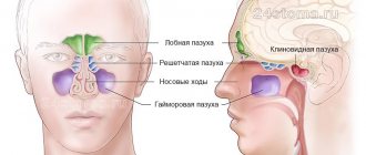
A person has several types of sinuses: frontal, sphenoid, maxillary and cells of the ethmoid labyrinth.
In each of these types, inflammation of the mucous membrane can occur. Then sinusitis will begin. Maxillary sinusitis is otherwise called sinusitis and is an inflammation of the paranasal sinuses. The maxillary sinuses were first illustrated by Leonardo da Vinci, and the disease itself was discovered by Nathaniel Highmore, a British surgeon and anatomist (he also described the maxillary sinuses in detail in his treatise of 1651). At that time, sinusitis was treated with home methods and heating.
Types of pathology
There are several types of maxillary sinus cysts:
- Odontogenic. The inflammatory process forms in the root system of an untreated dental unit located on the upper jaw. As the tumor increases in size, it destroys the bone and grows into the sinuses. The contents of the cyst are purulent.
- Retention. The reason for the development is dysfunction and obstruction of the glands responsible for the production of mucus.
False cysts are classified into a separate category. In such formations there are no epithelial cells. The appearance of such formations is due to a violation of the structure of the maxillofacial apparatus.
Types and types of maxillary sinusitis
Depending on the etiology (cause of occurrence) of the disease, rhinogenic, odontogenic, traumatic and allergic maxillary sinusitis are distinguished.
Rhinogenic sinusitis occurs against the background of rhinitis (when the nasal mucosa becomes inflamed). The mucous membrane is the main obstacle to infections. When bacteria gets on it, a runny nose or rhinitis develops. The causes of rhinitis are different: viruses, hyperthermia, allergies.
, decreased protective properties of the body, penetration of synthetic agents, influence of dry air, too long use of drugs with a vasodilator or vasoconstrictor effect.
Rhinitis is characterized by congestion, nasal discharge, impaired circulation in the nasal cavity, and the development of congestive blood phenomena. There are 4 types of rhinitis: allergic
, chronic, acute, vasomotor.
Odontogenic sinusitis occurs due to inflammation of the mucous membrane of the maxillary sinus due to infection from an unhealthy tooth, the tissues around it, and the communication formed between the sinus and the oral cavity after tooth extraction. Symptoms of this disease can be apathy, loss of appetite, headache, aching temples, discharge from the nose or ear, cough, runny nose and others. Depending on the course of the disease, different treatment methods are chosen: antibacterial therapy and washing of the maxillary sinuses, pumping out pus or surgery.
Injury to the maxillary sinus or jaw can also lead to sinusitis.
The cause of allergic sinusitis is the body's hypersensitivity to one of the irritants. The disease begins in the nasal cavity and then spreads to the maxillary sinuses. What usually serves as an allergen? This is pollen during the flowering period, fur and excrement of pets, dust mites, medicines, household chemicals, perfumes, cosmetics, chemicals, dirty city air.
According to the duration of the disease, sinusitis is divided into:
- acute (less than 3 months);
- recurrent acute (can repeat up to four times a year);
- chronic (more than 3 months);
- exacerbation of chronic sinusitis (adding new symptoms to existing ones).
According to the severity of symptoms, sinusitis is:
- lungs
- moderate severity
- heavy.
Symptoms of a maxillary sinus cyst
A cyst of the maxillary sinus does not have specific symptoms in the early stages; the first complaints appear when the tumor grows significantly (15 mm or more). The intensity of growth is an individual indicator for each patient, as is the severity of symptoms.
The main signs of a cystic formation in the maxillary sinus:
- Chronic nasal congestion on the affected side;
- Bursting sensations in the cheek area (closer to the eye);
- Persistent headaches, poorly controlled by analgesics;
- Accumulation of mucus at the back of the throat (especially in the morning);
- Intermittent mucous or clear nasal discharge;
- Frequent sinusitis with purulent discharge from the nasal passages;
- Pain when pressing on the causative tooth, redness and swelling of the gums;
- Facial asymmetry with pain in the anterior wall of the sinus.
Infection of the sinus by the odontogenic (that is, through the tooth) route most often develops in sinuses with a wide bottom and deep protrusions of the alveolar process into the jaw. The filling of the sinus with air is important - with moderate pneumatization, the risk of cyst formation is less.
During a diagnostic examination, the doctor identifies a hole in the bottom of the sinus. With an exacerbation of odontogenic sinusitis, sharp-smelling purulent masses are released from the nose, and upon rinsing, crumbly-granular white inclusions are found. An x-ray, as a rule, confirms the diagnosis of a maxillary sinus cyst, after which the doctor draws up a treatment plan.
Make an appointment
Types of operations
If a cyst of the maxillary sinus is detected, it will not be possible to limit oneself to therapeutic measures alone, but there is still a prospect of saving the tooth. The treatment of cysts is carried out by a dentist surgeon, who, based on the clinical picture and instrumental examination data, selects a surgical intervention technique:
- Cystectomy is an operation involving excision and curettage of the entire cyst cavity, followed by suturing;
- Cystotomy is an operation to excise the anterior wall of the tumor, and the doctor communicates the posterior wall with the oral cavity.
Indications for a specific technique are determined individually and depend on the type of cyst, the size of the neoplasm, and the number of teeth involved in the pathological process. Conservative treatment is possible only in the absence of obvious inflammation, it takes 3-4 months, but during this time the tumor can grow with the appearance of complications. That is why surgical opening of the sinus is considered the most reliable method of treating maxillary and other types of cysts.
Treatment of maxillary sinus cyst
Surgical activities carried out by a dentist in the clinic are aimed at:
- Relief of the inflammatory process;
- Elimination of the source of infection in the oral cavity;
- Removal of the root of a tooth with a cyst or extraction of the entire tooth;
- Sinus scraping, antiseptic treatment;
- Closing the communication between the oral cavity and the sinus;
- Creation of drainage for the outflow of liquid contents through the nasal passage.
First, the doctor determines which tooth caused the inflammatory process in the sinus, and the roots of several teeth may be affected. Using radiographs, the size of the sinus and the location of the cyst are analyzed, and the type of anesthesia is selected (usually local anesthesia with the latest generation of drugs). The surgeon will make every effort to save the tooth if possible. With shallow immersion into the cyst cavity, tooth-preserving operations (for example, root resection) show a good effect. When a tooth is immersed more than 1/3 of its length inside the tumor, it is recommended to remove it in order to prevent relapses of the disease.
The “Smile Factor” uses biocompatible modern materials to fill the cavity of the maxillary cyst, ensuring accelerated tissue regeneration. This tactic allows you to restore the jaw bone in a safe way; the prognosis for the intervention is favorable.
The protocol for working with the paranasal sinuses also includes a consultation with an ENT doctor. Treatment is considered successful when clinical symptoms of the disease disappear and there are no pathological changes in bone tissue on control radiographs.
Rehabilitation, features of care
After surgery, slight discomfort persists for 10-14 days. The turundas are removed from the nasal passages on the third day, and the sutures are removed after a week. Swelling, pain, difficulty breathing are temporary phenomena due to the specifics of the operation and do not require separate therapeutic measures. After removing the tampons from the nose, you should carefully follow the doctor’s instructions regarding rinsing the passages with antiseptics. Antibiotic therapy and instillation of vasoconstrictor drugs are prescribed.
During the first month after surgery it is recommended:
- Sneeze and cough gently with your mouth open;
- Follow a gentle diet with a predominance of soft foods;
- Limit intensive nose blowing;
- Avoid active facial movements;
- Postpone visiting the bathhouse, sauna, swimming pool;
- Do not go under water (even in the bathroom);
- Temporarily limit sports training;
- Sleep with the head of the bed raised on a comfortable pillow.
After treatment, you should visit the dental clinic once every 3-4 months throughout the year. During examinations, the attending physician monitors the progress of recovery, preventing relapses.
Possible complications
Treatment of a maxillary cyst should be carried out exclusively in a properly equipped clinic. Home measures to “resolve” the tumor are ineffective, and in many cases they contribute to the accelerated growth of the cyst due to improper actions by the patient.
Failure to contact a dental surgeon in a timely manner can result in serious consequences:
- Spread of the pathological process to healthy teeth followed by their loss;
- Penetration of infection into other air sinuses of the skull;
- Melting of the bone by purulent masses with the occurrence of osteomyelitis;
- Visual impairment, “double vision” due to compression of the eyeball by the cyst;
- Pronounced facial asymmetry with very large sinus tumors;
- Pathological fracture of the maxillary bone due to tissue thinning;
- Exhausting headaches, respiratory dysfunction, chronic malaise.
The most serious complication of a maxillary sinus cyst is considered to be the spread of inflammation to the membranes of the brain and the brain itself, which is extremely dangerous for the patient’s life.
Preventive actions
The following measures will help prevent the occurrence of a maxillary cyst and minimize the risk of complications with existing tumors:
- Sanitation of the oral cavity with competent treatment of teeth and gums;
- Timely treatment of ENT diseases (rhinitis, etc.);
- Correction of a deformed nasal septum;
- Treatment of allergic rhinitis;
- Early seeking medical help at the first symptoms of the disease.
Smile Factor doctors have modern techniques for treating jaw cysts of any size. Particular emphasis is placed on the painlessness and safety of the intervention, which is achieved by using proven drugs and consumables.
The patient receives competent advice during the rehabilitation period, so that recovery proceeds with maximum comfort. The exact price of treatment for a dental cyst is determined by the amount of work, type of surgery and other factors.
How is maxillary sinusitis diagnosed?
When diagnosing a disease, it is necessary to collect a number of indications: find out about complaints, write down symptoms, analyze the patient’s medical history and conduct an examination (computed tomography, x-ray).
Possible symptoms: headache, temporal pain, nasal and ear discharge, accumulation of mucus in the back of the throat, intoxication (in severe cases).
The medical history should contain information about past illnesses, injuries, hypothermia, etc.
The examination usually consists of palpation and percussion in the area of the paranasal sinuses, and also includes pharyngoscopy and rhinoscopy.
Puncture for sinusitis
If all conservative methods of treating sinusitis do not help, the ENT doctor will suggest a puncture of the maxillary sinus. This measure is necessary because pus accumulated in the sinus, as we already know, can lead to serious consequences, including inflammation of the brain.
During the procedure, the otolaryngologist releases the purulent contents of the sinuses and injects medication into the sinus. There is no need to be afraid of a puncture - before the procedure, anesthesia is performed: the ENT doctor inserts a cotton swab soaked in a lidocaine solution into the nasal passage of the patient sitting in a chair. It is completely safe and does not require patient preparation.
As soon as the anesthesia takes effect, the otolaryngologist, using a Kulikovsky needle, carefully inserts it into the sinus through the nasal cavity. Using a syringe, the purulent contents are sucked out. As soon as the purulent masses are completely removed, rinsing is carried out. The sinus should continue to be rinsed for several days after the procedure.
Treatment
Etiotropic therapy
Prescribing antibacterial agents (the main causative agents of acute sinusitis) helps to achieve rapid results in therapy:
- β-lactams: amoxicillin, clavulanate, cefaclor, cefuroxime axetil, sulbactam;
- fluoroquinolones: levofloxacin, gatifloxacin, moxifloxacin;
- macrolides: azithromycin, clarithromycin.
Puncture
This method is used if the disease cannot be cured with medication. It is the most famous method for removing pus from the maxillary sinuses. Quite painful, unlike other procedures.
"YaMiK"-catheterization
A fairly effective remedy in the fight against maxillary sinusitis, it does not cause complications. The procedure is quite painful, like puncture; patients cannot always tolerate it well.
Important: the operation is not advisable for isolated lesions of one paranasal sinus, since infection can be introduced into healthy paranasal sinuses.
Reasons for the development of sinusitis
Acute sinusitis can be caused by the following factors:
- colds;
- viral infections: ARVI, measles, influenza, etc.;
- allergic reactions;
- damage and injury to the nose;
- untreated teeth, tooth roots entering the cavity of the maxillary sinus, inflammation of the gums.
In medicine, it is customary to distinguish two ways of infection entering the maxillary sinuses: when the infection penetrates from the nasal mucosa into the maxillary sinus or when the infection develops directly in the maxillary sinus through blood flow and general inflammation.
Sinusitis (in addition to bacteria) can be provoked by factors that interfere with normal air circulation and the release of mucous masses from the sinuses. These include:
- deviated nasal septum;
- adenoids;
- cyst;
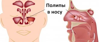
- polypous formations;
- and etc.
Unfavorable environmental conditions - dust, gas pollution, work in hazardous industries - can also disrupt the process of the release of mucous masses from the sinuses and further treatment.
Treatment of symptoms of maxillary sinusitis
Symptomatic treatment of sinusitis includes topical decongestants (such as xylometazoline and oxymetazoline) that improve sinus ventilation, mucolytics (such as carbocysteine) that improve mucus secretion, topical antiseptics (miramistin and others) and irrigation therapy (such as nasal douches). , before using which you need to use vasoconstrictor drugs), which flush out mucus and kill microbes in the sinus cavity, topical glucocorticosteroids, combined drugs (non-steroidal and anti-inflammatory drugs - paracetamol, ibuprofen).
Among the most effective drugs for treating the symptoms of sinusitis are
Sialor based on silver ions . It has an anti-inflammatory effect and prevents the proliferation of bacteria. Thanks to the mild action of the drug, the balance of microflora is maintained and favorable conditions are created for the regeneration of the nasal mucosa.
Surgical intervention
Drug treatments are not always effective; sometimes it is necessary to resort to surgery. At the same time, both approaches to diseased sinuses (extranasal, endonasal, combined), surgical technologies (magnifying devices and lighting devices), and surgical methods differ.
Important: after sinusitis, patients should be periodically (at least once every 3 months) observed by an otolaryngologist.
Treatment at home
An option for treating maxillary sinusitis at home is steam inhalation, which improves blood circulation, thins mucus accumulations, and also improves the flow of medications from the blood. But in acute cases of maxillary sinusitis, this method is dangerous; it can provoke generalization of the infectious process.
You can do inhalations with a decoction of herbs (calendula, celandine, bay leaf, string of sage, chamomile). Add alcohol tincture of propolis (1 teaspoon per 0.5 liter of decoction) and a couple of drops of iodine to boiling water.
In addition to such inhalations, you can also breathe over boiled potatoes. Important: you need to breathe for 10 minutes every day, the course is a week.
Warm the nose with table salt in a bag, boiled with an egg, and a blue lamp. Rinse the nose with the following solution: 1 teaspoon per glass, furatsilin (2 tablets), herbal decoctions (sage, celandine, chamomile). The procedure can be repeated up to 10 times a day.
Washing “cuckoo”: description of the method of treating sinusitis
The “cuckoo” method is a painless and, most importantly, effective procedure. Thanks to conservative treatment, purulent masses, mucous secretions along with pathogenic microorganisms are effectively washed out of the sinuses, the mucous membrane improves its function, nasal congestion decreases, and inflammation subsides. In some cases, thanks to Proetz lavage, puncture can be avoided. How is this procedure carried out?
The patient is positioned comfortably, lying on the couch, face up. The ENT doctor carefully pours an antiseptic into one nostril (Chlorhexidine, Furacilin, Miramistin, etc.). And at the same time, with the help of a special metal olive connected by a medical suction device, it sucks out this rinsing solution, but from the other nostril. The manipulation is repeated three times on each side, using a sterile plastic syringe with a volume of twenty ml. The entire procedure lasts about five minutes.

