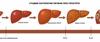
Congenital syphilis is one of the manifestations or clinical forms of syphilis.
A child becomes infected with Treponema pallidum during intrauterine development.
This form manifests itself from the prenatal period of the child’s development to the age of 12-18 years.
Congenital syphilis is extremely dangerous.
It can affect almost all connective tissues, the musculoskeletal system, the skin of epithelial tissues, mucous membranes, and tissues of internal organs.
It is especially dangerous when the damage spreads to the central nervous system (CNS).
Neurosyphilis can be fatal.
At the first sign of suspicion of congenital syphilis, various studies are carried out, the most common of which are:
- Examination of spinal fluid (CSF)
- Searching directly for pathogen cells in the blood
- PCR research
- Various serological studies
When treating congenital syphilis, various sets of measures are used.
Drug therapy includes the prescription of bismuth preparations, as well as medications aimed at strengthening and enhancing immunity.
Currently, there are two types of congenital syphilis:
- 1.Late congenital syphilis
- 2.Early congenital syphilis
Late manifestation of congenital syphilis
Occurs in children aged two years and older.
This type of congenital syphilis is most clearly observed in adolescents aged 12-15 years and is somewhat reminiscent of the clinical picture of the type of tertiary syphilis.
In children aged two or five years, the so-called secretive congenital syphilis occurs in a latent form.
Typically, late syphilis develops when early manifestations are not treated properly.
Or it was asymptomatic, i.e. secretly and therefore no appropriate treatment was prescribed.
If we talk about the clinical manifestations of late congenital syphilis, they have a number of specific symptoms:
- Reliable data accurately indicating the presence of late congenital syphilis
- Probable data that is 70% likely to indicate the presence of congenital syphilis in children. Complex signs, or clinical manifestations of other infectious diseases, similar to the symptoms of congenital syphilis, especially in children. They are not accurate indicators of the presence of syphilis, and only indicate confirmation of the disease.
- Dystrophic changes or stigmas.
Now let's talk about these clinical symptoms of congenital late syphilis in more detail.
Let us give an example of which of these manifestations are more accurate and which are not, and which of them carry more reliable information.
Characterized by dystrophic features of the teeth, hearing loss or so-called labyrinthine deafness, parenchymal keratitis.
Also, these data may include so-called tetrad diseases or specific gonitis, triad of congenital syphilis.
Bone damage
Syphilitic osteochondritis
Syphilitic osteochondritis (Wegener's osteochondritis), periostitis and isolated gummas are considered a constant symptom of early congenital syphilis. In children older than one year, osteochondritis is extremely rare, and after one and a half years of life it never occurs.
Osteochondritis develops from the fifth month of fetal development and is most often detected in the first three months of a child’s life. Osteochondritis is often the only manifestation of congenital syphilis. The disease is characterized by damage to long tubular bones (usually the upper limbs) at the border of the diaphysis and epiphysis, where there are disturbances in the formation of calcium and inhibition of the development of osteoblasts. Flat bones and phalanges are less commonly damaged. With the breakdown of the specific infiltrate, the epiphysis is separated from the diaphysis. In the second and third stages of syphilitic osteochondritis, pathological fractures may appear. The resulting pain becomes excruciating and occurs at the slightest movement of the child. This condition is called “Parrot’s pseudoparalysis.”
Under the influence of antisyphilitic treatment, the pathological process is suspended and does not subsequently affect bone growth.
Syphilitic periostitis
Together with osteochondritis, and sometimes independently with congenital syphilis, periostitis occurs - damage to the periosteum. Periostitis in the initial stage of its development remains unnoticed even on an x-ray. The first manifestations of periostitis can be noticed only with calcification of the periosteum. Periostitis is accompanied by pain in the area of long tubular bones—the sites of development of the pathological process.
Gummas in the bones
The gummous process in early congenital syphilis is rarely recorded. The ulna, tibia and flat bones are the most common sites for the development of syphilitic gummas. Gummas in bones are isolated, round-shaped lesions, single or multiple. They are located under the periosteum and often in the bone marrow. The periosteum in the gumma area thickens and becomes like a muff.
Syphilitic phalangitis (dactylitis)
With congenital syphilis, the phalanges of the upper extremities are most often affected. Couplings (thickenings of the periosteum) make the fingers look like barrels.
Rice. 8. Scheme of development of syphilitic osteochondritis. In the affected bone in the metaphysis, a rarefaction zone appears in the form of a white stripe (B). The pre-ossification zone has a jagged appearance.
Rice. 9. In the photo there is syphilitic osteochondritis (left) and periostitis (right).
Rice. 10. When bones are affected, even passive movements or careless touches cause severe suffering to the child.
Rice. 11. Damage to the skull bones with syphilis in children.
Possible signs of late congenital syphilis
Some of the most striking manifestations of late congenital syphilis are pathology of dental development, labyrinthine deafness and the manifestation of parenchymal keratitis.
These late manifestations of syphilis were first described by the famous surgeon, dermatologist, and ophthalmologist Jonathan Getchinson and A. Fournier.
The latter proposed calling such manifestations Hutchinson's triads.
He himself discovered the manifestations of late congenital syphilis, the symptom of tabes dorsalis.
Anomalies in congenital syphilis that appear directly on the teeth are described by scientists J. Getchinson, A. Fournier and Pfluger.
In their opinion, Treponema pallidum attacks the rudiments of teeth even during intrauterine development, thereby forming their further pathology in the future.
Each of their discoveries is fundamental:
- D. Getchinson described a special form of pathology of teeth, incisors, which are characterized by a moon-shaped notch. Although at that time two symptoms were needed to confirm his theory: parenchymal keratitis and labyrinthine deafness.
- Another scientist who studied this pathology, Pfluger, described that with the late development of congenital syphilis, damage to large molars or molars is characteristic. With this form of pathology, the tooth takes on a kidney-shaped appearance and significant underdevelopment of the tubercles (Carabelli tubercle) occurs.
Probable findings include changes in the structure of the skull (buttock-shaped skull), scars around the mouth (Robinson-Fournier), changes in the structure of the teeth, syphilitic chorioretinitis, damage to the central nervous system.
Changes in the structure of the legs are characteristic, namely the shins take on a saber-shaped appearance, becoming similar to a saber.
To recognize the likely signs of late congenital syphilis, the doctor must have extensive knowledge in this area.
After all, the symptoms of congenital syphilis are similar to the manifestations of other infectious diseases in their early development, but they do have some features, which will be discussed now.
Diffuse skin infiltration of Hochsinger
Diffuse thickening of the skin is a reliable sign of congenital syphilis. It is based on damage to small vessels. In the veins and arteries, perivascular infiltration of eosinophils, plasma and lymphoid cells is noted. The disease manifests itself at 8–10 weeks of a child’s life.
The palms and soles, the area around the lips and the chin are the most common places for syphilide to be localized. Somewhat less frequently, infiltration appears on the thighs, buttocks, around the anus, scrotum, labia, scalp, and on the skin of the elbows and knees. Initially, redness (erythema) occurs. Then the affected areas of the skin become denser, the folds are smoothed out, the lips thicken, swell and become yellowish-red in color, the red border of the lips becomes tense, and the skin on the soles and palms becomes “varnished” (“mirror soles”). When the process spreads to the scalp, hair loss is observed. When the process spreads to the area of the superciliary arches, hair loss on the eyebrows is noted.
Next, the affected surfaces peel off, macerate (tear) and become wet. Cracks occur at the slightest cry of the child and mechanical trauma, spread to the red border of the lips, bleed and quickly become covered with crusts. All elements of the damage contain a huge amount of pale treponema.
After 2 - 3 months, diffuse infiltration resolves even without treatment. At the site of inflammation, radial scars (Robinson-Fournier scars) remain in the corners of the mouth. Radial scars are a pathogonic symptom of congenital syphilis.
Rice. 6. In the photo, early congenital syphilis is a diffuse infiltration of the Gochsinger skin.
Damage to the lower leg areas (saber shins)
The pathology typically manifests itself in the child’s infancy.
It accounts for 70% of all pathologies associated with late syphilitic manifestations.
A large proportion of the damage occurs in the periosteum and tibia - osteoperiostitis, osteochondritis (damage to the cartilage itself involving a small area of the bone itself),
Due to the weight of the child, the bones simply bend forward.
The child grows and learns to walk, his legs resemble a saber blade.
The bones become thicker and longer.
At the same time, the child begins to sleep restlessly, as he is bothered by night pain in his legs.
In this case, damage to the bones of the forearm is not observed.
To confirm this diagnosis, an x-ray examination is necessary.
In addition to the saber-shaped manifestation of syphilis, there is a similar symptom with Paget's disease or rickets, when the bones bend outward.
Syphilitic drives
Initially, the lesion occurs on one joint, and after some time the lesion occurs on the second.
There is usually no increase in temperature, and there is practically no pain.
Despite the fact that the joints significantly increase in volume, the skin over them does not change color and remains physiological in color.
But in the joints themselves, or rather in their cavities, a significant amount of fluid accumulates, which can sometimes be purulent in nature.
It is during this course of the disease that the Wasserman reaction (WRR) always gives a positive result.
Another manifestation of syphilis is a saddle nose.
Usually manifests itself in cases where syphilitic rhinitis has been suffered in the past.
There is a change in the structure of the nose itself, the nostrils begin to protrude forward when the nose itself sinks inside the skull.
Due to changes in its mucous membrane, the manifestation of the so-called goat and lorgnette nose is observed.
In the very first months of life, the so-called buttock-shaped skull can form.
When the flat bones of the skull itself are damaged, due to osteoperiostitis and periostitis, respectively, the frontal lobes, namely their right and left tubercles, begin to protrude forward.
At the same time, they are separated by a small groove, reminiscent of a child’s buttock (hence the name).
It is during this period of a child’s life that manifestations of mental retardation due to congenital syphilis begin.
This should not be confused with the disease hydrocephalus of newborns, in which the entire skull increases in size.
The share of these manifestations of late congenital syphilis in children is 20%.
Here, small scars appear on the chin, frontal area, around the lips, including the corners of the mouth.
If a child suffers pyoderma, candidiasis or a burn in this area in childhood, the scars remain forever and do not heal.
The cause of this manifestation of the disease is usually diffuse papular infiltration of Hochsinger.
Complex data include mainly dystrophies and stigmas.
Characteristic changes in the structure of the little fingers of the hands.
They become short and thick, with no xiphoid process.
Thickening occurs at the end of the clavicle and other pathologies change the structure of the body during late congenital syphilis.
In most cases, complex data do not include lesions that directly involve the Treponema pallidum itself.
This is mainly due to pathologies that arose due to gross violations by future parents during pregnancy or when conceiving a child (dystrophy).
For example, if parents are alcoholic or if a child is conceived while drunk.
This may also be due to various intoxications or any infectious diseases.
Fetal syphilis
Treponema pallidum begins to penetrate into the fetal body with the development of placental circulation - from the fifth month of pregnancy. Diffuse inflammatory infiltration with subsequent proliferation of connective tissue leads to damage to internal organs. They become denser and increase in size.
The dead fetus remains in the amniotic fluid for 3 to 4 days before birth. During this time, its skin becomes saturated with liquid and swells (maceration). Stillborns have a small weight, they have damage to all vital organs, which contain a large number of pale treponema.
Signs of fetal syphilis
- Damage to the placenta leads to malnutrition of the fetus, which causes its death.
- The liver and spleen are sharply enlarged. They form miliary syphilomas.
- Often the cause of fetal death is “white pneumonia”, in which there is nested or diffuse small-cell interstitial infiltration, abundant desquamation of the epithelium, its fatty degeneration and filling of the alveoli with it, and proliferation of cells in the interalveolar space. The affected lungs lose their airiness, become heavy (sink in water), and have a grayish-white color when cut.
- Damage to the renal cortex is noted, where diffuse pinpoint infiltration also develops.
- In the submucosal layer of the stomach and intestines, flat infiltrates develop, some of them ulcerate.
- The heart is rarely affected.
- Of the endocrine glands, the adrenal glands are most often affected, and a little less often - the pancreas, gonads and pituitary gland.
- The vessels of the central nervous system are affected. Inflammation of the soft and arachnoid membranes of the spinal cord and brain (leptomeningitis), meningoencephalitis and ependymatitis develops. The medulla oblongata is often affected.
- There is intrauterine damage to the skeletal system in the form of osteochondritis and osteoperiostitis - reliable signs of fetal syphilis. Pathology is detected using an x-ray examination performed at 5 - 6 months of intrauterine development. A specific process develops in the growth zones of tubular bones.
- The skin of the fetus is damaged. The epidermis loosens, erodes and peels off in layers. Underdevelopment of subcutaneous fatty tissue leads to the skin becoming wrinkled and gathering in folds, especially on the face (“old man’s face”).
Rice. 3. The photo shows a child with syphilis. Underdevelopment of subcutaneous fatty tissue leads to the skin becoming wrinkled and gathering in folds, especially on the face (“old man’s face”).
Early congenital syphilis
Now let's look directly at the clinic of early congenital syphilis.
According to the XXIX World Health Assembly, it is divided into several periods:
- 1. Early congenital syphilis, which is characterized by the manifestation of syphilitic symptoms. In this case, symptoms can appear either as those that first appeared in children under the age of two years, or as those that already occur before this age:
- Syphilitic pathologies of the skin and mucous membranes
- Syphilitic symptom associated with visceral lesions of the internal organs
- Syphilitic manifestations such as laryngitis, pharyngitis, pneumonia, etc.
- 2. The clinical syphilitic picture does not manifest itself in any way, but the serological reaction gives positive results. Repeated examinations of cerebrospinal fluid do not give a positive effect.
- 3. Positive results of early syphilis can be judged only on the basis of histological and bacteriological examinations.
Syphilitic rhinitis
Syphilitic rhinitis most often appears immediately after birth or in the first month of a child’s life. The disease develops as a result of diffuse infiltration of leukocytes and round cell elements of the anterior part of the nasal mucosa, as a result of which it swells. Foul-smelling, purulent-bloody nasal discharge appears, and when it dries, massive crusts form. Swelling of the mucous membrane and dense crusts significantly complicate breathing, which is why the baby cannot breastfeed. Nosebleeds are noted.
Over time, the ulcerative process spreads to the cartilage and nasal bones. The nasal septum undergoes necrosis. The osteochondral skeleton is deformed and the nose takes on the shape of a saddle.
Sometimes diffuse infiltration affects the mucous membrane of the larynx. Developed ulcerative laryngitis is manifested by hoarseness of voice. The destruction of cartilage leads to organ stenosis.
Rice. 7. Syphilitic rhinitis and diffuse skin infiltration are symptoms of congenital syphilis.
What happens during the period of early congenital syphilis in the child’s body
According to research by many scientists, early congenital syphilis begins to form in the cells of the fetus.
Such a violation in the chromosomal apparatus leads to a genetic shift in development.
Depending on which gene chain was disrupted, different degrees of syphilitic manifestation are distinguished:
- Before the child is conceived
- Blastopathy – damage to the embryo during blastogenesis (formation of the embryo)
- Syphilitic embryopathy - damage to the fetus from four to five months of pregnancy
Syphilis of the brain
With syphilis of the brain, all vessels of the brain are damaged, with the capture of its mesodermal membranes.
It is this group that is characterized by the manifestation of meningism.
It occurs in the form of severe headaches, irritability, affective effects and constantly changing and fluctuating moods.
Hearing damage is also typical; deformation of the optic nerve occurs (deformation of the pupils), etc.
When diagnosing the Wasserman reaction (WRR), it is sharply positive, even when it is repeated.
Damage to the central nervous system
Hydrocephalus (dropsy of the brain) and chronic leptomeningitis (inflammation of the membranes of the spinal cord and brain, including the arachnoid and soft membranes) are reliable signs of congenital syphilis.
Hydrocephalus (excessive accumulation of cerebrospinal fluid in the ventricles of the brain) with congenital syphilis develops as a result of inflammation of the pia mater. More often, children are born with hydrocephalus; less often, hydrocephalus develops in the third month of a child’s life. The disease can have an acute or chronic course. With hydrocephalus, all sizes of the skull increase, it takes on an elongated shape, the parietal and frontal tubercles increase and protrude, the fontanel is tense, there is a divergence of the sutures, the eyeballs are protruded and displaced downward. There is an increased number of lymphocytes and protein in the cerebrospinal fluid.
In leptomeningitis, the soft meninges and vessel walls are thickened (infiltrated with fibroblasts, lymphoid and plasma cells). When the meninges are irritated, the child screams “for no reason.” Convulsions, paresis and strabismus often occur. Stiff neck, restlessness, short-term convulsions, paralysis and uneven pupils are the main symptoms of meningitis in syphilis.
Inflammation affects the substance of the brain, proliferation of glia and the formation of glial nodules are noted, and blood vessels become sclerotic.
Paresis and paralysis are the main signs of syphilitic meningoencephalitis. Sometimes asymptomatic syphilitic meningitis is recorded, the only sign of which is changes in the cerebrospinal fluid.
Rice. 12. Enlarged liver and spleen are a sign of congenital syphilis.
Diagnosis of congenital syphilis
It is worth noting that congenital syphilis in 90% of cases can manifest itself in the form of blisters or so-called syphilitic pemphigus and ulcers.
Diagnosis can be made on the basis of biomaterial taken from these blisters and the detection of pale treponema in them.
However, this syphilitic manifestation may not always “show itself.”
Therefore, this type of diagnosis is not of particular value.
To confirm the diagnosis of congenital syphilis, they resort to the so-called lumbar puncture.
The cerebrospinal fluid is taken and examined using a microscope.
But if congenital syphilis has a hidden course, this method may not be successful.
Serological examination methods occupy a special place in congenital syphilis.
Namely, a blood test using the Wasserman method, now MRP, which can give false positive results in 50% of cases.
When examining for congenital syphilis, studies such as RIF, RIBT, RPGA (highly specific, highly sensitive reactions to syphilis) are important.
With congenital syphilis, almost 98% accuracy is obtained by examining the blood plus scraping the skin elements of the patient.
If there is any doubt about the presence of congenital syphilis, a diagnosis can be made using a complex of serological reactions (CSR).
It includes the complement bundle of RSC with Treponema pallidum antigens.
This type of analysis today is called ELISA and RPGA in combination with MRP (blood microreaction to syphilis).
Despite all its advantages, it can be a false negative if the collection or preparation for blood collection was incorrect.
If you have congenital syphilis or suspect it, you should contact more than just one venereologist.
The examination may involve a number of specialists.
Since the damage occurs not only from the skin, but also from internal organs, bones, brain (pulmonologist, surgeon, traumatologist, general practitioner, nephrologist, hepatologist, neurologist, etc.)
EARLY CONGENITAL SYPHILIS: CLINIC, PATHOMORPHOLOGY, DIAGNOSIS, TREATMENT, PREVENTION
In connection with the increasing incidence of congenital syphilis in Russia, issues of its diagnosis, treatment and prevention are being discussed. Many forgotten pathomorphological data are presented. The article includes descriptions of problematic cases observed by the authors.
Due to the fact that there is an increase in the incidence of congenital syphilis in Russia, the paper discusses its diagnosis, treatment, and prevention. Many forgotten pathomorphological data are given. The paper includes accounts of the difficult cases observed by the authors.
K.K.
Borisenko - Dr. honey. Sciences, Professor, Head. Department of Syphilidology of the Central Research Dermatovenerological Institute, Moscow. OK. Loseva - Doctor of Medicine. Sciences, senior researcher of the same department, the same institute. O.V. Share - Candidate of Medical Sciences Sciences, researcher at the same department, at the same institute. E.L. Tumanova - Candidate of Medical Sciences Sciences, Associate Professor, Department of Pathological Anatomy, Faculty of Pediatrics, Russian State Medical University, Moscow. KK Borisenko - prof., MD, Head, Department of Syphilisology, Central Research Dermatovenereological Institute, Moscow OK Loseva - MD, Senior Researcher, the same Department, the same Institute OV Dolya - Candidate of Medical Sciences, Researcher, the same Department, the same Institute Ye.L.
Tumanova — Candidate of Medical Sciences, Associate Professor, Department of Pathological Anatomy, Pediatric Faculty, Russian State Medical University, Moscow В
Surveys on the diagnosis, treatment and prevention of congenital syphilis have now acquired particular relevance. Since 1990, the incidence of syphilis in the adult population has gradually increased, and in the last two years its growth has become epidemic. This could not but be followed by an increase in the incidence of congenital syphilis. According to the statistics department of the Ministry of Health of the Russian Federation, in 1992, 31 cases of congenital syphilis were registered in Russia, in 1993 - 46, in 1994 - 118, in 1995 - 221, in 1996 - 469, in 1997 - 714. Today there are three main problems associated with congenital syphilis: prevention, diagnosis and optimization of treatment methods. The basic principles for the prevention of congenital syphilis were proposed in our country back in the 20s by M.G. Mgebrov, G.I. Meshchersky, M.M. Reitz. In subsequent years they underwent changes and additions. Currently, scientifically based comprehensive measures are being carried out for antenatal and postnatal prevention of congenital syphilis. Antenatal prevention consists primarily of timely detection of the disease in a pregnant woman and its adequate treatment. The mandatory three-time serological examination of pregnant women adopted in our country - in the first and second half of pregnancy, as well as upon admission to the maternity hospital - is fully justified. Serological examination of pregnant women in the first and third trimesters of pregnancy is also recognized abroad as necessary [1].
| Gonorrhea. Typical purulent discharge from the urethra | Gonococcal proctitis (photo through a proctoscope) | Gonorrhea. Purulent discharge characteristic of gonococcal cervicitis. |
| Non-gonococcal urethritis. The discharge is more watery than with gonorrhea. | Trichomoniasis vulvovaginitis. | |
| Primary syphilis. Solid tanker. | ||
| Congenital syphilis. Hutchinson's teeth. | Congenital syphilis. Saddle nose. | |
However, at present, these measures are not carried out in some pregnant women due to a number of reasons: instability of social, economic and interpersonal relationships; a sharp increase in migration processes; growth of semi-legal prostitution; early onset of sexual activity, etc. [2]. All this leads to the fact that some pregnant women are not registered with the antenatal clinic and are not examined before birth. That is why children with congenital syphilis are more often born to young mothers, women leading an asocial lifestyle, planning to abandon the child and therefore not registered with the antenatal clinic.
Rice. 1. Classic “senile” appearance of a newborn; malnutrition, hydrocephalus.
Features of the clinical picture
, as well as the influence of syphilitic infection on the course and outcome of pregnancy, a significant number of studies conducted in our country, mainly in the 30–50s, are devoted. It is known that syphilitic infection has a toxic effect on the fetus, being a common cause of miscarriages, premature births, and stillbirths. Syphilis is characterized by late miscarriages and stillbirths in the 6–7th month of pregnancy. Miscarriages at earlier stages are less common, which is confirmed by numerous statistical data from both domestic and foreign authors [3–5]. It is believed that up to 89% of children born to mothers with untreated or inadequately treated syphilis die in utero or shortly after birth [6].
Rice. 2. Syphilitic pemphigus.
It is noted that currently the course and outcome of pregnancy with syphilis are characterized by the predominance of latent forms of the disease, a significant reduction in the frequency of late miscarriages, premature births and stillbirths [7]. Regarding pregnancy outcomes with seroresistance, literature data are contradictory. In women with a persistently positive Wasserman reaction who have received full treatment, there is still a significant number of spontaneous abortions, stillbirths, premature births, and there are also cases of congenital syphilis [8]. Other authors, having monitored pregnancy outcomes in women with persistently positive CSR and examined their children, came to the conclusion that such women can be guaranteed the birth of healthy offspring if they received antisyphilitic treatment during pregnancy [9].
Rice. 3. Massive fibrosis at the porta hepatis, inflammatory infiltrates of plasma cells and lymphocytes. Hematoxylin and eosin staining. Uv. 250.
Rice. 4. Kidney vessel. Fibrinoid necrosis of the vessel wall. Diffuse plasmatic infiltration. Hematoxylin and eosin staining. Uv. 160.
Rice. 5. Diffuse infiltration of the soft meninges by plasma cells. Severe vascular congestion. Hematoxylin and eosin staining. Uv. 63.
Rice. 6. Thymus gland. In the lobules there is no division into the cortical and medulla layers. Proliferation of interlobular connective tissue with diffuse plasmatic infiltrates. Hematoxylin and eosin staining. Uv. 63.
Rice. 7. Pancreas. Diffuse proliferation of connective tissue that walls up the islets of Langerhans. Hematoxylin and eosin staining. Uv. 160.
Rice. 8. Small intestine. Flattening of the villi. In the submucosa and villi there are infiltrates consisting of plasma cells and histiocytes. Hematoxylin and eosin staining. Uv. 160.
Rice. 9. Large intestine. No lint. In the submucosa and villi there are infiltrates consisting of plasma cells and histiocytes. Hematoxylin and eosin staining. Uv. 160.
It is generally accepted that the only way of infection with congenital syphilis is transmission of infection to the fetus by a mother with syphilis through the placenta [10]. It is known that Treponema pallidum does not penetrate the placenta and does not infect the fetus until the 4th month of pregnancy [3]. It is believed that before 16 weeks the fetus is not mature enough to react to the antigen by producing antibodies [11]. The pathogenesis of congenital syphilis depends largely on the immune response of the fetus and, to a lesser extent, on the cytodestructive effect of Treponema pallidum. That is why adequate treatment of syphilis in the mother before 16–19 weeks of pregnancy usually prevents damage to the fetus. For specific treatment
pregnant women are currently using penicillin preparations of varying degrees of duration in accordance with current instructions for the treatment and prevention of syphilis.
However, as can be seen from the example below, treatment, in particular with benzathine-penicillin preparations, carried out in the second half of pregnancy, may not provide the prevention of congenital syphilis. Patient P., 37 years old, was admitted for delivery on May 13, 1996. For latent early seropositive syphilis, from January 29, 1996 (at 25–26 weeks of pregnancy) she received specific and preventive treatment with benzathine penicillin at a dose of 2.4 million ED intramuscularly once a week, 6 injections in total. KSR dated January 29, 1996, 3+ 4 +3+, KSR dated April 2, 1996, neg. 2+ neg. On May 14, 1996, she gave birth to a boy weighing 3,040 g, height 52 cm. The general condition of the child after childbirth was moderate: chin tremor, muscular dystonia, tendency toward muscle hypertonia, liver enlarged by 1.5 cm, spleen at the edge of the costal arch, skin and visible mucous membranes are free from specific rashes.
The child was prescribed preventive treatment for early congenital syphilis. On the 1st day after the start of therapy, an exacerbation reaction was observed: temperature 37.9°C, papular rashes on the skin of the anterior abdominal wall, buttocks and extremities, and copious mucous discharge from the nasal passages. CSR dated May 14, 1996, 1+2+1+, against the background of ongoing therapy, these manifestations of the disease resolved on the 4th – 5th day. Diagnosis: early congenital manifest syphilis. The described case is not the only one in our practice. We recommend using durant penicillin preparations for the specific treatment of pregnant women only until the 18th week of pregnancy. After this period, specific and preventive treatment should be carried out with soluble penicillin or penicillin preparations of medium duration (procaine salt of penicillin, procaine penicillin), which penetrate the placenta in a higher concentration. In difficult conditions of increasing incidence, the role of early clinical and laboratory diagnosis of syphilis increases [12]. Recognition of congenital syphilis in the presence of pronounced clinical manifestations does not present any particular difficulties. Early congenital syphilis, according to the international classification, is usually called an intrauterine infection that manifests itself in a child under 2 years of age.
Features of the clinical picture make it possible to distinguish congenital syphilis in infancy - up to one year [10].
It most often appears in the first 2 months of life and is characterized by a number of typical symptoms. In “classical” cases, a newborn (often premature) has a peculiar “senile appearance”, primarily due to severe malnutrition (Fig. 1). The skin is wrinkled, flabby, dirty yellow. The head is enlarged, with a noticeably pronounced subcutaneous venous network, the skin is often covered with seborrheic scales. Children gain weight very slowly, develop poorly, and often cannot suck. They are restless, anxious, sleep poorly, cry almost constantly, and sometimes emit a sharp, piercing cry (Sisto's symptom). A similar condition in children with congenital syphilis is quite rare these days. However, with manifest forms of early congenital syphilis, a number of other characteristic signs are revealed. Skin changes
in children in the first months of life are characterized by unusual forms that are not found in acquired syphilis.
The earliest of them is syphilitic pemphigus (Fig. 2), which can be detected at birth or appear in the first days of life. Blisters with serous-purulent, sometimes bloody contents are most often localized on the palms and soles, and occasionally on other areas of the skin. At the base of the blisters there is a specific infiltrate, so the blisters are surrounded by a reddish-purple rim. They quickly burst, revealing an erosive surface surrounded by fragments of the epidermis. The second “special” symptom is diffuse infiltration of the skin, which most often occurs by the end of the 2nd month of life. It is preceded by diffuse erythema of the affected areas. Infiltration also appears primarily on the palms and soles; in addition, the face (often around the mouth), scalp, buttocks, thighs, and skin on the elbows and knees are also affected. The changed skin is thickened, its surface is shiny, and deep cracks easily form, especially around the mouth. In addition to the described skin changes for early congenital syphilis, specific rhinitis
is pathognomonic, which is detected already at birth: first there is a significant swelling of the mucous membrane, making breathing difficult, then a discharge from the nose appears - mucous, purulent and, finally, bloody, later profuse purulent-bloody fetid discharge is observed from the nose, nosebleeds.
Rhinitis is accompanied by deep ulcerations of the mucous membrane, damage to cartilage and bones. Wegener's osteochondritis
is quite specific - a lesion of long tubular bones at the border of the epiphysis and diaphysis - which is caused by impaired calcium formation and inhibition of the development of osteoblasts.
The second and third stages of osteochondritis (Parro's pseudoparalysis) occur only with congenital syphilis and are diagnosed by radiography of the tubular bones of the forearm and lower leg. In addition to the listed symptoms, other manifestations of the disease may be observed - from the skin (roseolous, papular rash), the central nervous system (convulsions, causeless screaming, meningeal symptoms, hydrocephalus), the organ of vision (chorioretinitis, lesions of the optic nerve), internal organs (hepatosplenomegaly ). Pathomorphological changes in congenital syphilis
are quite typical, but the rarity of such cases in the last 30 years has led to the fact that a qualified pathologist’s conclusion in the event of a child’s death cannot be obtained everywhere.
This makes it urgent to improve the professional level of pathologists and conduct training for specialists in this profile. Taking these circumstances into account, we dwell in some detail on the pathomorphological changes in congenital syphilis. Pathomorphological changes in the skin are manifested by damage to capillaries and small vessels in the form of productive endarteritis and phlebitis. In the early stages of the process, intimal cell proliferation occurs in the vessels, which often leads to narrowing and obliteration of the vessel lumen. At the same time, infiltrative changes in the walls of blood vessels occur, when the membrane itself and perivascular tissue are infiltrated with lymphoid and plasma cells, eosinophils. Such infiltrates are often localized around the sweat glands. In the skin itself, its papillary layer, significantly pronounced swelling, hyperemia, and infiltration of the above cells are found. The skin of the palms and soles is especially characterized by diffuse infiltration of plasmatic and lymphoid cells. Giant cells are rare. Syphilitic rhinitis is based on diffuse infiltration of the anterior part of the nasal mucosa with round cell elements and leukocytes. The process is accompanied by hyperplasia of the mucous membrane and subsequently spreads to cartilage and nasal bones, which leads to deformation of the osteochondral skeleton. Damage to internal organs in congenital syphilis is characterized by an infiltrative-productive process, and occasionally by the presence of gummous formations. According to various authors, syphilitic changes in 100% of cases are found in the liver and spleen, in 94% in the vessels, in 85% in the adrenal glands, the pancreas, kidneys, and bones are very often affected. Changes in the liver
are associated with the fact that the spirochete enters the fetal body through the umbilical vein.
Macroscopically, the liver is enlarged, very dense, yellow-brown or whitish-brown in color - the so-called “flint liver”. Histological changes in the liver are divided into three types. In the first, cellular infiltration of the capsule and periportal connective tissue, the walls of the hepatic veins and bile ducts is determined. The infiltrate consists of histiocytes, plasma and lymphoid cells, eosinophilic leukocytes. There is a slight proliferation of Kupffer cells. The second type of changes includes thickening and infiltration of the Glissonian capsule, proliferation of connective tissue in the lobules, and expansion of the portal tracts. Infiltration of periportal connective tissue is weak. The infiltrative process is sometimes concentrated around the bile ducts, where rings of proliferating connective tissue (syphilitic pericholangitis) and branches of the portal vein are formed (syphilitic periphlebitis; Fig. 3). In rare cases, the liver is reduced in size, dense, with a fine-grained surface. Microscopically, signs of portal cirrhosis are revealed. The connective tissue is infiltrated by lymphoid cells with an admixture of plasma cells, histiocytes and eosinophils. The third type of changes includes cases with the presence of miliary gummas (5% of sectional material). Macroscopically, gummas are defined as grayish dots. Microscopically, small foci of necrosis are detected, as well as diffuse inflammatory infiltrates in the growing connective tissue [13]. to the pancreas
is characterized by a sharp proliferation of intercellular tissue, which penetrates between the acini, ducts and islets.
The gland pattern may be completely erased. Due to the growth of connective tissue, the consistency of the gland sometimes reaches almost cartilaginous density [14]. The spleen
in early congenital syphilis is dense, its weight increases 10 times or more.
Fibrinous deposits on the capsule are often detected (syphilitic perisplenitis). Macroscopically, the thickening of the capsule is determined with the imposition of fibrin on it, sometimes the capsule is hyalinized. Usually the spleen is depleted of cellular elements, sometimes the light centers are sharply hyperplastic. Intrafollicular arteries and larger vessels are thickened with symptoms of productive endarteritis and obliteration of the lumen. The next place in frequency is vascular lesions.
Histological changes are represented by obliterating endarteritis.
These changes are the cause of ischemic necrosis. In the venous vessels there is infiltration of the wall with lymphoid elements with an admixture of plasma cells, neutrophils, and eosinophils (Fig. 4). Often the veins have a thickened adventitia. The vessels of the liver, myocardium, spleen, and kidneys are affected [13]. the kidneys
are also often affected .
The lesion is inflammatory in nature. Focal or diffuse interstitial nephritis occurs. Small round cell infiltration around the glomeruli and vessels and the proliferation of interstitial connective tissue with similar infiltration are specific [13]. Bone damage
in the form of syphilitic osteochondritis is considered one of the permanent manifestations of congenital syphilis.
However, many authors indicate that the presence and severity of Wegener's osteochondritis is variable. Macroscopically, there are 3 degrees of severity of the process. I degree - an uneven, winding strip of gray-yellow color with a reddish tint, 1 - 2 mm wide; II degree – the strip width increases to 5 mm; III degree – the diaphysis is separated from the epiphysis. Osteochondritis is observed in flat bones and phalanges, but more often in tubular bones. With treatment, osteochondritis quickly disappears and does not affect further bone growth. In recent years, grade III osteochondritis has practically never occurred [15]. Syphilitic periostitis can occur as an independent lesion of the periosteum or in combination with osteochondritis. The lungs
are rarely affected.
There is syphilitic, so-called white, Virchow's pneumonia. The process involves a lobe, sometimes the entire lung. Macroscopically, the lung is dense, airless, light gray in color. Microscopically, thickening of the interalveolar septa, peribronchial, and perivascular tissue, which are infiltrated with lymphoid, plasma, and epithelioid cells, is observed. In the lumen of the alveoli there is exudate consisting of neutrophils and alveolar macrophages. In the vessels, growth of the intima and narrowing of the lumen are noted. Damage to the central nervous system
in children with congenital syphilis is characterized by internal hydrocephalus and chronic syphilitic leptomeningitis.
With leptomeningitis, thickening of the soft meninges is observed, mainly at the base of the brain, extending to the cerebral hemispheres. Histological examination of the pia mater reveals infiltration of lymphoid, histiocytic, plasma cells, and fibroblasts (Fig. 5). In the walls of the vessels of the membranes, the same infiltration is observed, the lumen of the vessels is narrowed to the point of obliteration. The inflammatory process along the vessels spreads to the substance of the brain. Pathomorphological changes in syphilitic encephalitis are manifested by sclerosis of blood vessels with infiltration of their walls by plasma and lymphoid cells, proliferation of glia, formation of glial nodules, and perivascular infiltrates. Of the endocrine glands, the adrenal glands
.
Microscopically, capsule fibrosis is detected, the zona glomerulosa is destroyed in the cortex, and the zona fasciculata becomes fibrotic [16]. In the thymus gland,
there is a proliferation of interlobular connective tissue infiltrated with lymphohistoplasmocytes.
The lobules are small, they lack division into the cortical and medulla layers (Fig. 6). The number of lymphocytes is reduced in both layers. Hassall's corpuscles are small, some of them layered, located in both the cortical and medulla layers. Heart disease
with early congenital syphilis is very rare.
There is no convincing evidence of syphilitic intestinal lesions in infants [15]. Since congenital syphilis is transmitted to the fetus through the placental route, the placenta
is often involved in the pathological process. However, many authors indicate that with obvious syphilis in the mother and fetus, changes in the placenta may be absent. When the placenta is damaged, its weight increases 2–3 times. Macroscopically, the placenta is fleshy, with large lobules of grayish-yellow or pinkish color. Microscopically, villous hyperplasia is observed; villous sclerosis is often combined with vascular sclerosis and obliteration of their lumen. Thrombosis of the intervillous spaces, inflammatory infiltrates from round cell elements and leukocytes in the membranes are not specific to congenital syphilis. Treponemas are found very rarely in the placenta. It should be noted that the examination and visual assessment of the placenta is usually not given due attention, while the description of the placenta in congenital syphilis has diagnostic value. In the context of a syphilis epidemic, obstetrician-gynecologists who deliver babies and do not yet know the results of a serological examination of the mother should more carefully examine and describe the placenta. This is important for both clinical and pathological diagnosis of congenital syphilis. We present two cases of early congenital syphilis that resulted in death.
One child (A) died during the intrapartum period, the second (B) lived for 90 hours. The mothers were 27 and 20 years old, respectively; they were practically not examined during pregnancy and were not registered with the antenatal clinic. Upon admission to the maternity hospital, they underwent a DCS study, and positive results were obtained after delivery. During pregnancy, one woman was diagnosed with colpitis and vaginal condylomatosis, for which she was treated with Tsiprolet. Child A. was born at the 32nd week of pregnancy, birth weight 2250 g. Child B. was born at the 29th week of pregnancy, birth weight 1270 g. In both cases, polyhydramnios was noted from 1.5 to 2.0 l , the waters are cloudy and light brown in color. Child B. had an Apgar score of 4/4 points at birth; from the 5th minute of life he was intubated and transferred to artificial ventilation. At birth, a mild roseola rash was noted on the chest and abdomen, which disappeared on the 2nd day of life. From the 3rd day of life, the child released a large amount of light yellow sputum through the endotracheal tube. Bradycardia developed. There was an admixture of greenery in the stool. A general blood test revealed anemia and leukocytosis with a shift to the left.
The pathological examination revealed the following changes. Macroscopic examination. The skin of both children was slightly jaundiced and acrocyanosis was pronounced. The ossification nucleus of the femur is absent in both. Child B. has a small amount of dark liquid blood in the umbilical vein, and in the Arancia duct there is a red thrombus of dense consistency attached to the wall of the vessel. In both children, examination of the brain revealed expansion of the posterior horns of the lateral ventricles, and the ependyma was smooth and shiny. In one child, a softening focus measuring 1.0 x 0.5 cm was found in the periventricular zone, in the occipital region. In both children, macroscopic changes were noted in the lungs. The lungs are dense, completely occupy the pleural cavities; sections reveal multiple small and merging foci of light gray color. When examining the heart, fibromyxomatous growths on the tricuspid valve leaflets were discovered in one child. In both children, the liver is dense, its mass is increased by 1.5 - 2 times; in one child, a gray lesion with a hemorrhagic rim was found in the left lobe. The mass of the spleen was increased 5–6 times; perisplenitis was observed in one child. The pancreas in both children is dense, especially in the head area. In one child, a Wegener's strip was detected in the femurs. Microscopic examination. In the liver, vacuolar degeneration of hepatocytes, cholestasis, inflammatory infiltrates consisting of histiocytes, plasma cells and lymphoid cells, which are located along the portal tracts, penetrating into the lobules. Fibrosis is pronounced, especially at the porta hepatis. The liver capsule is thickened due to sclerosis and inflammatory infiltration. In the pancreas there is a diffuse proliferation of connective tissue with the accumulation of infiltrates in it, consisting of plasma cells, histiocytes and lymphocytes (Fig. 7). The lungs in both cases are immature - the bronchi are located under the pleura. Interstitial pneumonia is expressed with thickening of the interalveolar septa due to their infiltration by lymphocytes and plasma cells. Perivascular and peribronchial focal plasmacytic infiltrates are noted. In the adrenal glands - fibrosis of the capsule, disappearance of the glomerular and fibrosis of the zona fasciculata. In the substance of the brain there are perivascular infiltrates of plasma cells and mononuclear cells, the walls of the vessels are sclerotic. Diffuse gliosis of the brain substance is pronounced. The soft meninges are sclerotic, and there are plasma-histiocytic infiltrates. In both cases, damage to the cardiovascular system is noted. Perivascular sclerosis, as well as histioplasmacytic infiltrates with an admixture of lymphocytes, are observed in the Arancia duct and the vessels of all internal organs. Interstitial myocarditis was detected in the heart. In both cases, the small and large intestines were affected: there was a sharp flattening of the villi, sclerosis of the vessels of the submucosal membrane with the accumulation of plasma-histiocytic infiltrates in it (Fig. 8, 9).
Thus, as a result of a morphological study, we identified some features of pathomorphological changes in various organs (damages of the intestine, pancreas, cardiovascular system, thymus), which are poorly described in the literature.
It should be noted that congenital syphilis is most severe in premature infants, as well as in children with early clinical manifestations (from the 1st week of life); There is a high mortality rate in this group. Diagnosis of congenital syphilis,
compatible with life, or occurring in a latent or asymptomatic form, has its own characteristics.
| Currently, congenital syphilis is usually diagnosed on the basis of positive serological reactions (KSR, RIF, RIT), the presence of skin manifestations, bone changes (osteochondritis, periostitis), initial changes in the choroid of the eye (chorioretinitis), rhinitis, enlarged liver and spleen, pathology cerebrospinal fluid. |
Diagnosis of early latent congenital syphilis is difficult during 3 months of life, when it is difficult to determine whether the disease occurs in the child or transplacental transfer of antibodies from the mother. In these cases, it is necessary to take into account the mother’s medical history (the stage of syphilis, which characterizes how long ago the child was infected; incomplete, late treatment of the mother or its absence); the degree of positivity of serological reactions in the child in comparison with the results in the mother (more pronounced positivity in the child indicates his illness); positivity of serological tests for IgM (domestic test systems of this series are still unreliable, imported ones are at the testing stage). It is necessary to emphasize the importance of timely dynamic examination of the child, starting from the 1st month of life, otherwise the diagnosis of latent congenital syphilis may be late. Let's give an example.
Patient V., 27 years old.
She was admitted with a child aged 6 months on 03/04/96 for a clinical and serological examination. For early latent seropositive syphilis, detected on May 5, 1994, the woman received specific treatment with retarpen at a dose of 2.4 million units intramuscularly once a week, a total of 4 injections. Before the onset of this pregnancy, there was a persistent tendency towards negative CSR (DSR from 06.94: 2+3+ e/m 4+; DSR from 12.94: + neg. e/m 2+) – this was not observed further. During pregnancy, she did not receive preventive treatment and was not registered with the antenatal clinic. Weakly positive DSR results remained (DSR dated 04.10.95: e/m 3+ during labor). On 10/04/95 she gave birth to a boy weighing 3.200 g, length 52 cm. During examination of the woman on 03/04/96, seroresistance was determined (KSR dated 03/04/96 neg. 2+ e/m 4+, RIF 4+/3+, RIT 70%) and additional treatment was prescribed. The child was not examined after birth. Upon admission to the hospital in a child: CSR dated 03/04/96 2+ 2+ e/m 3+, RIF abs 4+, RIT 65%.
Objectively: the skin of the torso, limbs, scalp, visible mucous membranes are free from specific rashes. The liver is at the edge of the costal arch, the spleen is not enlarged. For organs and systems without pathology. The general condition of the child is satisfactory. Based on the positivity of serological reactions and test results of the mother, the child was diagnosed with early latent seropositive syphilis. Specific treatment with penicillin was carried out at the rate of 100,000 units per 1 kg of body weight 6 times a day for 28 days. Diagnosis of early congenital syphilis can be difficult in the presence of nonspecific clinical symptoms (enlarged liver, spleen, postnatal encephalopathy), when it is necessary to differentiate latent and manifest congenital syphilis. In these cases, in addition to the above factors, the therapeutic effect of the specific treatment is also of diagnostic value. In such situations, a somewhat “delayed” diagnosis is possible, taking into account the dynamics of clinical and serological data. Serological diagnostic methods
congenital syphilis are represented by non-treponemal and treponemal tests.
Abroad, to diagnose early congenital syphilis, the following serological examination is carried out: • RPGA [17]; • VDRL [18]; • RIFabs. In children with positive VDRL, RPGA and RIFabs, a 19S – IgM – RIFabs test is performed to confirm the diagnosis and decide on the need for treatment [19]. Recently, enzyme-linked immunosorbent assay (ELISA) has become one of the most common methods in various fields of medicine.
One of the reasons for the widespread use of ELISA for diagnostic purposes is the possibility of test automation [8].
ELISA refers to treponemal specific tests and is designed to detect specific antibodies in the blood serum and cerebrospinal fluid of patients with syphilis, as well as to differentiate false-positive results obtained in standard serological tests for syphilis [20]. Theoretically, antibodies against IgM are detected in the serum of a newborn, provided that Treponema pallidum is present in his tissues [21, 22]. According to various authors, IgM antibodies are detected in the patient’s body within 2 weeks after infection. Since, due to their large molecular weight, these antibodies do not penetrate the normal placenta from mother to fetus, the formation of treponeme-specific IgM antibodies in the child’s blood indicates the presence of infection in the newborn [17]. Methods for specific and preventive treatment of children
are constantly being improved. According to the current instructions for the treatment and prevention of syphilis, preventive treatment for children born to mothers with syphilis, poorly or insufficiently treated, is carried out with penicillin preparations for 2–4 weeks
Treatment of congenital syphilis, reasons for treatment failures
Syphilis is a very dangerous disease that has a very negative effect on an unborn or already born baby.
Therefore, all children must be preventively protected from this dangerous and formidable disease.
If suddenly this disease nevertheless overtakes your child, by order of the WHO there is a certain treatment regimen for congenital syphilis.
Attention! All recommendations and treatment regimens should be taken only after consultation with your doctor. This scheme is recommended for educational purposes only and is not intended to guide you and apply it in 100% of cases.
- 1. In most cases, penicillin, included in subgroup G (water-soluble), is used, 100,000-150,000 IU / kg / day (administer 50,000 IU / kg intravenously every 12 hours during the first seven days of life and then every nine hours). If this treatment was interrupted, the course is repeated again from the beginning.
- 2. The child is also prescribed benzathine penicillin G 50,000 units/kg intramuscularly once.
In general, treatment includes a whole series and complex of measures.
The doctor will tell you about them, who will prescribe the correct regimen, on which the effectiveness of the treatment itself will depend if you are suddenly overtaken by this disease.